Clinical Experience with Simultaneous MR-PET Acquisition: Developing Optimal Protocols for Anatomically Focused and Whole-Body Examinations
←
→
Page content transcription
If your browser does not render page correctly, please read the page content below
Clinical Body Imaging
Clinical Experience with Simultaneous
MR-PET Acquisition: Developing Optimal
Protocols for Anatomically Focused and
Whole-Body Examinations
Kathryn Fowler, M.D.1; Farrokh Dehdashti, M.D.1; Tammie L.S. Benzinger, M.D., Ph.D.1; Michelle Miller-Thomas, M.D.1;
Jonathan McConathy, M.D., Ph.D.1; Matthew Parsons, M.D.1; Vilaas Shetty, M.D.1; Constantine Raptis, M.D.1;
Perry Grigsby, M.D.1; Pamela K. Woodard, M.D.1; Richard Laforest, Ph.D.1; Robert J. Gropler, M.D.1; Vamsi Narra, M.D.1;
Barry A. Siegel, M.D.1; John Kotyk, Ph.D.1; Agus Priatna, Ph.D.2; Robert McKinstry, M.D., Ph.D.1
1
Mallinckrodt Institute of Radiology, Washington University School of Medicine, St Louis, MO, USA
2
R&D Collaborations, Siemens Healthcare, St Louis, MO, USA
1A 1B 1C
PET-CT
R L R L R L
1
1D 1E 1F
MR-PET
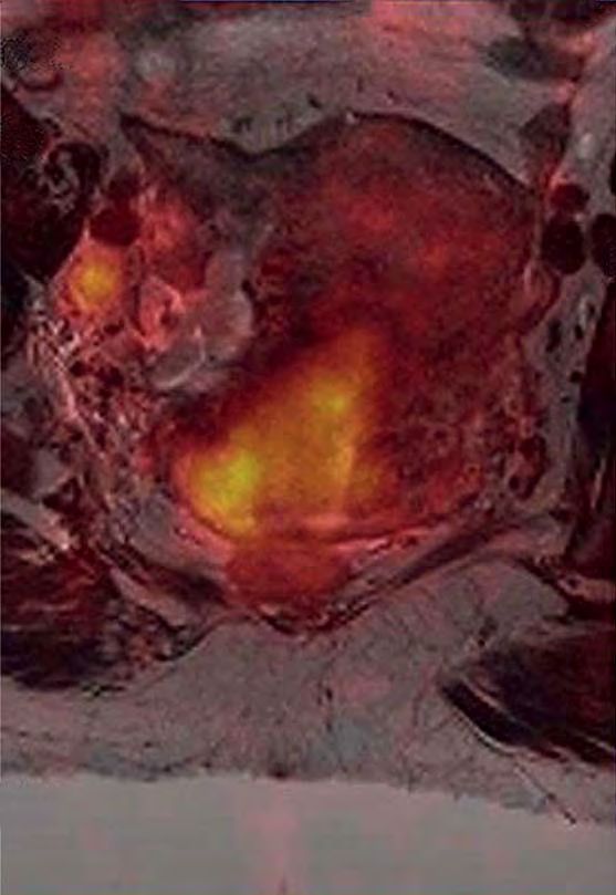
1 65-year-old male with a 1-year history of memory loss and episodic acute confusion; brain MRI demonstrated possible bilateral temporal
lobe hyper intensity on FLAIR, without associated volume loss. FDG PET recommended for further evaluation. (1A–C) conventional FDG
PET/CT shown: (1A) low-dose CT for attenuation correction, (1B) FDG PET, (1C) image fusion; (1D–F) simultaneous MR/PET of the same
patient: (1D) FLAIR, (1E) FDG PET data, (1F) overlay of metabolic information.
6 MAGNETOM Flash · 1/2012 · www.siemens.com/magnetom-world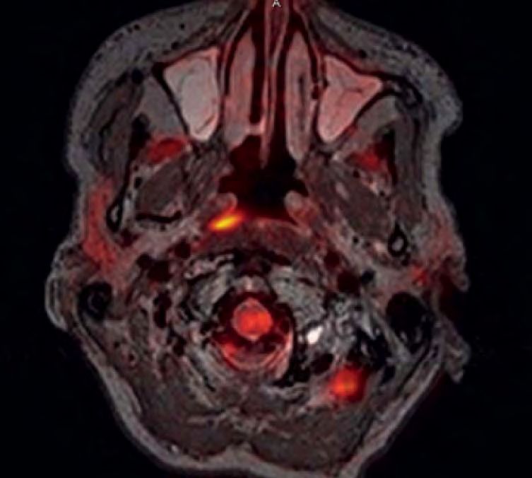
Body Imaging Clinical

                                              2A

Introduction
The new Biograph mMR offers simulta-
neous acquisition of PET and MR imag-
ing data of potential added benefit in
oncologic, neurological and other medi-
cal imaging [1–3]. The inherent benefit
of the simultaneous acquisition is
improved registration allowing optimal
localization of PET findings to anatomic
imaging and shortened overall imaging
times through acquisition of PET and
MRI in a single session. The purpose of
this article is to present initial clinical
experience with development of neuro-
logical protocols, anatomically focused
MR-PET imaging protocols of body
organs, including pelvic, thoracic, and
liver oncologic imaging, and cardiac
imaging experience. Initial experience,
validation with PET-CT, challenges, and
insights into protocol development
are presented through representative
examples.
                                              2B

Methods
Following IRB approval, patients sched-
uled for standard of care clinical FDG-
PET-CT were recruited and consented for
additional MR-PET imaging. The simul-
taneous MR-PET imaging was acquired
on a Biograph mMR system recently
installed in the Center for Clinical Imag-
ing Research (CCIR) at Washington Uni-
versity School of Medicine, with total
imaging matrix, with attenuation body
array and spine matrix coils. By incorpo-
rating Avalanche Photodiodes (APDs)
into the bore of a 3T magnet, the Bio-
graph mMR system fully integrates the
acquisition of state-of-the-art PET and
MR images. Attenuation correction
imaging was performed utilizing a dual
echo VIBE Dixon sequence that sepa-
rates water and fat with TE1/TE2 1.23
msec / 2.46 msec, TR 3.6 msec, left-right
field-of-view (FOV) 500 mm and ante-
rior-posterior FOV 300 mm. The acquisi-
tions were performed in either a single
station or a multiple station mode as
indicated. Depending on the applica-            2 Poorly differentiated nasopharyngeal carcinoma with bilateral cervical
                                               nodal (and liver) metastases is shown. Simultaneous MR-PET demonstrated
tions, PET images were simultaneously
                                               excellent MR image quality and excellent localization of FDG uptake.
acquired with the anatomical sequences         (2A) FDG PET overplayed on T1w (2B) corresponding PET/CT exam.

                                                         MAGNETOM Flash · 1/2012 · www.siemens.com/magnetom-world          7
3A                                                3B                                                3C

Clinical Abdominal Imaging

3D                                                3E                                                3F

  3A–F Patient with metastatic lung cancer; primary tumor is present in the left lower lobe (see arrow in 3H). Metastatic disease is shown in
 the mediastinum (lymph nodes), spine, and bony pelvis (osseous metastases). MR images include a STIR acquisition as well as diffusion-
 weighted imaging. Sagittal reconstructions: (3A) DWI, (3B) PET, (3C) MR/PET fused to DWI; coronal reconstructions: (3D) DWI, (3E) PET, (3F)
 MR/PET fused to DWI.
Body Imaging Clinical
3G 3H 3I
3G–I (3G) Coronal T2w HASTE for morphology, (3H) corresponding PET MPR, (3I) image overlay of PET and HASTE.
from MR such as HASTE for whole-body trated by this case. A 65-year-old male Head and neck oncology
acquisition, MPRAGE for brain imaging, with a 1-year history of memory loss Simultaneous head and neck MR-PET
SPACE or HASTE for pelvic application, and episodic confusion presented for performs well compared with PET-CT in
or delayed enhancement for cardiac workup at our institution (Fig. 1). A clini- our initial experience. In the head and
imaging. Additional examinations using cal MRI was interpreted as normal; how- neck, MR-PET combines the metabolic
high-resolution MR were added for the ever, there was a question of subtle and biochemical information from PET
focused examination such as high reso- FLAIR hyperintensity in the mesial tem- with high spatial resolution, anatomic
lution T2 TSE, diffusion-weighted imag- poral lobes. FDG-PET was recommended localization, and soft tissue contrast
ing or diffusion tensor imaging (DTI) and for further correlation of the MR findings from MR. The advantages of MR imaging
other sequences depending on the and clinical presentation. The PET acqui- in oral cavity and skull base neoplasms,
applications. sition was acquired simultaneously with the superior ability of MR to detect peri-
MRI. Brain MRI shows subtle bilateral neural spread of tumor, and improved
Clinical cases mesial temporal lobe hyperintensity on coregistration of PET imaging with MR
The following are some examples of the FLAIR. FDG-PET shows mild bilateral imaging during simultaneous acquisition
clinical cases acquired for anatomically mesial temporal lobe hypometabolism. make this a promising new technology
focused and whole-body examinations Further clinical workup was then for staging and follow-up of select head
with the Biograph mMR. ordered which generated a final diagno- and neck neoplasms.
sis of limbic encephalitis with voltage- Patient is a 51-year-old woman with
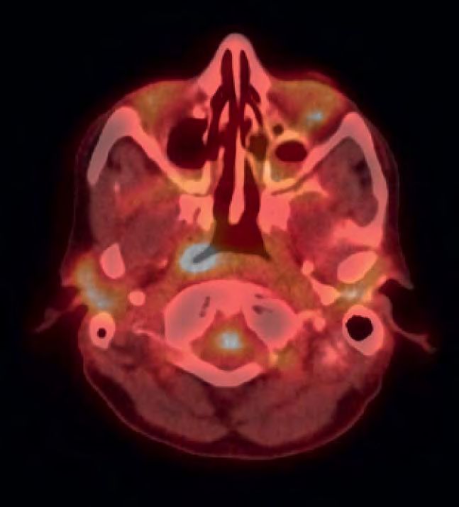
Brain imaging gated potassium channel antibodies poorly differentiated nasopharyngeal
Although fusion of separately acquired (VGKC). In this case, subtle findings on carcinoma with bilateral cervical nodal
brain MR and FDG-PET is readily avail- PET and MR performed independently, and liver metastases. Simultaneous MR-
able with offline software tools, a com- could be simultaneously reviewed and PET demonstrated excellent MR image
bined examination can allow for stream- confirmed with PET/MR, leading to quality and excellent localization of FDG
lined patient care and potentially diagnostic confidence and initiation of uptake. MR provided superior soft tissue
improved diagnostic specificity, as illus- the correct pathologic workup. resolution in the nasopharynx compared
MAGNETOM Flash · 1/2012 · www.siemens.com/magnetom-world 9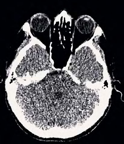
4A
Clinical Abdominal Imaging
4A Simultane- to PET-CT. Lesion detection based on
ous acquisition FDG uptake on the MR-PET was equiva-
of ECG-gated lent to PET-CT in this case (Fig. 2).
PET and
delayed con-
trast enhanced Lung cancer with metastatic disease
(DCE) cardiac While MRI can be used in the assessment
MR images of lung cancer, it has typically been
(simultaneous
reserved for specific cases in which CT
acquisition of
MR 2-point and PET-CT, the first-line modalities for
Dixon also the assessment of lung cancer patients,
acquired for are not able to answer a specific ques-
AC). PET data tion, such as whether a lesion is invad-
acquired in list
ing the chest wall or a vascular struc-
mode and
binned. DCE ture. One of the key reasons for the
MR images secondary role for MRI is the fact that
4B
acquired in whole-body MR imaging is cumbersome
diastole are and time consuming, thus preventing
fused with dia-
stolic PET data
assessment of the full extent of disease.
to create the MR-PET for lung cancer has the potential
center image. to foster the development of new imag-
Patient has a ing protocols that allow for detailed
normal heart.
assessment of the primary tumor while
(4A) MRI, (4B)
fused MR/PET, still providing the information regarding
(4C) FDG PET. more distant metastatic disease. In addi-
tion, simultaneously acquired diffusion
MR and PET data may prove useful both
in the initial evaluation of tumors as well
as in follow up after treatment.
Figure 3 shows a 50-year-old man with
metastatic lung cancer. Primary lesion is
present in the left lower lobe. Metastatic
disease is shown in the mediastinal
lymph nodes, spine, and bony pelvis.
MR images include a HASTE acquisition
4C as well as diffusion-weighted image
obtained at a b-value of 600.
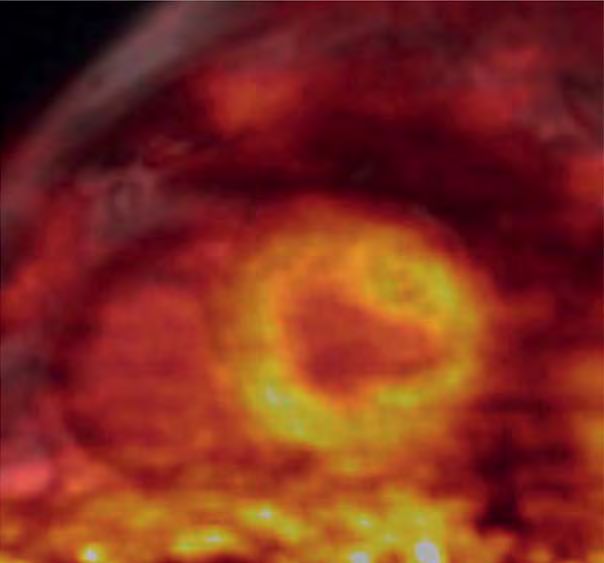
Cardiac imaging
Cardiac MR-PET imaging has the poten-
tial to play a role both in cardiac isch-
emia and viability assessment. Potential
clinical protocols include stable chest
pain assessment, playing on the
strengths of both modalities, performing
MR cine cardiac function assessment,
13
N-ammonia or rubidium-82 PET myo-
cardial perfusion and delayed contrast-
enhanced inversion recovery infarct
imaging in a single examination. Com-
bined FDG and delayed contrast-
enhanced inversion recovery cardiac
10 MAGNETOM Flash · 1/2012 · www.siemens.com/magnetom-world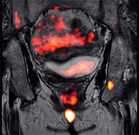
Body Imaging Clinical
imaging may play a role in co-localized, 5
simultaneously acquired functional
and anatomic viability assessment that,
theoretically, could play a role in imag-
ing-directed ventricular tachycardia
radiofrequency ablation or direct biven-
tricular pacing in dyssynchrony.
A 68-year-old man injected with FDG for
oncologic imaging was recruited imme-
diately after his whole-body PET-CT
examination to undergo a cardiac MR-
PET imaging for protocol development
(Fig. 4). Simultaneous acquisition of
ECG-gated PET and delayed contrast-
enhanced (DCE) cardiac MR images
(simultaneous acquisition of MR 2-point
Dixon also acquired for AC) allow for
precise fusion of imaging. PET data was
acquired in list mode, binned and recon-
structed into 3 phases. DCE MR images
acquired in diastole are fused with
diastolic PET data to create the center
image (Fig. 4B). This patient has a nor-
mal heart.
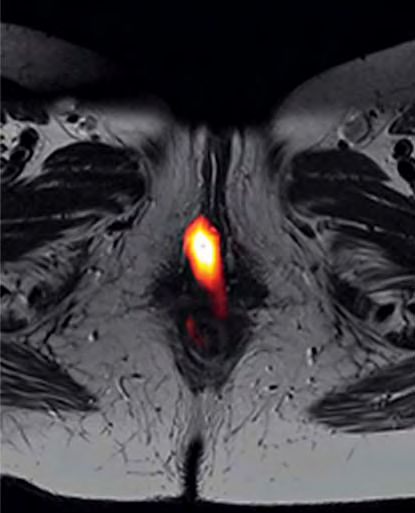
Cervical and vulvar cancers
PET-CT and MRI are established modali-
ties in initial staging and monitoring
treatment response in patients with cer-
vical cancer and other pelvic malignan-
cies. PET can provide estimated tumor
volumes for treatment planning and
diagnosis of nodal metastases. High res-
olution MR imaging of the pelvis can
detect parametrial spread of tumor, an
essential feature in determining surgical
5 Primary cervical cancer and involved adjacent right external iliac lymph
resectability. The combination of meta- node (arrow); overlay of PET on T2 TSE is shown.
bolic information derived from PET with
high resolution MR imaging of the pelvis
shows promise in both clinical manage-
ment and potential research opportuni-
ties in correlating functional MRI with interpolated breath-held examination vical malignancy and presence of meta-
tumor metabolism. (VIBE), and inversion recovery T2 fat bolically active nodal metastases, the
Given the complex anatomy of the pel- suppressed images complete a diagnos- patient subsequently underwent radia-
vis, high resolution T2-weighted imag- tic MR examination of the pelvis provid- tion therapy.
ing is best performed utilizing a 3D iso- ing additional staging information Figure 6 is a 31-year-old woman with
tropic dataset, such as SPACE. Isotropic regarding local invasion, tumor size, vulvar carcinoma. A small focus of
acquisition allows for infinite multipla- and regional metastases. residual tumor within the pelvis could be
nar reformations without loss of resolu- Figure 5 shows a 47-year-old woman missed on MR imaging alone and is in
tion. Diffusion-weighted imaging, pre- with biopsy-proven cervical cancer. a difficult location with PET alone given
and dynamic post-contrast volumetric Given the large size of the patient’s cer- the potential for contamination in this
MAGNETOM Flash · 1/2012 · www.siemens.com/magnetom-world 11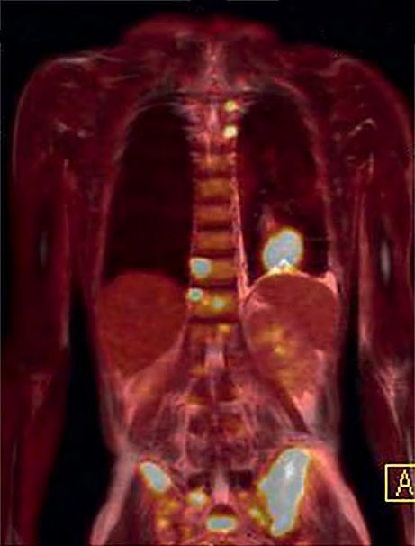
Clinical Body Imaging

6A                                                                        6B

   6 Patient with vulvar cancer; small focus of residual tumor within the pelvis could be missed on MR imaging alone. FDG PET overlay on
  transversal (6A) and coronal (6B) T2w TSE is shown.

                                                                                                        References
location from urine activity. The combi-          values may prove more specific in
                                                                                                      1 Pichler BJ, Judenhofer MS, Wehrl HF. Eur Radiol.
nation of MR-PET; however, nicely dem-            differentiating tumor from surrounding                2008; 18:1077-86.
onstrates the metabolically active soft           tissue than subjective assessment alone.            2 Antoch G, Bockisch A. Eur J Nucl Med Mol Imag.
tissue lesion.                                    Potential for benefit from simultaneous               2009; 36 Suppl 1:S113-20.
                                                  MR-PET acquisition also exists in recep-            3 Wehrl HF, Sauter AW et al. Technol Cancer Res
Discussion                                        tor-targeted oncologic imaging, demen-
                                                                                                        Treat. 2010;9:5-20.

MR-PET shows promise as a new onco-               tia assessment, and cardiac and athero-
logic imaging modality with inherent              sclerosis imaging.
                                                                                                        Contact
improved soft tissue contrast over CT,
lower radiation dose, and potential for           Acknowledgement                                       Professor Pamela K. Woodard, M.D.
                                                                                                        Washington University
better correlation of PET findings to             Jennifer Frye, Glenn Foster, Linda                    Mallinckrodt Institute of Radiology
anatomy given the simultaneous acqui-             Becker, Deb Hewing, Mike Harrod, Tim                  510 S. Kingshighway Blvd.
sition. Several challenges are evident in         Street, Betsy Thomas                                  St. Louis, MO, 63110-1076
                                                                                                        USA
developing optimal protocols, including
                                                                                                        Phone +1 314-362-7697
optimal MR sequence parameters,                                                                         woodardp@mir.wustl.edu
motion correction, and validation of
semiquantitative analysis using stan-
dardized uptake value (SUV) using MR
attenuation correction. In some cases,
the combination of PET SUV and MR ADC

12 MAGNETOM Flash · 1/2012 · www.siemens.com/magnetom-world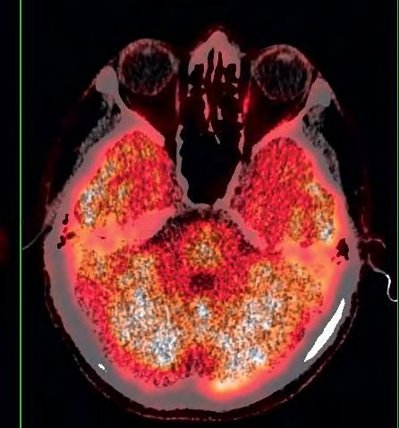
You can also read