Photoacoustic Imaging of Tattoo Inks: Phantom and Clinical Evaluation - MDPI
←
→
Page content transcription
If your browser does not render page correctly, please read the page content below
applied
           sciences
Communication
Photoacoustic Imaging of Tattoo Inks: Phantom and
Clinical Evaluation
Eftekhar Rajab Bolookat 1 , Laurie J. Rich 1,† , Gyorgy Paragh 2 , Oscar R. Colegio 2 ,
Anurag K. Singh 3 and Mukund Seshadri 1,4, *
 1    Laboratory for Translational Imaging, Center for Oral Oncology, Roswell Park Comprehensive Cancer
      Center, Buffalo, NY 14263, USA; Eftekhar.RajabBolookat@roswellpark.org (E.R.B.);
      Laurie.Rich@pennmedicine.upenn.edu (L.J.R.)
 2    Department of Dermatology, Roswell Park Comprehensive Cancer Center, Buffalo, NY 14263, USA;
      Gyorgy.Paragh@roswellpark.org (G.P.); Oscar.Colegio@roswellpark.org (O.R.C.)
 3    Department of Radiation Medicine, Roswell Park Comprehensive Cancer Center, Buffalo, NY 14263, USA;
      Anurag.Singh@roswellpark.org
 4    Department of Dentistry and Maxillofacial Prosthetics, Roswell Park Comprehensive Cancer Center, Buffalo,
      NY 14263, USA
 *    Correspondence: Mukund.Seshadri@roswellpark.org; Tel.: +1-716-845-1552
 †    Present affiliation: Center for Magnetic Resonance and Optical Imaging, Department of Radiology,
      University of Pennsylvania, PA 19104, USA.

 Received: 3 January 2020; Accepted: 31 January 2020; Published: 4 February 2020

 Abstract: Photoacoustic imaging (PAI) is a novel hybrid imaging modality that provides excellent
 optical contrast with the spatial resolution of ultrasound in vivo. The method is widely being
 investigated in the clinical setting for diagnostic applications in dermatology. In this report, we
 illustrate the utility of PAI as a non-invasive tool for imaging tattoos. Ten different samples of
 commercially available tattoo inks were examined for their optoacoustic properties in vitro. In vivo
 PAI of an intradermal tattoo on the wrist was performed in a healthy human volunteer. Black/gray,
 green, violet, and blue colored pigments provided higher levels of PA signal compared to white,
 orange, red, and yellow pigments in vitro. PAI provided excellent contrast and enabled accurate
 delineation of the extent of the tattoo in the dermis. Our results reveal the photoacoustic properties of
 tattoo inks and demonstrate the potential clinical utility of PAI for intradermal imaging of tattoos.
 PAI may be useful as a clinical adjunct for objective preoperative evaluation of tattoos and potentially
 to guide/monitor laser-based tattoo removal procedures.

 Keywords: photoacoustic imaging; tattoo; dermatology; ultrasound

1. Introduction
      Non-invasive in vivo imaging has been an integral part of the armamentarium in dermatology for
decades [1,2]. In addition to traditional dermoscopy and ultrasound (US), optical imaging methods such
as Raman spectroscopy, confocal microscopy, and optical coherence tomography have demonstrated
utility in the diagnosis of skin diseases [3–5]. Photoacoustic imaging (PAI) or optoacoustic imaging is
a relatively new imaging method that provides excellent optical contrast with the spatial resolution
of US [6,7]. Although administration of exogenous agents can enhance contrast on PAI, this is not
essential given the abundance of endogenous chromophores such as melanin and hemoglobin in
tissue. Clinical studies have demonstrated the usefulness of PAI as a label-free imaging tool for
assessment of melanomas and non-melanoma skin cancers [8–10] and to visualize microvascular and
inflammatory changes in the skin [11]. However, the ability of PAI to visualize tattoos has not been
previously reported. Since tattoo dyes are known to absorb and reflect light, we hypothesized that

Appl. Sci. 2020, 10, 1024; doi:10.3390/app10031024                                 www.mdpi.com/journal/applsci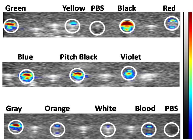
Appl. Sci. 2020, 10, 1024 2 of 5
Appl. Sci. 2020, 10, x FOR PEER REVIEW 2 of 5
PAI canpigments.
tattoo visualize andTo determine the three-dimensional
test this hypothesis, localizationthe
we first examined of tattoo pigments.
optoacoustic To test this
properties of
commercially available tattoo inks in a tissue-mimicking phantom. Subsequently, we examined thea
hypothesis, we first examined the optoacoustic properties of commercially available tattoo inks in
tissue-mimicking
ability phantom.
of PAI to visualize anSubsequently, we examined
intradermal tattoo the ability
in a healthy of PAI to visualize an intradermal
volunteer.
tattoo in a healthy volunteer.
2. Materials and Methods
2. Materials and Methods
In vitro examination of the photoacoustic properties of the inks was performed using hollow
In vitro
channels examination
created within a of the photoacoustic
tissue-mimicking properties
phantom of the of
composed inks was performed
agarose using hollow
(Bio Rad, Hercules, CA,
channels created within a tissue-mimicking phantom composed of agarose (Bio
USA) and intralipid (Sigma, St. Louis, MO, USA) . The synthesis of the phantom has been previously Rad, Hercules, CA,
USA) and intralipid
described [12]. Ten (Sigma,
samplesSt.ofLouis, MO, USA).
commercially The synthesis
available (Scream of the
Ink;phantom
Worldwide has been
Tattoopreviously
Supply)
described [12]. Ten samples of commercially available (Scream Ink; Worldwide Tattoo
tattoo inks (black, pitch black, blue, gray, green, violet, orange, red, yellow, and white) (Figure Supply) tattoo
1a)
were used. The samples were diluted in phosphate buffered saline (1:1) and injected intoused.
inks (black, pitch black, blue, gray, green, violet, orange, red, yellow, and white) (Figure 1a) were the
The samples
channels. wereblood
Whole diluted in phosphate buffered saline
andphosphate-buffered saline(1:1)
(PBSand injected
)were into the channels.
also injected Whole
into separate blood
channels
andphosphate-buffered saline (PBS) were also injected into separate channels for
for comparison (Figure 1b). The PA images were obtained using a 21 MHz probe with the followingcomparison (Figure 1b).
The PA images were obtained using a 21 MHz probe with the following settings:
settings: 2D multi-wavelength PA mode: 680–900 nm; Gain: 40 dB, Depth: 20.00 mm, Width: 23.04 2D multi-wavelength
PA mode:
mm, 680–9003.nm; Gain: 40 dB, Depth: 20.00 mm, Width: 23.04 mm, Persistence: 3.
Persistence:
(a)
(b)
Figure 1. (a)
Figure 1. (a) Photograph
Photographshowing
showingthethe
tenten commercially
commercially available
available tattoo
tattoo ink samples
ink samples evaluated
evaluated in
in vitro.
vitro.
(b) Set(b)
up Set up for evaluation
for evaluation of photoacoustic
of photoacoustic propertiesproperties of tattoo
of tattoo inks using inks using
hollow hollow
channels channels
created in a
created in a tissuephantom.
tissue mimicking mimicking phantom.
An institutional
institutionalreviewreview board
board (IRB(IRB Protocol
Protocol #48917)#48917)
approvedapproved pilotstudy
pilot clinical clinical
wasstudy was
conducted
conducted at Roswell
at Roswell Park Park Comprehensive
Comprehensive Cancer Center. Cancer Center.volunteer
A healthy A healthy volunteer
with withtattoo
an existing an existing
on the
tattoowas
wrist on the wristto
enrolled was enrolledintothe
participate participate in the imaging
imaging study. study. Written
Written informed consentinformed
was obtainedconsent was
prior to
obtained prior to imaging examination. Prior to image acquisition, the volunteer
imaging examination. Prior to image acquisition, the volunteer was comfortably seated, and acoustic was comfortably
seated, and to
gel applied acoustic
the skin geloverlying
applied to thethe skin overlying
tattoo. Combinedthe tattoo.US
B-mode Combined
and PAI B-mode US and
of the tattoo PAIleft
on the of
the tattoo
wrist onhealthy
in the the left volunteer
wrist in thewashealthy volunteer
performed usingwasa performed
commercially using a commercially
available available 21
21 MHz linear-array
MHz linear-array
transducer transducer of
system (bandwidth system (bandwidth
13–24 MHz; of 13–2475MHz;
axial resolution axial resolution
µm, lateral resolution165 75 µm
μm,imaging;
lateral
resolution
maximum FOV 165 23 μm× imaging;
30 mm, focal maximum
zone of 15 FOVmm23 × 30
(Vevo ® LAZR;
mm, focal zone of Inc.,
VisualSonics 15 mm (Vevo®
Toronto). For LAZR;
ease of
VisualSonics Inc.,
visualization, pseudo Toronto).
colorized ForPAease
maps of were
visualization,
generated.pseudo colorized
Contrast (50) andPA maps were
brightness generated.
(90) levels were
Contrast (50)
set prior to and brightness
application of the(90) levels
color mapwere setimages.
on the prior to The
application
tattoo onofthe
theleft
color mapofon
wrist thethe images.
volunteer
The tattoo
was examinedon thein left
the wrist of theand
transverse volunteer was examined
longitudinal planes. in thetransducer
The transversewas andgently
longitudinal
placedplanes.
on the
The transducer was gently placed on the skin surface and US-PAI images
skin surface and US-PAI images were obtained. The imaging procedure took approximately 15 min. were obtained. The
imaging
Following procedure
completion tookof approximately
imaging, imaging 15 min. Following
datasets completiontoofthe
were transferred imaging,
imagingimaging datasets
workstation for
were
offlinetransferred
processing to the imaging
(Vevo LAB 3.2.0, workstation
VisualSonics,forToronto,
offline processing (Vevo LAB 3.2.0, VisualSonics,
Canada, 2019).
Toronto, Canada, 2019).
3. Results
Among the tattoo inks tested, black/gray, green, violet, and blue colored pigments provided
higher levels of PA signal (Figure 2a–c) in vitro. Yellow, red, and orange had a comparable signal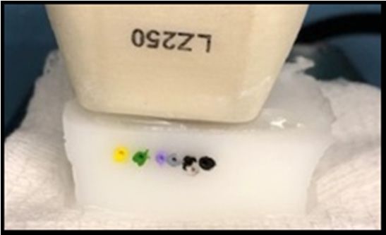
Appl. Sci. 2020, 10, 1024 3 of 5
3. Results
Among the tattoo inks tested, black/gray, green, violet, and blue colored pigments provided higher
Appl. Sci. 2020, 10, x FOR PEER REVIEW 3 of 5
levels of PA signal (Figure 2a–c) in vitro. Yellow, red, and orange had a comparable signal to blood
and
to whiteand
blood hadwhite
the lowest
had thesignal detected
lowest signal by PAI (Figure
detected by PAI2a,d). In the
(Figure volunteer,
2a,d). the skin overlying
In the volunteer, the skin
the tattoo appeared normal without any evidence of inflammatory change on clinical
overlying the tattoo appeared normal without any evidence of inflammatory change on clinical examination
(Figure 3). In(Figure
examination vivo PAI3).provided excellent
In vivo PAI providedcontrast and enabled
excellent contrast accurate delineation
and enabled accurateofdelineation
the extent of
of
the extent
the tattoo in
of the
the dermis.
tattoo inThe
the reconstructed MIP image allowed
dermis. The reconstructed for visualization
MIP image of the tattoo of
allowed for visualization in the
the
dermis in an unambiguous manner. PAI allowed for visualization of the tattoo in the
tattoo in the dermis in an unambiguous manner. PAI allowed for visualization of the tattoo in thepigmented skin
and the surrounding
pigmented skin and theregion (Figure 3).region (Figure 3).
surrounding
30
Black Pitch Black
25
20
PA signal
15
10
5
0
680 700 750 800 850 900
Wavelength (nm)
(a) (b)
25 3.0
Green Violet PBS White Yellow
Gray Blue 2.5
20 Orange Red Blood
2.0
PA signal
PA signal
15
1.5
10
1.0
5 0.5
0 0.0
680 700 750 800 850 900 680 700 750 800 850 900
Wavelength (nm) Wavelength (nm)
(c) (d)
Figure 2. PAI
Figure 2. of tattoo
PAI of tattoo inks
inks in
in vitro.
vitro. (a)
(a) Pseudo-colorized
Pseudo-colorized PA
PA signal
signal maps
maps (680
(680 nm)
nm) of
of the
the ten
ten tattoo
tattoo
inks
inks along
along with
with blood
blood and
and PBS
PBS filled
filled channels.
channels. (b–d)
(b–d) Plots
Plots showing
showing PA
PA signal
signal from
from 680–900
680–900 nm
nm from
from
the ten tattoo ink samples along with blood and PBS.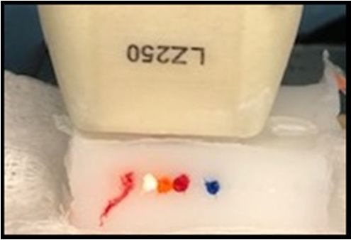
Wavelength (nm) Wavelength (nm)
(c) (d)
Figure 2. PAI of tattoo inks in vitro. (a) Pseudo-colorized PA signal maps (680 nm) of the ten tattoo
inks along with blood and PBS filled channels. (b–d) Plots showing PA signal from 680–900 nm from
Appl. Sci. 2020, 10, 1024 4 of 5
the ten tattoo ink samples along with blood and PBS.
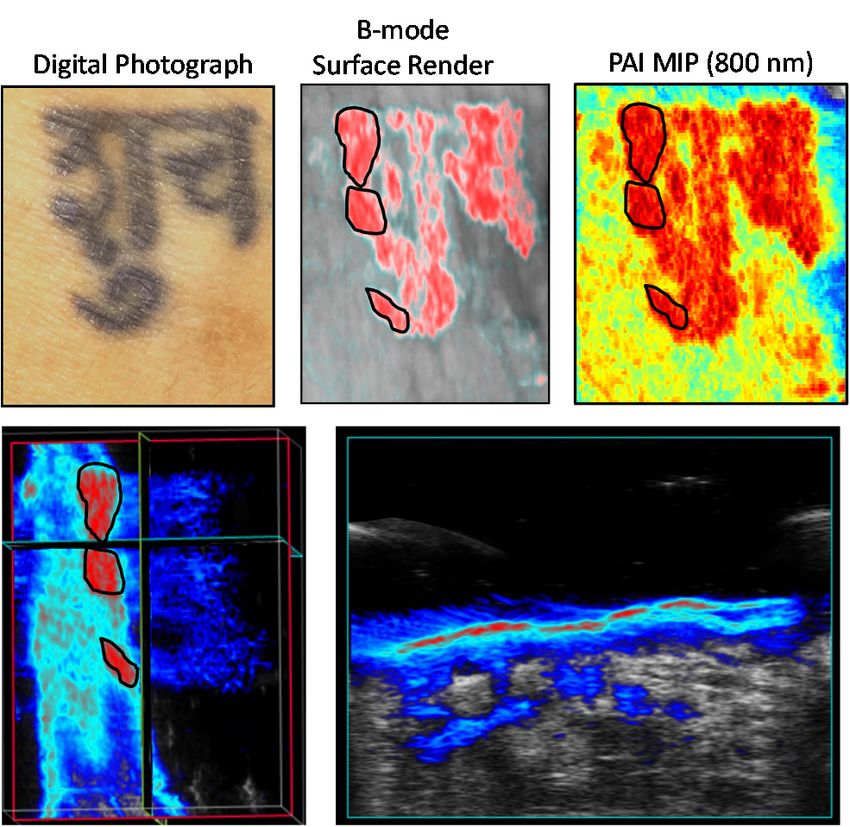
Figure 3. PAI of an intradermal tattoo. The panel of images on the top represents digital photograph
(left), a B-mode surface rendering (middle), and a reconstructed maximum intensity projection (MIP;
right) image (800 nm) of a black tattoo on the wrist of a healthy volunteer. Corresponding longitudinal
(left) and transverse axial (right) images of a part of the tattoo (outlined in black) is shown. The images
have been pseudo-colorized for enhanced visualization.
4. Discussion
In this report, we used PAI a relatively new optical imaging method for imaging tattoo inks in vitro
and for in vivo visualization of a tattoo in the wrist of a healthy volunteer. Given its portability, low cost
and high resolution, US is widely used in dermatology [13]. Clinical studies have also demonstrated
the usefulness of US for imaging musculoskeletal anatomy [14,15]. Tattoo inks exhibited strong yet
distinct photoacoustic properties that can be exploited for non-invasive visualization using PAI. Our
phantom study demonstrated that the PA signal varies greatly among tattoo inks. Lighter colors
like white mostly reflect laser light so there is less absorption and low PA signal. Darker tattoo inks,
especially the black color generates more robust signals. Dermal tattoo pigmentation was successfully
imaged in vivo using PAI. Our study demonstrates the feasibility of PAI to spatially map tattoos in
human skin. To the best of our knowledge, this is the first report on the use of PAI for visualization of
an intradermal tattoo.
While tattooing is relatively cheap, tattoo removal is an expensive procedure that involves use of
laser light and relies on photolysis of the tattoo pigment. Currently, laser removal of tattoos including
the wavelength of the laser light and fluence is based on qualitative, visual examination. As a result,
the outcome following tattoo removal procedures is dependent on the experience of the clinician.
A non-invasive imaging method that can provide an objective assessment of the extent of the tattoo
in all three dimensions could be valuable in guiding tattoo removal. In this regard, PAI could serve
as a relatively inexpensive, non-invasive, clinical tool for preoperative evaluation of tattoos and for
objective monitoring of laser removal procedures.
Author Contributions: Conceptualization: M.S., A.K.S., G.P.; Data curation: E.R.B., L.J.R., A.K.S., M.S.; Formal
analysis: E.R.B., L.J.R., M.S.; Funding acquisition: A.K.S., M.S.; Investigation: E.R.B., L.J.R., A.K.S., M.S.;
Methodology: E.R.B., L.J.R., A.K.S., M.S.; Project administration: A.K.S., M.S.; Resources: A.K.S., M.S.; Supervision:
A.K.S., M.S.; Validation: E.R.B., L.J.R., G.P., O.R.C., A.K.S., M.S.; Visualization: E.R.B., L.J.R., O.R.C., M.S.;
Writing—original draft preparation: E.R.B., L.J.R., M.S.; Writing—review and editing: E.R.B., L.J.R., G.P., O.R.C.,
A.K.S., M.S. All authors have read and agreed to the published version of the manuscript.
Funding: This work was supported by grants from the National Cancer Institute 1R01CA204636, National Center
for Research Resources S10OD010393-01 and the Alliance Foundation of Western New York (to MS), and utilizedAppl. Sci. 2020, 10, 1024 5 of 5
shared resources supported by Roswell Park Cancer Institute Cancer Center Support Grant from the National
Cancer Institute P30CA06156. The funding sponsors had no role in the design of the study, collection, analyses,
or interpretation of data, writing of the manuscript, and in the decision to publish the results.
Conflicts of Interest: The authors declare no conflict of interest.
References
1. Hamblin, M.R.; Avci, P.; Gupta, G.K. Imaging in Dermatology, 1st ed.; Academic Press: London, UK, 2016.
2. Hibler, B.P.; Qi, Q.; Rossi, A.M. Current state of imaging in dermatology. Semin. Cutan. Med. Surg. 2016, 1,
2–8. [CrossRef] [PubMed]
3. Patil, C.A.; Kirshnamoorthi, H.; Ellis, D.L.; van Leeuwen, T.G.; Mahadevan-Jansen, A. A Clinical Instrument
for Combined Raman Spectroscopy-Optical Coherence Tomography of Skin Cancers. Lasers Surg. Med. 2011,
2, 143–151. [CrossRef] [PubMed]
4. Dubois, A.; Levecq, O.; Azimani, H.; Siret, D.; Barut, A.; Suppa, M.; Del Marmol, V.; Malvehy, J.; Cinotti, E.;
Rubegni, P.; et al. Line-field confocal optical coherence tomography for high-resolution noninvasive imaging
of skin tumors. J. Biomed. Opt. 2018, 10, 1–9. [CrossRef] [PubMed]
5. Tkaczyk, E.R. Innovations and developments in dermatologic non-invasive optical imaging and potential
clinical applications. Acta derm Venereol. 2017, 97 (Suppl. S218), 5–13. [CrossRef] [PubMed]
6. Kruger, R.A. Photoacoustic ultrasound. Med. Phys. 1994, 21, 127–131. [CrossRef] [PubMed]
7. Wang, L.V. Ultrasound-mediated biophotonic imaging: A review of acousto-optical tomography and
photo-acoustic tomography. Dis. Markers. 2004, 19, 123–138. [CrossRef] [PubMed]
8. Zeitouni, N.C.; Rohrbach, D.J.; Aksahin, M.; Sunar, U. Preoperative ultrasound and photoacoustic imaging
of nonmelanoma skin cancers. Dermatol. Surg. 2015, 41, 525–528. [CrossRef] [PubMed]
9. Schwarz, M.; Buehler, A.; Aguirre, J.; Ntziachristos, V. Three-dimensional multispectral optoacoustic
mesoscopy reveals melanin and blood oxygenation in human skin in vivo. J. Biophotonics 2016, 9, 55–60.
[CrossRef] [PubMed]
10. Zhou, Y.; Tripathi, S.V.; Rosman, I.; Ma, J.; Hai, P.; Linette, G.P.; Council, M.L.; Fields, R.C.; Wang, L.V.;
Cornelius, L.A. Noninvasive Determination of Melanoma Depth using a Handheld Photoacoustic Probe.
J. Investig. Dermatol. 2017, 137, 1370–1372. [CrossRef] [PubMed]
11. Hindelang, B.; Aguirre, J.; Schwarz, M.; Berezhnoi, A.; Eyerich, K.; Ntziachristos, V.; Biedermann, T.;
Darsow, U. Non-invasive imaging in dermatology and the unique potential of raster-scan optoacoustic
mesoscopy. J. Eur. Acad. Dermatol. Venereol. 2019, 33, 1051–1061. [CrossRef] [PubMed]
12. Rich, L.; Seshadri, M. Photoacoustic Imaging of Vascular Hemodynamics: Validation with Blood Oxygenation
Level-Dependent MR imaging. Radiology 2015, 275, 110–118. [CrossRef] [PubMed]
13. Kleinerman, R.; Whang, T.B.; Bard, R.L.; Marmur, E.S. Ultrasound in dermatology: Principles and applications.
J. Am. Acad. Dermatol. 2012, 67, 478–487. [CrossRef] [PubMed]
14. Wu, W.T.; Chang, K.V.; Mezian, K.; Naňka, O.; Lin, C.P.; Özçakar, L. Basis of shoulder nerve entrapment
syndrome: An ultrasonographic study exploring factors influencing cross-sectional area of the suprascapular
nerve. Front. Neurol. 2018, 9, 902. [CrossRef] [PubMed]
15. Chang, K.V.; Yang, K.C.; Wu, W.T.; Huang, K.C.; Han, D.S. Association between metabolic syndrome and
limb muscle quantity and quality in older adults: A pilot ultrasound study. Diabetes Metab. Syndr Obes. 2019,
12, 1821–1830. [CrossRef] [PubMed]
© 2020 by the authors. Licensee MDPI, Basel, Switzerland. This article is an open access
article distributed under the terms and conditions of the Creative Commons Attribution
(CC BY) license (http://creativecommons.org/licenses/by/4.0/).You can also read