Microstructure of metatitanic acid and its transformation to rutile titanium dioxide
←
→
Page content transcription
If your browser does not render page correctly, please read the page content below
High Temperature Materials and Processes 2020; 39: 627–632
Research Article
Xiaoping Wu* and Yong Liu
Microstructure of metatitanic acid and its
transformation to rutile titanium dioxide
https://doi.org/10.1515/htmp-2020-0097
received January 16, 2020; accepted October 01, 2020
1 Introduction
Abstract: The microstructure of metatitanic acid and its Titanium dioxide is an important inorganic material that
transformations to titanium dioxide during calcination have has a wide range of applications such as pigment and
been investigated previously. However, the detailed micro- functional materials in energy and environmental areas
structure of metatitanic acid has not been elucidated. Herein, [1–6]. One of the most common methods for the commer-
we report the high-resolution scanning electron microscopy cial production of titanium dioxide is the sulphate pro-
and X-ray powder diffraction determinations of the micro- cess [7,8]. In the sulphate process, ilmenite (FeTiO3)
structure of metatitanic acid and its transformation to tita- or slag reacts with the concentrated sulphuric acid in
nium dioxide during calcination. It is the first time that the the reaction: FeTiO3(s) + 2H2SO4(aq) = TiOSO4(aq) +
detailed microstructure of metatitanic acid and its transfor- FeSO4(aq) + 2H2O. The resulting solution contains titanyl
mation to rutile titanium dioxide during calcination have sulphate (TiOSO4) and ferrous sulphate (FeSO4). The ad-
been observed and elucidated. A mechanism of the transfor- dition of iron into the solution is necessary to reduce Fe3+
mation from metatitanic acid to crystalline titanium dioxide to Fe2+, so that Fe2+ will remain in the solution. After a
during calcination is described. The basic building blocks of series of precipitation, removal of sulphate heptahydrate
metatitanic acid are the ultrafine crystals with an averaged (FeSO4·7H2O), and solution concentration steps, the ti-
diameter of a few nanometres, and these ultrafine crystals tanyl sulphate solution is hydrolysed by heating and di-
aggregate to form the porous primary particles. The primary luting with water, and a hydrated titanium dioxide (gen-
particles further agglomerate to form the porous secondary eral formula TiO2·nH2O) precipitates. The hydrolysis is
particle. During the calcination, metatitanic acid undergoes accelerated by adding the small amount of seeds to the
size enlargement of ultrafine crystals, anatase–rutile trans- feed solution. The most common form of hydrated tita-
formation, merge of primary particles, and the crystal growth nium dioxide produced by the hydrolysis of the titanyl
of titanium dioxide. sulphate in the sulphate process is metatitanic acid
Keywords: microstructure, metatitanic acid, titanium (TiO2·H2O or H2TiO3). Metatitanic acid is an important
dioxide inorganic precursor for making a number of important
materials such as titanium dioxide white pigment, tita-
nium dioxide nanomaterials, and other functional ma-
terials [9–11]. The precipitated metatitanic acid is be-
lieved to consist of ultrafine particles with the large
surface area. However, despite a large number of stu-
dies, the detailed structure of metatitanic acid has not
yet been elucidated [9,12–18].
* Corresponding author: Xiaoping Wu, Department of Titanium The transformation of metatitanic acid to rutile tita-
Chemical Engineering, Ansteel Research Institute of Vanadium &
nium dioxide is through a calcination process. The trans-
Titanium (Iron & Steele), State Key Laboratory of Vanadium and
Titanium Resources Comprehensive Utilization, Sichuan, formation usually goes through a number of stages:
Panzhihua, 617000, China, dehydration (100–500°C), desulfurisation (600–800°C),
e-mail: 13308173290@163.com and the conversion of anatase phase of titanium dioxide
Yong Liu: Research Center for Environmental Science & Technology,
to rutile phase (800–1,000°C) [19–21]. Rutile seeds are
Institute of Fundamental and Frontier Sciences, University of
Electronic Science and Technology of China, Chengdu, 611731, used to produce rutile pigment. The optimised particle
China size of pigmentary rutile titanium dioxide particles is
Open Access. © 2020 Xiaoping Wu and Yong Liu, published by De Gruyter. This work is licensed under the Creative Commons Attribution 4.0
International License.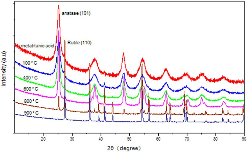
628 Xiaoping Wu and Yong Liu
around 250 nm or between 200 and 300 nm, as these pattern fitting [22]. The mass fractions of anatase phase
particles have optimised light-scattering property of tita- (wra) and rutile phase (wrr) were first determined using
nium dioxide pigments [7,8]. To a large extent, the mor- the Rietveld whole pattern fitting method, and the con-
phology, the structure, and the particle size of metati- tents of anatase TiO2 (wa) and rutile TiO2 (wr) were then
tanic acid determine the structure and the pigmentary calculated by using equations
quality of calcined titanium dioxide. wa = wra/(wra + wrr) × 100% and wr = 100 − wa
Herein, we report a scanning electron microscopy
The average size of the ultrafine crystals was deter-
(SEM) and X-ray powder diffraction (XRD) observation
mined based on the Scherrer equation [23]:
of the microstructure of metatitanic acid and its trans-
formation to titanium dioxide during heating treatment Dhkl = 0.9λ /(β1 / 2 cos θ ),
from ambient temperature to 1,000°C. It is the first time
that the detailed microstructure of metatitanic acid has where Dhkl = crystallite dimension normal to the hkl re-
been observed using a high resolution of SEM. The mor- flecting planes, λ = X-ray wave length, β1/2 = half-max-
phological changes, the anatase-rutile transformation, imum line breadth, and θ = Bragg angle.
and the crystal growth of titanium dioxide during cal-
cination have been investigated using SEM and XRD
techniques.
3 Results and discussion
3.1 Microstructure of metatitanic acid
2 Experimental
Figure 1 shows a SEM image of metatitanic acid powder
2.1 Materials
(30k× magnifications). The particles in the image are
secondary particles of metatitanic acid with the particle
Metatitanic acid was obtained from an industrial source. size of a few hundreds of nanometres. This type of
The metatitanic acid generally contains about 40–60% images of metatitanic acid has been observed in a
water, a few percent of rutile seeds, and a small amount number of previous studies [24–26]. However, when
of inorganic compounds such as potassium compound the metatitanic acid was observed under exceptional
(K2O of 0.40 wt% to TiO2), phosphate compound (P2O5 of magnifications (over 400k× magnifications), the fine mi-
0.20 wt% to TiO2), and zinc compound (ZnO of 0.20 wt% crostructure of metatitanic acid is revealed, as shown in
to TiO2). Metatitanic acid was air dried at ambient Figure 2.
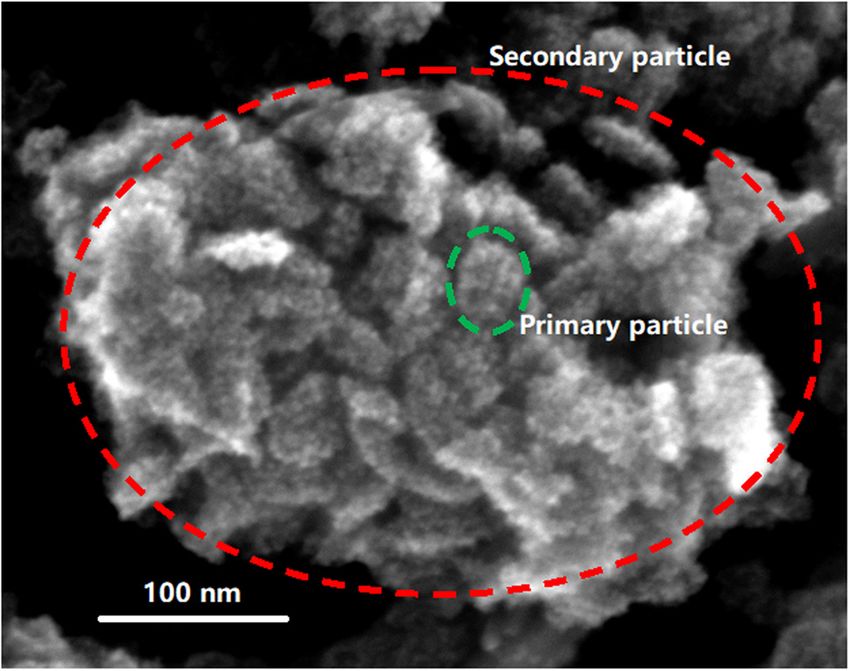
temperature. It is shown in Figure 2 that metatitanic acid consists
of secondary particles with a particle diameter of a few
hundred nanometres (red cycle in Figure 2). A secondary
2.2 Methods
The dried metatitanic acid is grinded into fine powder for
SEM and XRD measurement. The calcination of metati-
tanic acid was carried out in a furnace. After calcination
of metatitanic acid at different temperatures, the samples
were grinded into fine powder. The microstructure and
morphologies of the metatitanic acid and its calcined
samples were characterised by SEM (SEM model: ZEISS
Sigma 500). The particle size of crystallites and crystal
phases were determined by XRD (diffractometer model:
PANalytical/EMPYREAN, The Netherlands) operating in a
transmission mode with Cu Kα radiation (λ = 1.5418 Å).
The quantitative analysis of anatase and rutile phases
was conducted by using the method of Rietveld whole Figure 1: SEM image of metatitanic acid (30k× magnifications).Microstructure of metatitanic acid and its transformation to rutile titanium dioxide 629
Table 1: Size changes of particles inside metatitanic acid
100°C 400°C 600°C 800°C 900°C
Du (nm) 6.0 9.0 23.0 62.0 —
Dp (nm) 72.0 75.0 56.0 — —
Ds (µm) 1.0 1.0 0.9 — —
Du: particle size of ultrafine crystals in metatitanic acid (by XRD);
Dp: particle size of primary particles (by SEM); Ds: particle size of
secondary particles (by SEM).
Figure 2: SEM image of metatitanic acid (419.78k× magnifications).
Figure 4: Primary particles of metatitanic acid after heating treat-
ment at 750°C.
metatitanic acid) is due to the addition of rutile seeds
for producing rutile titanium dioxide. The particle size
of anatase ultrafine crystals is determined to be 6 nm. As
the calcination temperature increases, the particle size of
anatase ultrafine crystals increases (Table 1), and the ana-
Figure 3: XRD patterns of metatitanic acid and its calcined samples tase–rutile transformation become significant at 800°C.
at different temperatures. The interactions between particles that constitute the
secondary and primary particle are also observed to be
particle is the agglomeration of the primary particles that
different. As shown in Figure 4, after heating at 750°C for
have the averaged diameters of tens of nanometres (green
an hour, the secondary particles largely disintegrated into
cycle in Figure 2). The internal structure of a primary
primary particles, but the integrity of primary particles
particle under the high resolution of SEM can be further
remains. This confirms that the secondary particles are
observed to consist of ultrafine particles with a diameter
the agglomeration of the primary particles, and the inter-
less than 10 nm. A XRD determination shows that ultra-
actions between primary particles in a secondary particle
fine particles inside primary particles are anatase crystals
are physical and relatively weak. The interactions between
with a size of around 5 nm.
microcrystals inside a primary particle are of a degree of
chemical interaction and relatively strong.
Table 1 summaries the size changes of particles,
3.2 Transformation of metatitanic acid which constitute metatitanic acid in the heating treat-
to TiO2 ment at different temperatures. As the temperature in-
creases over 400°C, the particle size of ultrafine crystals
Figure 3 shows the XRD pattern of metatitanic acid after increases rapidly. At 800°C, the particle size increases to
heating treatment at different temperatures. At 100°C, the 62 nm. The sizes of the primary and secondary particles
XRD pattern is the same as metatitanic acid at ambient remain fairly constant up to 400°C. After 600°C, these
temperature. The small peak of the rutile phase (in sizes become smaller.630 Xiaoping Wu and Yong Liu
Figure 5: Morphology changes and crystal growth of metatitanic acid during calcination. (a) 800°C, (b) 830°C, (c) 845°C, (d) 850°C, (e)
900°C, (f) 1000°C.
Table 2: Averaged particle size and anatase–rutile transformation
rate of metatitanic acid during calcinations Park and Shin studied the specific surface areas and
pore volumes of metatitanic acid after heating treatment
SEM Temperature (°C) Averaged A–R at different temperatures [27]. The BET-specific surface
image particle transformation areas (ABET, m2/g) and pore volumes (V, cm3/g) of heat-
size (nm) rate (%)
treated metatitanic acid at 100°C are around 377 and
(a) 800 146 51.7 0.297, respectively. This is consistent with our observa-
(b) 830 224 90.7 tion that a primary particle of metatitanic acid consist of
(c) 845 231 95.7
ultrafine crystals and is porous. The previous thermal
(d) 850 252 99.1
(e) 900 382 100.0
analysis and XRD studies [24–26] have demonstrated
(f) 1,000 772 100.0 that after heating over 100°C, metatitanic acid begin to
loss water: initially, free water, H2O(l) → H2O(g), and
Ultrafine Primary Enlarged
crystal parcle TiO2
Ultrafine Primary
crystal parcles parcles
TiO2 parcles
4
3
Pore
1 Secondary 2 Secondary
parcle parcle
Figure 6: The mechanism of the transformation from metatitanic acid to crystalline titanium dioxide during calcination.Microstructure of metatitanic acid and its transformation to rutile titanium dioxide 631
then hydrated water at higher temperature up to 500°C: titanium dioxide particles to form larger particles. A me-
TiO(OH)2(s) → TiO2(s) + H2O(g). In this study, the com- chanism of the transformation from metatitanic acid
bined SEM and XRD investigation show that at 800°C, me- to crystalline titanium dioxide during calcination is
tatitanic acid has already transformed into a mixture of described.
anatase and rutile titanium dioxide particles (see Figure
5(a) and Table 2). At the temperature over 800°C, smaller Acknowledgments: This work was financially supported
particles coalesce further to form larger particles, and at the by the Pangang Group under a Basic Research Grant. We
same time, anatase continues to transform into the rutile thank Dr Zhaohua Liu, Mr Zhixin Shi, and Mr Xibin Liu for
phase (Figure 5(b, c) and Table 2). The agglomerated sec- SEM and X-ray diffraction analyses, experimental help,
ondary particles of metatitanic acid have disintegrated, and and discussion.
the size of primary particles of metatitanic acid increase
rapidly. At the same time, rutile content of titanium dioxide Conflict of interest: There are no conflicts to declare.
increase. At 850°C, the anatase–rutile transformation is al-
most complete, and the particles of rutile titanium dioxide
reach an optimum averaged size of around 250 nm (Figure
5(d) and Table 2). At the higher temperatures, rutile tita- References
nium dioxide particles coalesce into even larger particles
(Figure 5(e, f) and Table 2). [1] O’Regan, B., and M. Gratzel. A low-cost, high-efficiency solar
From the aforementioned studies and analysis, the cell based on dye-sensitized colloidal TiO2 films. Nature,
Vol. 353, 1991, pp. 737–740.
mechanism of the transformation from metatitanic acid
[2] Kay, A., and M. Gratzel. Artificial photosynthesis. 1.
to crystalline titanium dioxide during calcination is shown Photosensitization of titania solar cells with chlorophyll deri-
as in Figure 6: 1, the original microstructure of metatitanic vatives and related natural porphyrins. The Journal of Physical
acid; 2, dehydrated intermediate, with the increased size Chemistry, Vol. 97, 1993, pp. 6272–6277.
of ultrafine crystals; 3, TiO2 pigment particles after anata- [3] Fujishima, A., and K. Honda. Electrochemical photolysis of
se–rutile transformation and crystal growth; 4, further water at a semiconductor electrode. Nature, Vol. 238, 1972,
pp. 37–38.
merged and grew TiO2 particles after prolonged heating
[4] Fujishima, A., T. N. Rao, and D. A. Tryk. Titanium dioxide
at high temperature. photocatalysis. Journal of Photochemistry and Photobiology C,
Vol. 1, 2000, pp. 1–21.
[5] Tryk, D. A., A. Fujishima, and K. Honda. Recent topics in
photoelectrochemistry: achievements and future
4 Conclusion prospects. Electrochimica Acta, Vol. 45, 2000,
pp. 2363–2367.
[6] Chen, X., and S. S. Mao. Titanium dioxide nanomaterials:
We have observed and determined the detailed micro- synthesis, properties, modifications, and applications.
structure of metatitanic acid using the high-resolution Chemical Reviews, Vol. 107, 2007, pp. 2891–2959.
SEM and XRD. The basic building blocks of the metati- [7] Winkler, J. Titanium dioxide, production, properties and
tanic acid are the anatase ultrafine crystals with an effectives usage, 2nd edn. Vincentz Network, Hanover,
2013.
average size of around 5 nm. These ultrafine crystals ag-
[8] Lakshmanan, V. I., A. Bhowmick, and M. A. Halim. In Titanium
gregate to form the porous primary particles with an dioxide, chemical properties, applications and environmental
average diameter of a few tens of nanometres. The pri- effects, J. Brown, Eds, Nova Science Publishers, New York,
mary particles further agglomerate to form the secondary 2014, pp. 75–130.
porous particles with an average diameter of a few hun- [9] Gesenhues, U. Calcination of metatitanic acid to titanium
dioxide white pigments. Chemical Engineering & Technology,
dreds of nanometres. During calcination, the particle
Vol. 24, 2001, pp. 685–694.
size of anatase ultrafine crystals increases continuously. [10] Song, H., H. B. Liang, L. Lu, P. Wu, and C. Li. Effect of hydrolysis
After reaching 800°C, the primary particles begin to conditions on hydrous TiO2 polymorphs precipitated from a
merge together, and at the same time, the anatase–rutile titanyl sulfate and sulfuric acid solution. International Journal
transformation become significant. At 850°C, the merge of Minerals, Metallurgy and Materials, Vol. 19, 2002,
pp. 642–650.
of primary particles complete, and anatase–rutile trans-
[11] Xu, R., J. Li, Z. Tang, and Z. Zhang. Ultrafine metatitanic acid
formation reach about 100%. This point represents an
electrode for ultrafast lithium ion batteries. Electrochimica
optimised particle size of pigmentary titanium dioxide. Acta, Vol. 56, 2011, pp. 6330–6335.
Furthermore, prolonged calcination at high temperature [12] Li, Z., Z. Wang, and G. Li. Preparation of nano-titanium dioxide
leads to coalesce and increases the chances of rutile from ilmenite using sulfuric acid-decomposition by liquid632 Xiaoping Wu and Yong Liu
phase method. Powder Technology, Vol. 287, 2016, pp. [20] Ginsberg, T., M. Modigell, and W. Wilsmann. Thermochemical
256–263. characterisation of the calcination process step in the sul-
[13] Jalava, J., L. Heikkila, O. Hovi, R. Laiho, E. Hiltunen, A. phate method for production of titanium dioxide. Chemical
Hakanen, et al. Structural investigation of hydrous TiO2 pre- Engineering Research and Design, Vol. 89, 2011,
cipitates and their aging products by X-ray diffraction, atomic pp. 990–994.
force microscopy, and transmission electron microscopy. [21] Tian, C., Y. Yang, and H. Pu. Effect of calcination temperature
Industrial & Engineering Chemistry Research, Vol. 37, 1998, on porous titania prepared from industrial titanyl sulfate
pp. 1317–1323. solution. Applied Surface Science, Vol. 257, 2011,
[14] Jalava, J., E. Hiltunen, H. Kähkönen, H. Erkkilä, H. Härmä, and pp. 8391–8395.
V. Taavitsainen. Structural investigation of hydrous titanium [22] Dong, W., C. Gilmore, G. Barr, C. Dallman, N. Feeder, and
dioxide precipitates and their formation by small-angle X-ray S. Terry. A Quick method for the quantitative analysis of mix-
scattering. Industrial & Engineering Chemistry Research, tures. 1. Powder X-ray diffraction. Journal of Pharmaceutical
Vol. 39, 2000, pp. 349–361. Technology, Vol. 97, 2008, pp. 2260–2276.
[15] Bavykin, D. V., V. P. Dubovitskaya, A. V. Vorontsov, and V. N. [23] Chung, F. H., and D. K. Smith. Industrial Application of X–Ray
Parmon. Effect of TiOSO4 hydrothermal hydrolysis conditions Diffraction. Marcel Dekker, New York, 2000.
on TiO2 morphology and gas-phase oxidative activity. [24] Sathyamoorthy, S., G. D. Moggridge, and M. J. Hounslow.
Research on Chemical Intermediates, Vol. 33, 2007, Particle formation during anatase precipitation of seeded
pp. 449–464. titanyl sulfate solution. Crystal Growth & Design, Vol. 1, 2001,
[16] Denisova, T. D., G. Maksimova, E. V. Polyakov, N. A. Zhuravlev, pp. 123–129.
S. A. Kovyazina, O. N. Leonidova, et al. Metatitanic acid: [25] Santacesaria, E., M. Tonello, G. Storti, R. C. Pace, and S. Carra.
synthesis and properties, Russian Journal of Inorganic Kinetics of titanium dioxide precipitation by thermal hydro-
Chemistry, Vol. 51, 2006, pp. 691–699. lysis. Journal of Colloid and Interface Science, Vol. 111, 1986,
[17] Grzmil, B. U., D. Grela, and B. Kic. Hydrolysis of titanium sul- pp. 44–53.
phate compounds. Chemical Papers, Vol. 62, 2008, pp. 18–25. [26] Zhang, W., C. Ou, and Z. Yuan. Precipitation and growth be-
[18] Nosaka, Y., and A. Nosaka. Introduction to photocatalysis, haviour of metatitanic acid particles from titanium sulfate
from basic science to application. Royal Society of Chemistry, solution. Powder Technology, Vol. 315, 2017, pp. 31–36.
Cambridge, 2016. [27] Park, S., and H. Shin. Microstructural evolution of metatitanic
[19] Sullivan, W. F., and S. S. Cole. Thermal chemistry of colloidal acid with temperature and its photosensitization property.
titanium dioxide. Journal of the American Ceramic Society, Reaction Kinetics, Mechanisms and Catalysis, Vol. 101, 2013,
Vol. 42, 1959, pp. 127–133. pp. 237–249.You can also read