STRUCTURAL CONSEQUENCES OF VARIATION IN SARS-COV-2 B.1.1.7
←
→
Page content transcription
If your browser does not render page correctly, please read the page content below
https://www.scientificarchives.com/journal/journal-of-cellular-immunology

 Journal of Cellular Immunology                                                                                      Commentary

Structural Consequences of Variation in SARS-CoV-2 B.1.1.7

David A. Ostrov*
Department of Pathology, Immunology and Laboratory Medicine, University of Florida College of Medicine, Gainesville, FL,
USA

*Correspondence should be addressed to David Ostrov; ostroda@pathology.ufl.edu

Received date: February 06, 2021, Accepted date: March 03, 2021

Copyright: © 2021 Hao Q, et al. This is an open-access article distributed under the terms of the Creative Commons Attribution
License, which permits unrestricted use, distribution, and reproduction in any medium, provided the original author and source
are credited.

Abstract
 New globally circulating SARS-CoV-2 strains are causing concern about evolution of virus transmissibility, fitness and immune
evasion mechanisms. A variant emerging from the United Kingdom called SARS-CoV-2 VUI 202012/01, or B.1.1.7, is thought to exhibit
increased transmissibility that results from replication 4-10 times faster than the original Wuhan virus (Wuhan-Hu-1). Although this
property is suspected to result from a specific mutation in the spike glycoprotein, D614G, there are 9 mutations that distinguish the
UK variant B.1.1.7 from Wuhan-Hu-1 yet to be evaluated for functional effects. We asked if mutated positions fixed in UK variant
B.1.1.7 may be involved in the virus life cycle, or evasion of the immune response, by modeling the UK variant spike protein and
conducting structural analysis of mutations on the spike glycoprotein trimer (protomer) complexed to ACE2. Importantly, 4 out of 9
differences between the UK variant B.1.1.7 and Wuhan-Hu-1 spike protein alter direct intermolecular interactions. N501Y increased
affinity between the spike protein and ACE2. The mutations at A570D, D614G and S982A reduced contact between individual chains
of the trimeric spike protomer, potentially enhancing cleavage into S1 and S2 subunits, dynamic structural rearrangement and host
cell fusion mechanisms. These data suggest that combined characteristics of mutations unique to UK variant B.1.1.7 enable high
affinity binding to ACE2 and enhanced replication properties. The D614G mutation, associated with enhanced virus transmissibility,
opens a potentially druggable structural pocket at the interface between spike glycoprotein subunits S1 and S2.

Keywords: SARS-CoV-2; Angiotensin Converting Enzyme-2; Mutation; Drug discovery

Results and Discussion                                              between subunits of the trimeric protomer. Unlike the
                                                                    mutation at position 501 that increased affinity for ACE2,
 There are 9 sites that differ between SARS-CoV-2 spike             the 3 substitutions at spike trimer interfaces likely reduce
glycoproteins from the original Wuhan strain (Wuhan-                intermolecular binding affinity. The mutations likely
Hu-1) and UK variant B.1.1.7 [1] (Figure 1, Table 1). There         increase spike protein lability in a manner that enhances
are 2 sites with deletions (positions 69-70, 144-145) and           dynamic virus processes that include spike protein
7 sites with amino acid substitutions (4 positions in the           cleavage, structural rearrangement and host cell fusion
S1 subunit and 3 positions in S2). Most substitutions in            mechanisms.
the spike protein that distinguish UK variant B.1.1.7 are
located at sites of intermolecular interaction (4 out of             Intermolecular interactions between individual chains
7 substitutions). One mutation (N501Y) enhanced the                 of the SARS-CoV-2 spike glycoprotein were observed
affinity of the spike protein with ACE2 [2]. N501Y was              at positions A570 (Figure 3A), D614 (Figure 3B), and
modeled based on the cryoEM structure of the Wuhan-                 S982 (Figure 3C) in the original Wuhan strain (Wuhan-
Hu-1 spike protein/ACE2 complex [3] (Figure 2A, PDB                 Hu-1). The A570D substitution in the UK variant B.1.1.7
6M17), indicating that the gain in affinity likely results          variant introduces steric clash with the backbone amide of
from aromatic interactions (π stacking) between Tyr501              K964 (Figure 3D). The D614G substitution results in the
and Tyr41 of ACE2 (Figure 2B).                                      formation of a distinctive cavity at the interface of spike
                                                                    protein subunits in the UK variant B.1.1.7 trimer (Figure
 Strikingly, 3 mutations in the spike protein that                  3E). The S982A in UK variant B.1.1.7 lacks intermolecular
distinguish UK variant B.1.1.7 are located at interfaces            hydrogen (H) bonding potential between spike protein

J Cell Immunol. 2021
Volume 3, Issue 2                                                                                                                        103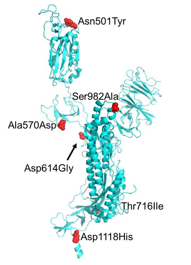
Ostrov DA. Structural Consequences of Variation in SARS-CoV-2 B.1.1.7. J Cell Immunol. 2021; 3(2): 103-108.

    Figure 1
                A.

                 B.                                                                   C.

 Figure 1: The location of mutated positions that distinguish the United Kingdom variant SARS-CoV-2 B.1.1.7
 from Wuhan-Hu-1. A. Sites that differ between B.1.1.7 and Wuhan-Hu-1 indicated on the primary structure of the coronavirus
 spike protein. NTD: N-terminal Domain; RBD: Receptor Binding Domain; FP: Fusion Peptide; HR1: Heptad Repeat 1; HR2:
 Heptad Repeat 2; TM: Transmembrane anchor; IC: Intracellular tail. B. Ribbon diagram model of B.1.1.7 spike glycoprotein based
 on the structure of the trimeric protomer (PDB 6VSB). Sites that differ between B.1.1.7 and Wuhan-Hu-1 are shown as red spheres.
 One chain of the spike trimer is shown in blue with one RBD in the up conformation. C. Model of the B.1.1.7 spike protein trimer
 complexed to ACE2. ACE2 is shown in yellow. The spike protein trimer is shown in blue, green and grey. The modeled interaction
 between ACE2 and the RBD was based on the 2019-nCoV RBD/ACE2 complex (PDB 6M17).

J Cell Immunol. 2021
Volume 3, Issue 2                                                                                                           104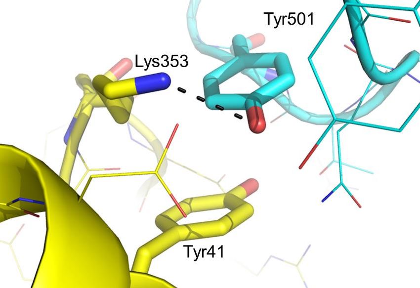
Ostrov DA. Structural Consequences of Variation in SARS-CoV-2 B.1.1.7. J Cell Immunol. 2021; 3(2): 103-108.
Figure 3
A.
Wuhan-Hu-1
B.
B.1.1.7
Figure 2: A mutation at position 501 in SARS-CoV-2 B.1.1.7 increases affinity for ACE2 by altering intermolecular
interactions. A. Asn501 in the spike glycoprotein from Wuhan-Hu-1 (blue) forms a H bond (black dashes) with Tyr41 in ACE2
(yellow). B. Tyr501Figure
in SARS-CoV-2
2 VUI 202012/01 form an aromatic stack with Tyr41 and a H bond with Lys353.
A. B. C.
Wuhan
-Hu-1
B.1.1.7
D. E. F.
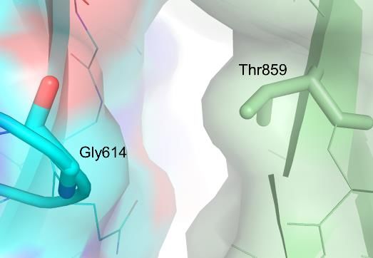
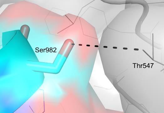
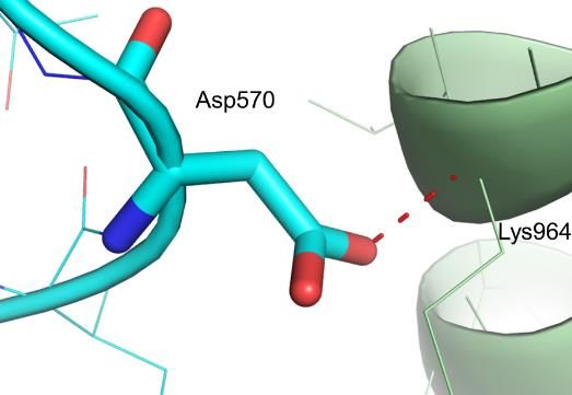
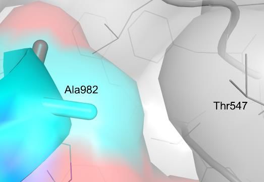
Figure 3: Mutations in the SARS-CoV-2 B.1.1.7 hinder intermolecular interactions between subunits of the spike
glycoprotein. A. Ala570 in the spike glycoprotein from Wuhan-Hu-1 (blue) forms an intermolecular van der Waals (green
dashes) contact with the main chain amide of Lys964 from the neighboring chain (grey). B. Asp570 in the SARS-CoV-2 UK variant
is expected to form a repulsive interaction (red dashes) because of a potential clash with the main chain of the neighboring chain
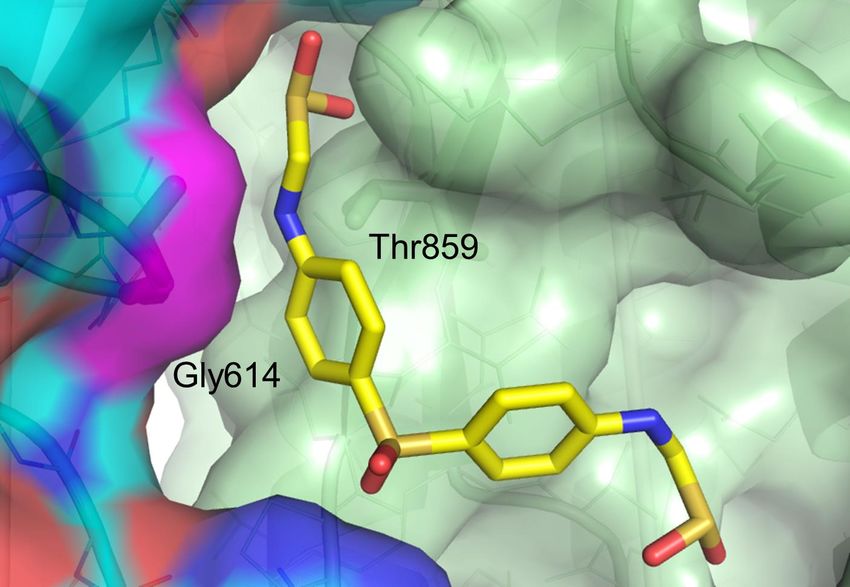
(grey). C. Asp614 in the spike glycoprotein from Wuhan-Hu-1 (blue) forms an intermolecular H bond with Thr859 a neighboring
chain (green). D. Gly614 in the SARS-CoV-2 UK variant results in a loss of a H bond with Thr859 at the interface between two
spike protein subunits (blue and green) of the trimeric protomer forming a potentially druggable structural pocket. E. Ser982 in
the spike glycoprotein from Wuhan-Hu-1 (blue) forms an intermolecular H bond with Thr547 of a neighboring chain (grey). F.
Ala982 in the spike protein of the UK variant (blue) prevents H bonding with Thr547 of a neighboring chain (grey).
J Cell Immunol. 2021
Volume 3, Issue 2 105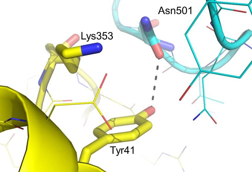
Ostrov DA. Structural Consequences of Variation in SARS-CoV-2 B.1.1.7. J Cell Immunol. 2021; 3(2): 103-108.

subunits at this site (Figure 3F). Collectively, these data      between position D614G and T859 of neighboring chains
suggest that: 1) the UK variant B.1.1.7 exhibits a change        in the spike protein trimer. Drugs were estimated to bind
that enhances affinity for the coronavirus receptor ACE2         the UK variant B.1.1.7 at the D614G site, such as the anti-
(N501Y), and 2) mutations may enhance dynamic virus              leprosy drug sulfoxone, ΔG -24.4 kcal/mol (Figure 4).
fusion mechanisms by reducing intermolecular stability of        These data suggest that drugs may be developed to target
spike protein subunits (A570D, D614G, S982A).                    highly transmissible SARS-CoV-2 strains including the UK
                                                                 variant B.1.1.7.
 P681H represents a potentially important difference
between Wuhan-Hu-1 sequence and UK variant B.1.1.7.               As mutations that distinguish the UK variant B.1.1.7
Position 681 is located adjacent to the RRAR proprotein          may influence immune responses, we analyzed solvent
convertase motif considered a hallmark of high                   accessibility of positions that differ with Wuhan-Hu-1
pathogenesis (PRRAR in Wuhan-Hu-1 [4], HRRAR in                  (Table 1). Although N501Y has the potential to influence
B.1.1.7). This site is cleaved by furin and other proteases to   neutralizing antibody binding, this semiconservative
separate the S1 and S2 subunits of the spike protein, which      difference is located at the edge of the ACE2/spike protein
undergo structural rearrangement and fusion with host            interface, therefore not expected to dramatically alter
cell membranes mediated by heptad repeat domains in              neutralizing antibody responses. T cells recognize SARS-
S2. Since endosomal S1/S2 cleavage occurs in an acidified        CoV-2 peptides in the context of multiple HLA molecules
environment, a protonated histidine at position 681 of the       [5], suggesting that differences between peptides derived
UK variant B.1.1.7 has the potential to influence the rate of    from the Wuhan-Hu-1 spike protein or UK variant B.1.1.7
spike protein cleavage and subsequent membrane fusion            are not expected to radically influence the overall function
mechanisms to gain cell entry.                                   of polyclonal T cell responsiveness to infection. However,
                                                                 surveillance of neutralizing antibody responses and T
  Since SARS-CoV-2 variants with D614G, such as UK               responses to peptides derived from the UK variant B.1.1.7
variant B.1.1.7, currently predominate globally, we asked        in COVID-19 patients will be required to understand
if the interface between individual chains of the spike          functional effects on immune recognition.
protein trimer at position 614 may be druggable. A model
of the spike protein trimer of the UK variant B.1.1.7             Over the past year, the virus has adapted variant forms
was used as the basis for molecular docking. 1,207 FDA           with improved fitness. Mutations that distinguish UK
approved drugs were docked to the interface site formed          variant B.1.1.7 increase virus fitness through mechanisms

                 Figure 4

 Figure 4: Highly transmissible strains of D614G SARS-CoV-2 in the global human population exhibit a unique,
 potentially druggable structural pocket. A model of the B.1.1.7 spike protein was used as the basis for molecular docking
 screening of 1,207 approved drugs. Sulfoxone was predicted by DOCK6.7 (UCSF) to bind the interface between subunits of the
 spike protomer at position 614 with an estimated ΔG value of -24.4 kcal/mol.

J Cell Immunol. 2021
Volume 3, Issue 2                                                                                                      106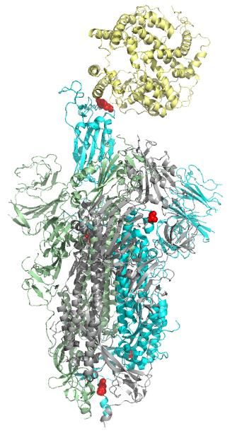
Ostrov DA. Structural Consequences of Variation in SARS-CoV-2 B.1.1.7. J Cell Immunol. 2021; 3(2): 103-108.

                                                                       Solvent accessibility
  Mutation            Location               Interaction              in protomer (PDB            Potential effect
                                                                      6VSB)

  Deletion 69-70      N-terminal domain      None                      None                       Loss of T and B cell epitopes
  Deletion 144-145    N-terminal domain      None                      None                       Loss of T and B cell epitopes

                                                                                                  Enhance ACE2 binding affinity
                                            Lys 353 in ACE2 PDB
  Asn501Tyr           RBD                                              Exposed
                                           6M17
                                                                                                  Altered T and B cell epitopes

                                                                                                  Protomer stability, dynamics of
                                            Intermolecular contact
                      Between RBD and                                                            cleavage and fusion
  Ala570Asp                                with Lys 964, (chain B      Exposed
                     S1/S2 boundary
                                           6VSB)
                                                                                                  Altered T and B cell epitopes

                                                                                                  Protomer stability, dynamics of
                                            Intermolecular contact
                      Between RBD and                                                            cleavage and fusion
  Asp614Gly                                with Thr 859 (chain B       Exposed
                     S1/S2 boundary
                                           6VSB)
                                                                                                  Altered T and B cell epitopes

                                                                                                  Altered S1/S2 cleavage in
                                                                                                 endosomes from protonated His
                      Adjacent to furin
  Pro681His                                  None                      Exposed
                     cleavage site
                                                                                                  Alteration of T and B cell
                                                                                                 epitopes

                      S2 between fusion
                                                                                                  Minor alteration of T and B cell
  Thr716Ile          peptide and Heptad      None                      Exposed
                                                                                                 epitopes
                     Repeat 1

                                                                                                  Protomer stability, dynamics of
                                            Intermolecular contact                               cleavage and fusion
                      Heptad Repeat 1
  Ser982Ala                                with Thr 547 (chain C       Exposed
                     in S2
                                           6VSB)                                                  Alteration of T and B cell
                                                                                                 epitopes

                                            Intramolecular contact
  Asp1118His         S2                                                Buried                     Alteration of T cell epitopes
                                           with Val 951
Table 1: Locations, surface accessibility and interactions of mutations that distinguish the B.1.1.7 and Wuhan-Hu-1 spike proteins.

that likely include: 1) increased affinity for binding to         Assessment Brief, December 20, 2020, was used to define
the coronavirus receptor ACE2, and 2) alteration of               mutations that distinguish the UK variant B.1.1.7 (https://
intermolecular contacts between subunits of the spike             www.ecdc.europa.eu/en/publications-data/threat-
protein trimer. Changes in intermolecular contacts are            assessment-brief-rapid-increase-sars-cov-2-variant-
expected to improve dynamic mechanisms involved in                united-kingdom).
spike protein cleavage, structural rearrangement and host
cell membrane fusion. Emerging mutations such as D614G             Modeling mutations in the UK variant B.1.1.7
can serve as the basis for drug discovery efforts to target       spike glycoprotein
specific highly transmissible variants such as UK variant
B.1.1.7.                                                           The cryoEM structure of the SARS-CoV-2 (Wuhan-Hu-1)
                                                                  RBD/ACE2-B0AT1 complex [3] (PDB 6M17) was used as
Methods                                                           the basis for modeling N501Y. The prefusion SARS-CoV-2
                                                                  (Wuhan-Hu-1) spike glycoprotein with a single receptor-
Sequences of UK variant B.1.1.7 and reference                     binding domain up [6] (PDB 6VSB) was used as the basis
Wuhan-Hu-1                                                        for modeling D570A, D614G, T716I, S982A and D1118H.
                                                                  Side chains were mutated in COOT [7] using rotamers that
 The Wuhan-Hu-1 sequence was used as a reference                  represent a local energy minimum of torsional angles.
(GENBANK accession number MN908947). A Threat

J Cell Immunol. 2021
Volume 3, Issue 2                                                                                                              107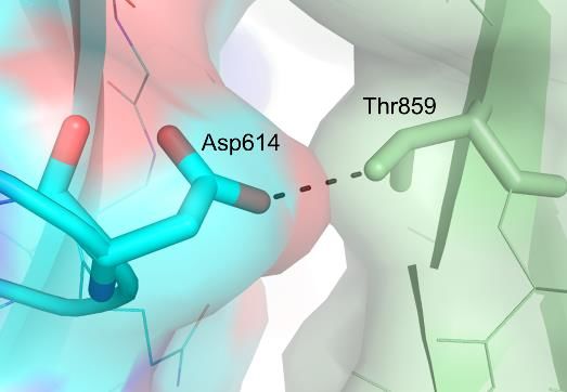
Ostrov DA. Structural Consequences of Variation in SARS-CoV-2 B.1.1.7. J Cell Immunol. 2021; 3(2): 103-108.

 Molecular docking                                            Nature. 2020 May;581(7807):221-4.

 We used the atomic model of the UK variant B.1.1.7 spike      5. Grifoni A, Weiskopf D, Ramirez SI, Mateus J, Dan JM,
protein as the basis for molecular docking. To prepare the    Moderbacher CR, et al. Targets of T cell responses to SARS-
site for docking, all water molecules were removed. We        CoV-2 coronavirus in humans with COVID-19 disease and
explored the molecular surface of the structure using sets    unexposed individuals. Cell. 2020 Jun 25;181(7):1489-
of spheres to describe potential binding pockets at the UK    501.
variant B.1.1.7 interface between the equivalent of chains
A and B of PDB 6VSB. The sites selected for molecular          6. Wrapp D, Wang N, Corbett KS, Goldsmith JA, Hsieh
docking were defined using the SPHGEN program in              CL, Abiona O, et al. Cryo-EM structure of the 2019-nCoV
DOCK [8,9], which generates a grid of points that reflect     spike in the prefusion conformation. Science. 2020 Mar
the shape of the selected site, then filtered through         13;367(6483):1260-3.
CLUSTER. The CLUSTER program groups the selected
spheres to define the points that were used by DOCK6.7         7. Emsley P, Lohkamp B, Scott WG, Cowtan K. Features
(UCSF) to match potential ligand atoms with spheres.          and development of Coot. Acta Crystallographica Section
Intermolecular AMBER energy scoring (van der Waals            D: Biological Crystallography. 2010 Apr 1;66(4):486-
plus columbic), contact scoring, and bump filtering were      501.
implemented in the DOCK program algorithm. Atomic
coordinates for 1,207 FDA approved small were positioned       8. Kolb P, Ferreira RS, Irwin JJ, Shoichet BK. Docking
in a structural pocket located at position G614 of chain A,   and chemoinformatic screens for new ligands and targets.
and position T859 in chain B. Each drug was docked in         Current Opinion in Biotechnology. 2009 Aug 1;20(4):429-
1,000 different orientations and scored on the basis of       36.
predicted polar (hydrogen bond) and nonpolar (van der
Waals) interactions. The most favorable orientation and        9. Shoichet BK, McGovern SL, Wei B, Irwin JJ. Lead
scores (contact and electrostatic) were calculated. PyMOL     discovery using molecular docking. Current Opinion in
(https://pymol.org/2/) was used to generate molecular         Chemical Biology. 2002 Aug 1;6(4):439-46.
graphic images and animation (Supplemental material).

Acknowledgement
 The Global Virus Network and Christian Brechot are
thanked for providing the UK variant B.1.1.7 sequence.
Supported by a COVID-19 Rapid Response Pilot Project,
University of Florida Clinical and Translational Science
Institute, which is supported in part by the NIH National
Center for Advancing Translational Sciences under award
number UL1TR001427.

References
 1. Kirby T. New variant of SARS-CoV-2 in UK causes surge
of COVID-19. The Lancet Respiratory Medicine. 2021 Feb
1;9(2):e20-1.

 2. Starr TN, Greaney AJ, Hilton SK, Ellis D, Crawford
KH, Dingens AS, et al. Deep mutational scanning of SARS-
CoV-2 receptor binding domain reveals constraints on
folding and ACE2 binding. Cell. 2020 Sep 3;182(5):1295-
310.

 3. Yan R, Zhang Y, Li Y, Xia L, Guo Y, Zhou Q. Structural
basis for the recognition of SARS-CoV-2 by full-length
human ACE2. Science. 2020 Mar 27;367(6485):1444-8.

 4. Shang J, Ye G, Shi K, Wan Y, Luo C, Aihara H, et al.
Structural basis of receptor recognition by SARS-CoV-2.

J Cell Immunol. 2021
Volume 3, Issue 2                                                                                                  108
You can also read