Molecular Imaging: from cell to man - Italy-BioImaging
←
→
Page content transcription
If your browser does not render page correctly, please read the page content below
Molecular Imaging: from cell to man
                           Torino, 24 November 2011

Advances in PET/SPECT technology
 for pre-clinical molecular imaging
             applications

                     Alberto Del Guerra

                 Dipartimento di Fisica "E.Fermi'
           Università di Pisa and INFN, Sezione di Pisa

               e-mail: alberto.delguerra@df.unipi.it
               http://www.df.unipi.it/~fiig/

Torino, 24 Novembre 2011               e-mail: alberto.delguerra@df.unipi.it
CONTENTS

    • MicroPET Technology

    • MicroPET Tomographs

    • MicroSPECT Technology

    • MicroSPECT Tomographs

    • MicroCT imaging

    • Multimodality imaging
Torino, 24 Novembre 2011      e-mail: alberto.delguerra@df.unipi.it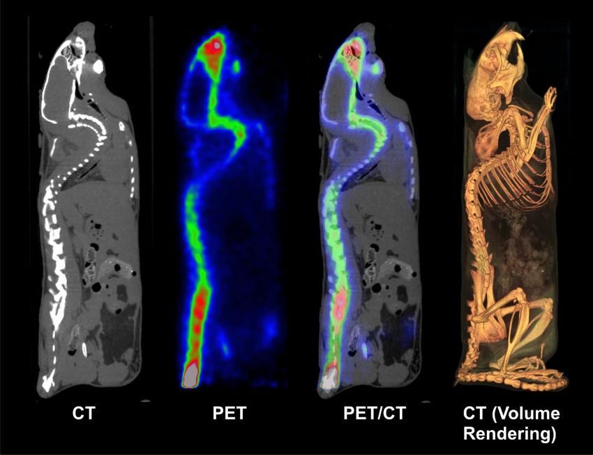
PET spatial resolution / 1

                              2                                       2
FWHM 1.2               d2               b2          0.0022D                   r2        p2
                                                         Non              Positron Parallax
                   Crystal size      Coding           collinearity         Range    error

       1.2     :    Degradation factor due to reconstruction
       d       :    Crystal pitch
       b       :    Coding error (range: 0-2 mm)
       D       :    Detector separation (i.e. gantry diameter)
       r       :    effective source size (including positron range)
       p       :    Parallax error

      * Derenzo & Moses, "Critical instrumentation issues for resolution
“From men to monkeys , to rats …..
                    to mice”
 Human PET
                                                               human

                                    *Images courtesy of Simon Cherry, UCLA
microPET

                            mouse
                                                                         rat

                             rat

                                                                     infant
                            mouse                                   monkey      4
 Torino, 24 Novembre 2011                   e-mail: alberto.delguerra@df.unipi.it
Spatial resolution requirements

                                                               5
Torino, 24 Novembre 2011   e-mail: alberto.delguerra@df.unipi.it
From the block detector to PSPMT’s

              “Block detector”       “1st generation” PSPMT

                                  Hamamatsu
                                  PS-PMT R2486.
                                  •50 mm Ø active area
                                  • 16 x + 16 y anodes

                                                  Small crystals can be used
                                                  (down to d = 1mm)

                                                                      Used in the
                                                                      YAP-(S)PET
                                                                      (Univ of Ferrara
                                                                      Italy,1993)

                                     Flood field irradiation (511 keV)
    •Large “b”                      of a matrix of scintillator YAP:Ce,
    •Limitations on minimum “d”       read by a Hamamatsu R2486
                                           (resistive readout)                    6
Torino, 24 Novembre 2011            e-mail: alberto.delguerra@df.unipi.it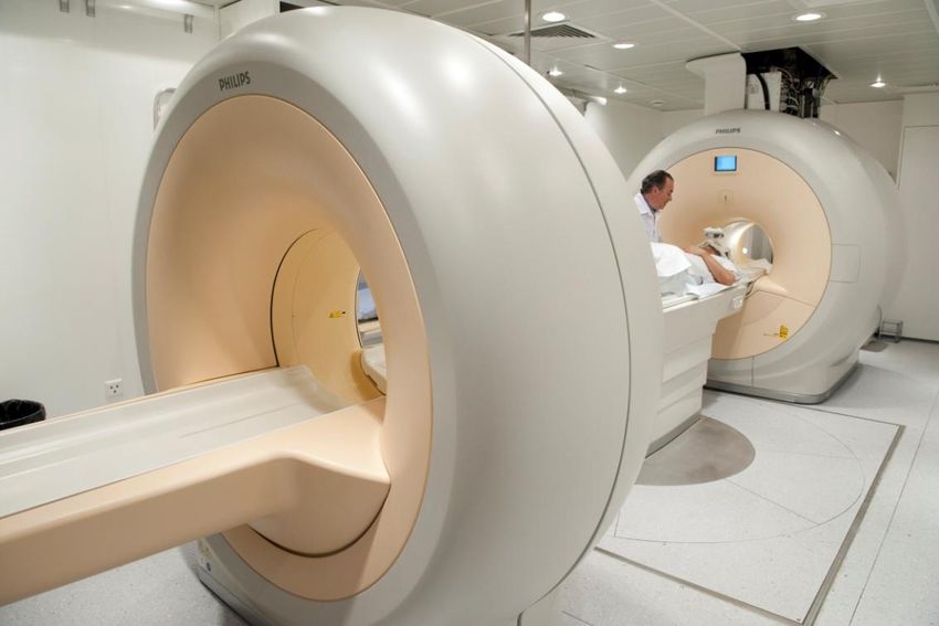
Coding problem:
                         “Individual coupling” with APD’s
                            or light sharing with PSPMT’s
  APD                                              2nd generation PSPMT
             APD array                              Hamamatsu
                                                    PS-PMTR8520-C12
                                                    • Active area 22 mm × 22mm
  Scintillator matrix                               • 6 x + 6 y anodes
    (BGO/LSO)

                                                   + Few channels to readout
                                                       (resistive chain)
                                                   + High gain and stability
                                                   - Non negligible coding error
                                                   - Pile-up increases with area
                                                   -
+ High spatial resolution (b=0)                                           PS-PMT Hamamatsu
                                                                          R7600-C8
+ No Pile-up
+ No scattering
   in the crystals   The detector module is composed
                       by a matrix of 8×4 LSO crystals
- Expensive
                      readout by a Hamamatsu S8550
- Many channels       (Pichler B., IEEE TNS 45 (1998)          Matrix 8 × 8 square
- Difficult tuning               1298-1302)                    fibres           8×8 LSO matrix

      Torino, 24 Novembre 2011                             e-mail: alberto.delguerra@df.unipi.it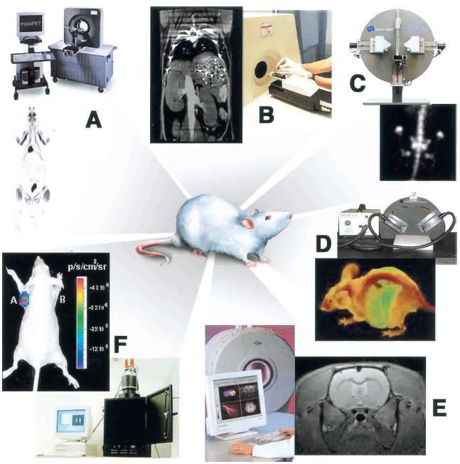
Today: advanced light sharing
                                        photodetectors
                         “light sharing” technique:                              “light sharing” technique:
PET History           Hybrid position sensitive APD                             Multi Anode flat panel PMT

PET Physics
and technology

PSDs in PET:
PMT

PSDs in PET:
solid state                                                                           MA-PMT (8×8 ch’s)
                                                                                      Hamamatsu H8500
Advanced PET                                                                      Active area 49 mm x 49 mm
                         Picture of a HPS-APD with four output
detectors: DOI
                                       connectors
and TOF

SiPMs for PET

Conclusions

                 Flood field image (241Am, 60 keV ) obtained with a 4x4   Flood field image (511 keV ) obtained with a 20x20
                           and 8x8 CsI(Tl) scintillating matrices            YAP:Ce scintillating matrix (resistive readout)
                                                                                                                               8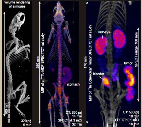
Spatial resolution of
                    of commercial scanners

                                           Commercial
                                           scanners do not
                                           show large
                                           differences in spatial
                                           resolution

       YAP-(S)PET          1.5   2.00

Torino, 24 Novembre 2011            e-mail: alberto.delguerra@df.unipi.it
Sensitivity requirements
Requirements
Imaging of low activity sources
low uptake processes such as in gene research
Possibility to study fast metabolic processes
with characteristic time comparable with the scanning time
Limitations
Brain receptor saturation
usually a maximum of 100 Ci can be injected to a mouse
Limitation on the volume
a maximum of 300 l can be injected to a mouse
Solutions
Utilization of radionuclides with a very high specific activity
such as PET short half-life radioisotopes:
15O (122 s), 13N (10 min), 11C (20 min), 18F (110min)
High geometry efficiency (large solid angle covered by detectors)
High detection efficiency (e.g. for crystals: high/medium Z, high density)
10
Torino, 24 Novembre 2011 e-mail: alberto.delguerra@df.unipi.itAbsolute sensitivity
of commercial scanners
Larger variations
can be observed
in the sensitivity
figure of merit.
YAP-(S)PET 50-850 2.3
Torino, 24 Novembre 2011 e-mail: alberto.delguerra@df.unipi.itSIEMENS
microPET Focus 220 / Inveon PET
18F-Paclitaxel biodistribution in rat MicroPET Focus 220 is a
PET only scanner using the
fiber technique
Rat heart 18F-FDG
The SIEMENS Inveon is dockable with a CT scanner 12
Torino, 24 Novembre 2011 e-mail: alberto.delguerra@df.unipi.itYAP-(S)PET II small animal scanner
Scanner configuration
Configuration: Four rotating heads
Scintillator: YAlO3:Ce (YAP:Ce)
Crystal size: 27 x 27 (1.5 x 1.5 x 20 mm3 each)
Photodetector: Position Sensitive PMT
Readout method: Resistive chain (4 channels)
FoV size: 40.5 mm axial 40.5 mm Ø
Collimators (SPECT): Lead (parallel holes) Scanner installed at the “Institute of
Head-to-head distance: 10-15 cm clinical Physiology (IFC-CNR)” within
the framework of the Center of
Excellence AmbiSEN of the
University of Pisa, Italy
Torino, 24 Novembre 2011 e-mail: alberto.delguerra@df.unipi.itHeart and bone metabolism in mouse
with 18F-FDG and18F-
Mouse with 18F-FDG Mouse with 18F- (post-mortem)
Horizontal slices: Gray and color scale
injection of 11 MBq of 18F-, 120 min. uptake time
Step-and-shoot acquisition 128 views/180°
(Acquisition time 60 min)
Transaxial sections
Total body (MIP)
120 min. uptake time
(Acquisition time 100 min)
Horizontal section
Voxel size 375 m × 375 m × 750 m
3D ML-EM reconstruction
14
Torino, 24 Novembre 2011 e-mail: alberto.delguerra@df.unipi.itSmall animal SPECT
instrumentation development
• The use of pinhole-collimators allows a large magnification
obtaining a high spatial resolution on medium-small field of view.
• The implementation of multi-pinholes increases the sensitivity
• Tipically based on large area NaI gamma camera similar to
clinical one.
================
• Other solutions based on solid state detectors are available.
• In this case the detector has a high intrinsic spatial resolution
(smaller magnification allowed) and are characterized by a high
energy resolution (multi-isotope imaging allowed)
Torino, 24 Novembre 2011 e-mail: alberto.delguerra@df.unipi.itSmall animal SPECT
collimator geometry
Parallel hole pinhole
D
h
L d
System spatial resolution System spatial resolution
2 2 2 2 2
D2 1 d L Rint De 1 d L Rint d L
System sensitivity System sensitivity
2 2 2
D D De
sin 3
L D h 4d
16
Torino, 24 Novembre 2011 e-mail: alberto.delguerra@df.unipi.itSmall animal SPECT
Effect of collimator-to-target distance
Geom. Resol.
Sensitivity
High sensitivity of
pinholes only at small d
(small FOV)
d d
FOV size
Parallel hole
Pinhole
d
17
Torino, 24 Novembre 2011 e-mail: alberto.delguerra@df.unipi.itMILabs – U-SPECT II
Bone scan: rat
Bone scan: mouse
Detector active area 510 x 381 mm
Crystal NaI(Tl), 9.5 mm thick
Continuous
Number of detectors 3 stationary
Based on a standard three heads NaI gamma
Number and size of the 75 / 0.15 -1.5 mm
pinholes camera (no rotation) equipped with multi-pinhole
FOV 28 mm x 140 mm
collimators.
60 mm x 240 mm
Spatial resolution 0.35-0.45 mm FWHM
Sensitivity >1500 cps/MBq
18
Torino, 24 Novembre 2011 e-mail: alberto.delguerra@df.unipi.itBioscan NanoSPECT
Features
Detector active area 230 x 215 mm
Crystal NaI(Tl), 9.5 mm thick
continuous
Number of detectors 4 (1,2 or 4)
Number and size of the 36 / 1.0 mm
pinholes
Also available as
HiSPECT: FOV 26 mm x 20 mm
transforms a Spatial resolution 0.8 mm FWHM
clinical SPECT Sensitivity 1640 cps/MBq
camera into an
Animal Imager
Multi-pinhole with elical scanning 19
Torino, 24 Novembre 2011 e-mail: alberto.delguerra@df.unipi.itSiemens Inveon SPECT
Detector active area 150 x 150 mm
Crystal NaI(Tl), 10 mm thick
Pixilated (2.2 mm pitch)
Mouse bone scan
Number of detectors 2 or 4
Number and size of the 1 or more / 0.5 – 3.0
pinholes mm
FOV variable
Spatial resolution variable
Sensitivity variable
20
Torino, 24 Novembre 2011 e-mail: alberto.delguerra@df.unipi.itGE eXplore speCZT
• Full-ring solid-state detector small animal SPECT
• Utilizes a cadmium zinc telluride (CZT) detector
• High-energy resolution to enable dual or triple radio-
nuclide imaging.
Stationary, full-ring, 10 detector design
Interchangeable, rotating cylindrical collimators
- Multi-slit: 80mm axial FOV, full 360-degree coverage
- Multi-pinhole: high resolution, full 360-degree coverage
Detector active area 124 x 124 mm
Crystal eV-CZT pixilated
Number of detectors 10 full ring stationary
Number and size of Multi pinhole
the pinholes Multi slit
FOV 80 mm axial with
multi slit
21
Torino, 24 Novembre 2011 e-mail: alberto.delguerra@df.unipi.itCT imaging @ Dipartimento di Fisica e
IFC-CNR
22
Torino, 24 Novembre 2011 e-mail: alberto.delguerra@df.unipi.itPET/CT image fusion
PET Co-registration
CT Advantages
Anatomical repere:
• CT provides high resolution
morphological information
map Better quantification:
• Attenuation correction of PET data
• CT may provide the shape and size of
the target for recovery coeff. correction
Additional information w.r.t. PET
• Using CT as a stand alone modality for
The map is scaled at 511 keV and blurred stem cell imaging
at the YAP-(S)PET spatial resolution 23
Torino, 24 Novembre 2011 e-mail: alberto.delguerra@df.unipi.itImaging PET/CT nel ratto Torino, 24 Novembre 2011 e-mail: alberto.delguerra@df.unipi.it
Whole body PET/CT (topo)

Torino, 24 Novembre 2011   e-mail: alberto.delguerra@df.unipi.it
Multimodality systems
                   Bioscan NanoSPECT/CT
         The BIOSCAN NanoSPECT is available also
               in combination with a CT module

                                                                       26
Torino, 24 Novembre 2011            e-mail: alberto.delguerra@df.unipi.it
Multimodality systems
SIEMENS Inveon
Available as integrated PET/SPECT/CT or dockable PET + SPECT/CT
(only 2 SPECT heads available when in combination with CT)
27
Torino, 24 Novembre 2011 e-mail: alberto.delguerra@df.unipi.itGE Triumph (Gammamedica)
X-SPECT®: CZT based SPECT Sub-System
LabPET™: APD based PET Sub-System
X-O™: Fast, Low Dose CT Sub-System
Triple isotope 18F-FDG CT
SPECT + CT PET + CT
28
Torino, 24 Novembre 2011 e-mail: alberto.delguerra@df.unipi.itRationale for PET/MR
PET
High sensitivity (10-11/10-12 mol/l)
Good spatial resolution (4mm for clinical system)
Functional info and quantitation
MRI
Good sensitivity (10-3/10-5 mol/l)
Excellent spatial resolution (1mm isotropic for clinical system)
Better soft tissue contrast with respect to CT
Anatomical info (but also functional)
No radiation dose
COMBINED PET/MR
29
Torino, 24 Novembre 2011 e-mail: alberto.delguerra@df.unipi.itTechnical Challenges in PET/MRI
Interference on PET (photomultiplier and electronics)
– Static magnetic field
– Electromagnetic interference from RF and gradients
Interference on MR (homogeneity and gradients)
– Electromagnetic radiation from PET electronics
– Maintaining magnetic field homogeneity
– Eddy currents
– Susceptibility artifacts
General Challenges
– Space
– Environmental factors (temperature, vibration…)
– Cost
30
Torino, 24 Novembre 2011 e-mail: alberto.delguerra@df.unipi.itEffect of magnetic field on positron range
2
A) B)
0
0 Tesla
-2
-4
4
-4 -2 0 2
4
2
9 Tesla (X-Y
plane)
0
-2
86Y (Emax = 3.15 MeV)
-4
4
-4 -2 0 2
4 Distance
A). Influence of the magnetic field on positron range, for (mm)
86Y (Emax=3.15 MeV) in water,illustrated by Monte Carlo B). Simulated positron range reduction
simulations obtained at 0 Tesla (top) and 10 Tesla for I-124 (Emax=2.14 MeV) in a 0 Tesla
(bottom) field. The 3-D tracks are projected onto a plane (top) and 9 Tesla (bottom) magnetic field.
perpendicular to the direction of the magnetic field. 31
Torino, 24 Novembre 2011 e-mail: alberto.delguerra@df.unipi.itPET/MRI solutions
Artistic cross-view of various potential designs of combined PET-MRI systems
a) tandem: The two scanners are mounted together back-to-back allowing sequential (like PET/CT)
rather than simultaneous acquisition,
b) insert: The PET scanner is inserted between the RF-coil and gradient set of the MR system,
c) full integration: the two systems are fully integrated within the same gantry 32
Torino, 24 Novembre 2011 e-mail: alberto.delguerra@df.unipi.itPhilips tandem PET/MRI
PET MRI
PET
MRI
PET-MRI
Photograph of the Philips whole-body Ingenuity TF PETMR system design installed at Geneva
University Hospital. A turntable patient handling system facilitates patient motion between the
Achieva X-series 3T MRI system on the right and the time-of-flight PET system on the left. 33
Whole-body MRI, PET and fused PET-MRI images are also shown.
Torino, 24 Novembre 2011 e-mail: alberto.delguerra@df.unipi.itThe advent of Solid State Photodetectors
CdZnTe
Used succesfully in SPECT by GE and used in
SPECT/MR prototype
APD = Avalanche Photodiode
Criticity: High performance Amplifier
Variation with T particularly relevant
SiPM (Silicon Photomultiplier)
Geiger-Muller APD
DSiPM = Digital SiPM
34
Torino, 24 Novembre 2011 e-mail: alberto.delguerra@df.unipi.itPrototype combined solution
Combined small animal
PET/MRI developed by
the University of
Tuebingen (Germany).
The PET insert is fully
integrated into a 7 Tesla
MRI system (ClinScan,
Bruker):
(a) Drawing of PET/MRI combination, showing the PET insert placed inside the MRI scanner, matching
the centers of both fields of view.
(b) Photograph of the MRI compatible PET insert, consisting of ten detector modules.
(c) Single PET detector module showing the LSO scintillator block, APD-array, and preamplifier built into
a MRI compatible copper shielding. 35
Torino, 24 Novembre 2011 e-mail: alberto.delguerra@df.unipi.itA Whole body PET/MRI solution from
SIEMENS
B
(A) Showing the basic components of the system where the PET detector ring is placed between
the RF coil and the RF body coil.
(B) Configuration of the detector block consisting of 8×8 LSO crystals readout by a matrix of 3×3 APDs.
(Courtesy of Siemens Medical Solutions). 36
Torino, 24 Novembre 2011 e-mail: alberto.delguerra@df.unipi.itSilicon Photomultiplier (SiPM) as a the most
promising solid state photodetector
The SiPM has all of the
SiPM are p-n diodes operating in Geiger mode,
characteristics: speed, QE,
which means that the bias voltage is above the
granularity, flexibility,
diode breakdown voltage.
robustness for a successful
implementation in small
In this way output is independent from input:
animal instrumentation.
the surface is divided into -cells (~1000/mm2)
LSO slab
Light
guide
Signal Ncell of
SiPM array
hit cells
+ High gain Triple layer detector block
+ Low noise
+ Good proportionality if Nphoton < Ncell
SiPM are insensitive to magnetic fields
compatible with MRI 37
Torino, 24 Novembre 2011 e-mail: alberto.delguerra@df.unipi.itPET/MRI
38
Torino, 24 Novembre 2011 e-mail: alberto.delguerra@df.unipi.itThank you! Torino, 24 Novembre 2011 e-mail: alberto.delguerra@df.unipi.it
Torino, 24 Novembre 2011 e-mail: alberto.delguerra@df.unipi.it
MRI based Attenuation Correction

Whole-body MRI            MR- map                CT- map          MRAC PET             CTAC PET

From left to right:
- whole-body T1 weighted gradient echo MRI sequence co-registered to CT image of the same patient,
- derived three-segment (soft tissue, lung and air) attenuation map (MRAC),
- CT-based attenuation map (CTAC),
- attenuation corrected PET images using MRAC
-- attenuation corrected PET images using CTAC.                                               41
           Torino, 24 Novembre 2011                      e-mail: alberto.delguerra@df.unipi.it
MRI based Attenuation Correction
MR- map Modified MR- map CT- map
LEFT: Attenuation correction maps derived from segmentation of T1 weighted
MRI followed by assignment of known linear attenuation coefficients to the lung
and soft tissue and addition of the scanner table template
MIDDLE: same image shown on the left after non-rigid alignment to the CT
attenuation map following removal of the PET-MR bed and addition of the CT
scanner bed CT
RIGHT: the CT-based attenuation map of the same patient.
42
Torino, 24 Novembre 2011 e-mail: alberto.delguerra@df.unipi.itCombined and simultaneous
PET/SPECT with the YAP-(S)PET II
One PET pair One SPECT pair
Low energy
shielding foil
Thanks to the planar detectors Removable
and the YAP:Ce scintillator the collimators
YAP-(S)PET can perform SPECT Cross contamination is reduced by:
imaging too on the same gantry
by adding parallel hole collimators • shielding the low energy single photons with a thin
lead slab in front of the PET detectors
• With a dual window subtraction technique for
selecting 99mTc gamma’s only in the SPECT data
Torino, 24 Novembre 2011 e-mail: delguerra@df.unipi.itPerformance: Transaxial resolution
Derenzo Phantom (SPECT) with 99mTc
1.2 mm
FBP (ramp filter) reconstruction was used on a
0.375 0.375 1.5 mm3 voxel space.
Sinograms were build using 140-250 keV energy
window (37 cps/MBq).
1.5 mm thick slices
Torino, 24 Novembre 2011 e-mail: delguerra@df.unipi.itDual tracer SPECT study
(Myoview-Annexin) on rat heart
99mTc-Annexin V
(Apoptosis)
99mTc-Myoview
(Perfusion)
Fusion
45
Torino, 24 Novembre 2011 e-mail: delguerra@df.unipi.itSimultaneous PET/SPECT imaging
with YAP-(S)PET
SPECT (99mTc) PET (18F) SPECT PET
5:1
10:1 Images of a section of an image quality
phantom: the phantom is filled with 99mTc
while the two holes are filled with 18F.
(99mTc/18F activity ratio 30:1).
30:1
50:1
Transaxial and coronal sections of a
Images of two small cylinders simultaneous PET/SPECT acquisition of two
simultaneously present in the FOV with capillaries. The left one was filled with 18.5
different SPECT/PET isotopes activity MBq of 99mTc, while the right with 370 kBq of
concentration ratio 18F (99mTc/18F activity ratio 50:1).
Torino, 24 Novembre 2011 e-mail: delguerra@df.unipi.itMolecular imaging technique for small
animals
A. PET Imaging on rats using 18F-FDG
showing glucose metabolism
B. CT Imaging of a mouse abdomen
after the injection of a contrast agent
C. SPECT Imaging of a mouse abdomen
after the injection of 99mTc “methylene
diphosphonate” showing the
accumulation of the tracer in bones.
D. Optical Imaging of a mouse showing
the fluorescence of GFP from liver,
abdomen, spinal chord and brain due
to the presence of cancer cells.
E. MRI image T2-weighted of a mouse
brain.
F. Bioluminescence optical imaging of a
mouse superimposed to the picture of
the animal.
MULTIMODALITY
Torino, 24 Novembre 2011 e-mail: alberto.delguerra@df.unipi.itYou can also read