Neonatal Major Thromboembolic Event: Challenging in Diagnosis and Treatment - Research Infotext
←
→
Page content transcription
If your browser does not render page correctly, please read the page content below
Journal of Clinical Research
            and Reviews [JCRR]
                Volume 2020 Issue 01
Case Report

Neonatal Major Thromboembolic Event: Challenging
in Diagnosis and Treatment
Najd S. Musibeeh*1 and Abdulrahman M. AlNemri2
1
 Pediatric Resident, Department of Pediatrics, College of Medicine King Saud University Kingdom of Saudi Arabia
2
 Professor of Pediatrics & Consultant Neonatologist, Department of Pediatrics, College of Medicine King Saud University Kingdom
of Saudi Arabia

Received: September 02, 2020; Accepted: September 15, 2020; Published: September 17, 2020

R-Infotext Citation: Najd S. Musibeeh NS, AlNemri AM (2020) Neonatal Major Thromboembolic Event: Challenging in Diagnosis
and Treatment. J Clinc Res & Rev 01(01): 12–16.

    Abstract
Neonatal purpura fulminans is a rare and rapidly progressive life-threatening disease that occurs secondary to acute disseminated
intravascular coagulation and hemorrhagic necrosis of the skin. Here we described a newborn that developed purpura fulminans of the
left leg at 16 hours of age with a normal thrombophilia screen. The infant was transferred to the neonatal intensive care unit at King
Saud University Medical City (King Khaled University Hospital) and treated with Heparin bolus followed by infusion along with other
supportive measures. With aggressive management, the left leg improved dramatically, and the baby was discharged home in good health
on low molecular weight heparin. The left foot underwent autoamputation one-month post-discharge from the intensive care unit.
Keywords: Newborn; Purpura; fulminans; Congenital; Protein deficiency; thrombosis; coagulopathy; anticoagulant

    Introduction                                                       screen, which constitutes a challenge in diagnosis and
                                                                       management.
Thromboembolic events are being more recognized and
diagnosed in the pediatric age group [1]. Among children,
neonates are more susceptible to develop symptomatic                    Case Report
venous thrombosis, with an incidence of 5.1 cases per
                                                                       A Saudi male was born at term by an elective cesarean
100,000 live births [2].
                                                                       section with an APGAR score of 6 and 8 at 1st & 5th
    Newborns have a distinct hemostatic system in terms                minutes, respectively, following a smooth antenatal
of quantity and quality compared to older children and                 course. This baby was the second child for healthy parents
adults [3]. The tendency to develop thrombosis in the                  with third-degree consanguinity. There was no history
neonatal period may arise from inherited or acquired                   thrombophilic event or any hematological disease in
factors and may vary in presentation from being                        the family. The baby developed secondary apnea, so he
asymptomatic to being life-or limb-threatening [4].                    was given positive pressure ventilation then admitted
Moreover, with the lack of evidence- based guidelines, it              to the Neonatal Intensive Care Unit (NICU) on non-
is challenging for the neonatologist to manage such cases.             invasive mechanical ventilation. His respiratory distress
   Here we are reporting a case of Saudi neonate who                   was worsening, so he was intubated and given two doses
developed neonatal purpura fulminans in the left lower                 of surfactant. Partial sepsis screen done then started
limb shortly after birth with a normal thrombophilia                   on broad-spectrum antibiotics. No umbilical arterial

June 10, 2020                                                                            J Clinc Res & Rev, Volume 01(01): 12–16, 2020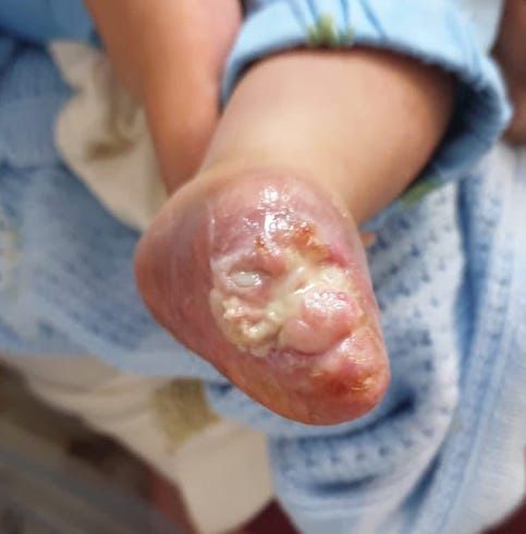
Neonatal Major Thromboembolic Event
catheterization was reported and no inotropic support. At the femoral and popliteal artery. Repeated thrombophilia
16 hours of age, the baby developed a pale and pulseless screen showed normal platelets with normal coagulation
left leg. Urgent doppler US done showed echogenic profile. Protein C level was 53%; Protein S Total 51% and
thrombus with lumen occlusion at the distal aspect of Antithrombin III 66%, all of which were normal for his
the external iliac artery & femoral artery. He was given age. Factor V Leiden & Factor II Mutation by molecular
Immediately Heparin Bolus 260 IU over 10 minutes, genetics was negative. Other laboratory tests, including
followed by continuous Infusion 28 unit/kg/hr. No focal serum electrolytes, renal functions, liver functions, blood
neurological deficits or seizures and no hematuria. Both cultures, and virology screening all were normal. The
cranial & abdominal ultrasound was reported normal. baby also underwent echocardiography and CT brain to
rule out any abnormality, and both were normal. Doppler
Over the next two days, the color changed to bluish
ultrasound showed no blood flow in the left popliteal
involving distal 1/3 left foot with mild ecchymosis of
artery, posterior tibial artery, and dorsalis pedis. The baby
the left buttock (Figure 1). The vascular surgeon was
was initially started on heparin infusion then shifted to
consulted and advised for urgent referral to a higher
Low Molecular Weight Heparin (LMWH) subcutaneous
surgery center for catheterization and thrombectomy, and
injections 1 mg/kg BID by a pediatric hematologist.
further thrombophilia and genetic workup.
Vascular Surgery, at this point, recommendedno surgical/
vascular intervention and continues conservative
management till the foot undergoes auto-amputation.
The patient was on broad-spectrum antibiotics, and a
nitroglycerin patch was applied over the distribution of
the posterior tibial artery of the left foot to improve the
perfusion.
Figure 1: Shows a well-defined gangrenous plaque over the
dorsum of the left foot and gangrenous toes
The patient was transferred to our center at King
Saud University Medical City (KSUMC) & King Khaled
University Hospital (KKUH). Laboratory values from the
referring hospital reported normal platelet count, Protein
C 27% (normal range 20–64%), and Antithrombin III
22% (normal range 39–87%).
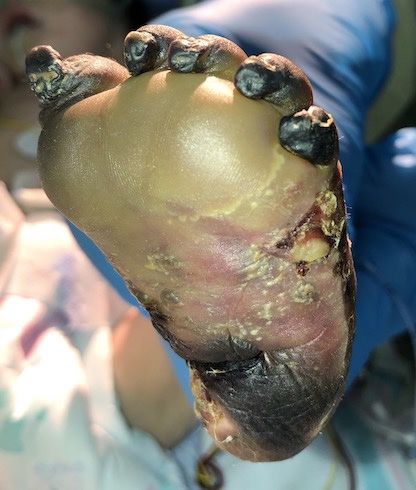
Figure 2: Shows total necrotic left foot upon admission to our NICU
The baby was admitted in our NICU connected to
mechanical ventilation, with stable gas and vital signs, After five days of therapy, the left foot showed dramatic
and a physical examination showed a gangrenous left foot improvement and healed without any signs of infection
(Figure 2). He was managed by a multidisciplinary team and no new infarcts (Figure 3). The baby was discharged
as left lower limb major thromboembolic phenomena in after 30 days of hospitalization on a prophylactic dose
June 10, 2020 J Clinc Res & Rev, Volume 01(01): 13–16, 2020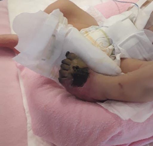
Neonatal Major Thromboembolic Event

of LMWH 4.5 mg once daily for three months. After                  Discussion
four weeks of discharge, the left foot underwent self-
amputation (Figure 4).                                            Thromboembolism is relatively rare in the pediatric
                                                                  population, but neonates are at the highest risk of
                                                                  developing such events [5]. The incidence of neonatal
                                                                  thrombophilia is trending up, especially in the critically
                                                                  ill neonate, due to the prematurity of their hemostatic
                                                                  mechanisms and interaction with external factors such as
                                                                  sepsis and invasive medical procedures [6].
                                                                      The causes could be inherited or acquired. Inherited
                                                                  causes are commonly due to homozygous protein
                                                                  C/S deficiency (quantitative) or functional protein
                                                                  defect (qualitative). Others such as Factor V Leiden or
                                                                  antithrombin deficiency [7,8]. Acquired causes are more
                                                                  frequent and often associated with sepsis and secondary
                                                                  consumptive coagulopathy or procedures such as
                                                                  complicated central catheterization [9].
                                                                      Here we report the case of a Saudi male newborn that
                                                                  developed a left lower limb thromboembolic event soon
                                                                  after birth while having a thrombophilia screen within
                                                                  normal ranges for his age. It raises the matter of having
                                                                  a functional anticoagulant protein defect rather than
                                                                  an actual deficiency, or a possible unreported central
Figure 3: shows improvement after five days of therapy
                                                                  catheterization.
                                                                      Worldwide, there are several cases of purpura
                                                                  fulminans reported during 1985 and 2019. All patients
                                                                  reported presented with necrotic lesions in the scalp
                                                                  and extremities during the first few days of life [10–
                                                                  17]. Homozygous protein C mutation was detected in
                                                                  patients with a history of consanguineous parents [10],
                                                                  whereas heterozygous mutation was found in newborns of
                                                                  unrelated parents [11,14,17,18]. Findley T et al. reported
                                                                  a newborn who had similar presentation and outcome
                                                                  as in our case but found to have undetectable levels of
                                                                  protein C with negative genetic testing [16].
                                                                      The prevalence of neonatal thrombophilia is still
                                                                  unknown in the Middle East and Arab countries. In
                                                                  Saudi Arabia, there are 6 case reports of neonatal purpura
                                                                  fulminans from 2002 to 2017. Two of the reported cases
                                                                  were secondary to hereditary homozygous protein C
Figure 4: Shows left foot after undergoing auto-amputation four
                                                                  gene mutation, while the rest of the cases didn’t undergo
weeks post-discharge                                              genetic evaluation [19,20]. Purpura Fulminans most
                                                                  commonly presented as intracranial hemorrhage and
                                                                  blindness secondary to retinal detachment and vitreous

June 10, 2020                                                                      J Clinc Res & Rev, Volume 01(01): 14–16, 2020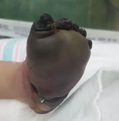
Neonatal Major Thromboembolic Event

hemorrhage [19–22]. Another case was secondary to                      purpura fulminans. Pediatr Hematol Oncol 20: 1–5.
group B streptococcus sepsis [23]. In all cases, there were            [View]
positive consanguineous marriages. All cases reported            9.    Hermansen MC, Hermansen MG (2005) Intravascular
unfavorable outcomes (blindness, seizure, hydrocephalus,               catheter complications in the neonatal intensive care
                                                                       unit. Clin Perinatal 32: 141–156. [View]
cerebral palsy, and death). On the contrary, our patient
                                                                 10.   Marciniak E, Wilson HD, Marlar RA (1985) Neonatal
has a good outcome, and he survived until discharge.
                                                                       purpura fulminans: a genetic disorder related to the
    In conclusion, it is essential to acknowledge that                 absence of protein C in blood. Blood 65(1):15–20.
healthy term neonates show reduced and highly variable                 [View]
physiological levels of natural anticoagulants such as           11.   Sen K, Roy A (2006) Management of neonatal purpura
                                                                       fulminans with severe protein C deficiency. Indian
protein C and protein S [24]. Some neonates may become
                                                                       Pediatrics 43: 542–545. [View]
symptomatic and develop purpura fulminans while others
                                                                 12.   Saeidi R, Gharaee R, Nobakht Z (2014) Neonatal
don’t, which could be attributed to a functional defect in
                                                                       Purpura Fulminans. Iranian Journal of Neonatology IJN
a natural anticoagulant protein rather than a decrease in              5: 34–37. [View]
its number. Reporting such cases from the Arab region            13.   Sharma S, Anbazhagan J, Plakkal N (2015) Neonatal
where consanguineous marriage rate is high is crucial in               purpura fulminans due to protein C deficiency Arch Dis
detecting and treating such a disease.                                 Child Fetal Neonatal Ed 100: F453. [View]
                                                                 14.   Shah R, Ferreira P, Karmali S, Le D (2016) Severe
                                                                       Congenital Protein C Deficiency: Practical Aspects of
 References                                                            Management. Pediatr Blood Cancer 63: 1488–1490.
                                                                       [View]
1.   Nowak-Gottl U, During C, Kempf-Bielack B, et al.            15.   Kizilocak H, Ozdemir N, Dikme G, Koc B, Celkan T
     (2003)Thromboembolic diseases in neonates and                     (2018) Homozygous protein C deficiency presenting
     children. Pathophysiology Haemost Thromb 33: 269–274.             as neonatal purpura fulminans: management with fresh
     [View]                                                            frozen plasma, low molecular weight heparin and protein
2.   Corrigan J (1992) Normal hemostasis in fetus and                  C concentrate. J Thromb Thrombolysis 45: 315–318.
     newborn: coagulation. In: Fetal and neonatal physiology.          [View]
     Saunders WB; Pg No: 1368–1371.                              16.   Findley T, Patel M, Chapman J, Brown D, Duncan AF
3.   Andrew M (1995) Developmental Hemostasis: Relevance               (2018) Acquired Versus Congenital Neonatal Purpura
     to thromboembolic complications in pediatric patients.            Fulminans: A Case Report and Literature Review. J
     Thromb Haemost 74: 415–425. [View]                                Pediatr Hematol Oncol 40: 625–627. [View]

4.   Saracco P, Bagna R, Gentilomo C, Magarotto M, Viano         17.   Zhang H, Bi X, Su Z, Tu X, Wang L, Shen B (2018) A
                                                                       novel compound heterozygous mutations in protein C
     A, et al. (2016) Clinical data of neonatal systemic
                                                                       gene causing neonatal purpura fulminans. Blood Coagul
     thrombosis. J Pediatr 171: 1. [View]
                                                                       Fibrinolysis 29: 216–219. [View]
5.   Raffini L, Huang YS, Witmer C, Feudtner C (2009)
                                                                 18.   Watanabe K, Arakawa Y, Yanagi M, Isobe K, Mori M, et
     Dramatic increase in venous thromboembolism in
                                                                       al. (2019) Management of severe congenital protein C
     children’s hospitals in the United States from 2001 to
                                                                       deficiency with a direct oral anticoagulant, edoxaban: A
     2007. Pediatrics 124:1001–1008. [View]                            case report. Pediatr Blood Cancer 66: 27686. [View]
6.   Andrew M, David M, Adams M, Ali K, Anderson R, et al.       19.   Abu-Amero KK, Al-Hamed MH, Al-Batniji FS (2003)
     (1994) Venous thromboembolic complications (VTE) in               Homozygous protein C deficiency with purpura
     children: first analyses of the Canadian Registry of VTE.         fulminans: report of a new case and a description of a
     Blood 83: 1251–1257. [View]                                       novel mutation. Blood Coagulation & Fibrinolysis 14:
7.   Reitsma PH, Bernardi F, Doig RG, Gandrille S, Greengard           303–306. [View]
     JS, et al. (1995) Protein C deficiency: a database of       20.   Al-Hamed MH, Albatniji F, Aldakheel GA, El-Faraidi H,
     mutations, 1995 update. Thromb Haemost 73: 876–889.               Al-Zahrani A, et al. (2016) Molecular characterization of
     [View]                                                            novel splice site mutation causing protein C deficiency.
8.   Dogan Y, Aygun D, Yilmaz Y, Kanra G, Secmeer G, et                Blood Coagulation & Fibrinolysis 27: 585–588. [View]
     al. (2003) Severe protein S deficiency associated with      21.   M Abdellatif, M Al Bakrah, M Qureshi, A Al-Nemri
     heterozygous factor V Leiden mutation in a child with             (2007) Protein C deficiency in a neonate with purpura

June 10, 2020                                                                      J Clinc Res & Rev, Volume 01(01): 15–16, 2020
Neonatal Major Thromboembolic Event
fulminans. The Internet Journal of Pediatrics and
Neonatology 8: 2. [View]
22. Al Hawsawi ZM, Alhejaili AA, Mohy Uddin M,
Abdelkarim ME (2016) Hereditary protein C deficiency
in a Saudi neonate with bilateral adrenal gland
hemorrhages: A rare case report. J Taibah Univ Med Sc
11:485–488. [View]
23. AlBarrak M, Abdulrahman Al-Matary (2013) Neonatal
purpura fulminans manifestation in early-onset group
B Streptococcal infection. Am J Case Rep 14: 315–317.
[View]
24. Williams MD, Chalmers EA, Gibson BE (2002)
Hemostasis and Thrombosis Task Force, British
Committee for Standards in Hematology. The
investigation and management of neonatal hemostasis
and thrombosis. Br J Haematol 119: 295–309. [View]
Corresponding author: Najd S. Musibeeh, Pediatric
*
Resident, Department of Pediatrics, College of Medicine,
PO Box. 2925, Riyadh 11461, Kingdom of Saudi Arabia;
Phone: 00966-11-4671099;
Fax: 00966-11-4671709;
E-mail: nmusibeeh@ksu.edu.sa
June 10, 2020 J Clinc Res & Rev, Volume 01(01): 16–16, 2020You can also read