AFFINITY SEPARATION AND CHARACTERIZATION OF IGG SUBFRAGMENTS BY FAST PROTEIN LIQUID CHROMATOGRAPHY WITH HITRAP_R PROTEIN A COLUMN
←
→
Page content transcription
If your browser does not render page correctly, please read the page content below
G. Ertürk et al. / Hacettepe J. Biol. & Chem., 2011, 39 (2), 133–138

Affinity Separation and Characterization of
IgG Subfragments by Fast Protein Liquid
Chromatography with HiTrap_r Protein A
Column
HiTrap_r Protein A Kolon İçeren Hızlı Protein
Sıvı Kromatografisi ile IgG Altbirimlerinin Afinite
Ayrılması ve Karakterizasyonu
Research Article / Araştırma Makalesi

Gizem Ertürk1, Nilay Bereli2, Lokman Uzun2, M. Aşkın Tümer1*
1
    Hacettepe University, Department of Biology, Beytepe, Ankara, Turkey
2
    Hacettepe University, Department of Chemistry, Beytepe, Ankara, Turkey

                                                               ABSTR AC T

I t has been previously reported that the antibody molecules can be cleaved into fragments by the proteolytic
  enzymes and these fragments provide information to determine an antibody’s structure and function.
Antibody fragments offer several advantages over intact antibodies for use in experimental applications and
immunochemical techniques. These fragments have smaller sizes and higher chemical and physical resistances.
In this study, papain was used for the digestion of human IgG and the resulting fragments were separated by
Fast Protein Liquid Chromatography (FPLC). For this purpose, GE Healthcare HiTrap_r Protein A FF column was
used, the resolution (Rs) and theoretical plate numbers (N) of the column were calculated. The unbound and
bound fragments were collected by the Frac 920 fraction unit of the FPLC system and the collected fragments
were analysed by enzyme-linked immunosorbent assay (ELISA) and sodium dodecyl sulphate polyacrylamide
gel electrophoresis (SDS-PAGE).

Key Words
Antibody fragmentation, Fab fragment, Fc fragment, FPLC, Protein A column.

                                                                    ÖZET

A   ntibadi moleküllerinin proteolitik enzimlerle alt birimlerine parçalanabildiği bilinmektedir. Bu alt birimler,
    antibadilerin yapısı ve işlevlerinin belirlenmesi çalışmalarında önemli veriler sunmaktadır. Deneysel
uygulamalarda ve immünokimyasal teknikler için antibadi alt birimleri, parçalanmamış bütün antibadi
moleküllerine göre önemli avantajlar sunmaktadır. Alt birimlerin boyutları daha küçüktür, kimyasal ve fiziksel
dirençleri daha yüksektir. Sunulan çalışmada, insan immunoglobulin G parçalanması için papain kullanılmıştır.
Elde edilen alt birimler, hızlı protein sıvı kromatografisi (FPLC) sistemi ile ayrılmıştır. Bu amaç için GE Healthcare
HiTrap_r Protein A FF kolon kullanılmış, ayırıcılık (Rs) ve teorik plaka sayısı (N) hesaplanmıştır. Kolona tutunan
ve tutunmayan alt birimler, FPLC sisteminin Frac 920 fraksiyon toplama birimi ile toplanmış ve enzim-bağlı
immünosorbent kit (ELISA) ve sodyum dodesil sülfat-poliakrilamid jel elektroforez (SDS-PAGE) yöntemleri ile
karakterize edilmiştir.

Anahtar Kelimeler
Antibadi parçalanması, Fab fragmanı, Fc fragmanı, FPLC, Protein A kolon.
Article History: Received October 15, 2010; Revised February 15, 2011; Accepted February 19, 2011; Avaliable Online: April 5, 2011.

Correspondence to: M. Aşkın Tümer, Hacettepe University, Department of Biology, Beytepe, Ankara, Turkey
Tel: +90312 297 8082                               Fax: +90312 299 2028                               E-Mail: tumer@hacettepe.edu.tr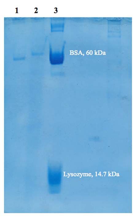
134    G. Ertürk et al. / Hacettepe J. Biol. & Chem., 2011, 39 (2), 133–138

      INTRODUCTION                                                            and have also been searching for alternative
                                                                              ways to obtain antibody-like receptors [16]. Thus,
                                                                              bioengineered antibody fragments that are smaller
      A    ntibodies are complex biomolecules that
           show specific binding capabilities for specific
      antigens [1]. Antibodies have a growing role in
                                                                              than antibodies and have the ability to bind antigen
                                                                              bivalently have been promoted by the researchers
      biomedical research and development. Antibody                           [17].
      based in vivo diagnostics and therapeutics are
      gaining wider approval from regulatory agencies                              In this study, Fab fragments were obtained by
      around the world [2]. Antibodies have been used                         the enzymatic hydrolysis of IgG molecule. For this
      for a long time for in vivo passive immunization                        purpose, papain was used and digestion process
      for bacterial and viral infections or contamination                     was performed at 37°C for 24 h. IgG fragments were
      with toxins. Owing to the high specificity of                           separated by Fast Protein Liquid Chromatography
      antibodies and the advanced techniques for the                          (FPLC). GE Healthcare rProtein A column was used
      production of specific antibodies, treatment                            for separation, the resolution (Rs) and theoretical
      strategies based on targeting to malignant                              plate numbers (N) of the column were calculated.
      cells are developing. Furthermore antibodies                            Fab and Fc fragments were collected by the Frac 920
      are important tools for the purification of their                       fraction unit of the FPLC system and in the last part,
      specific antigens for pharmaceutical applications                       ELISA and SDS-PAGE analysis were performed.
      and for research purposes [3]. The specific
      interactions between antibody and antigen allow                         MATERIALS AND METHODS
      the functional manipulation of target antigens.
      The most common applications of antibodies                              Materials
      are in immunosensors [4], immunoassays [5-7]                            Human immunoglobulin G (IgG), papain, ethylene-
      and immunoaffinity separations [8-10]. Common                           diamine tetraacetic acid (EDTA), iodoacetamide
      point of all of these techniques is the binding of                      were ob-tained from Sigma Chemical Co. (St.
      an analyte to an antibody. An antigen specific                          Louis, USA). All other chemicals were of reagent
      antibody fits its unique antigen in a highly specific                   grade and purchased from Merck A.G. (Darmstadt,
      manner. This unique property of antibodies is the                       Germany). All water used in the experiments was
      key to their usefulness in all applications where                       purified using a Barnstead (Dubuque, IA) ROpure
      only the specific antigen fits into the antibody                        LP® reverse osmosis unit with a high flow cellulose
      binding site [1].                                                       acetate membrane (Barnstead D2731) followed by
                                                                              a Barnstead D3804 NANOpure® organic/colloid
          Proteolytic digestion provides important                            removal and ion exchange packed-bed system.
      information to determine an antibody molecule’s
      structure and function. Antibody molecules can be                       Preparation of Fab fragments
      cleaved into fragments and each of these fragments                      Papain digestion of IgG
      have a distinct activity. A number of enzymes like                      Digestion of IgG to produce Fab fragments was
      papain, pepsin, ficin, bromelain and elastase can                       performed in 20 mM sodium phosphate buffer
      be used for this purpose, papain being the most                         (pH: 7.0) containing 20 mM EDTA. The digestion
      frequently used [11]. Because of their smaller size                     was initiated by the addition of papain to the
      as functional components of the whole molecule,                         digestion solution containing 0.5 mg/mL IgG
      antibody fragments offer several advantages over                        at an enzyme to antibody ratio of 1:20. The
      intact antibodies for use in certain immunochemical                     resulting solution was incubated at 37ºC for 24
      techniques and experimental applications. While Fab                     h. Reaction was stopped by adding 30 mM fresh
      fragments have the advantage of being the smallest                      iodoacetamide to the digestion solution.
      antigen binding fragments, F(ab) ’ fragments derive
                                        2
      special utility from having no Fc domain and                            Analysis of digestion products by fast protein
      somewhat smaller size than whole IgG [12-15]. For                       liquid chromatography (FPLC)
      this purpose, researchers have attempted to use                         The digestion mixtures were analyzed by Fast
      smaller, more stable counterparts of antibodies                         Protein Liquid Chromatography (FPLC). FPLC
G. Ertürk et al. / Hacettepe J. Biol. & Chem., 2011, 39 (2), 133–138 135
separation was performed using an AKTA-FPLC on a 10% SDS-PAGE at a constant current of 23
(Amersham Bioscience, Uppsala, Sweden) system mA for 6 h and then stained with Coomasie Blue.
equipped with a UV detection system. The system
includes M-925 mixer, P-920 pump, UPC-900 ELISA analysis
monitor, INV-907 injection valve and Frac920 Intact IgG and unbound fraction of papain-
fraction collector. Separation was achieved on digested IgG were analysed by an enzyme-linked
a 2.5 cm x 0.7 cm Hi_Trap_r Protein A_FF (GE, immunosorbent assay method (ELISA). Human
Healthcare) column. FPLC mobile phases A and anti-IgG (Sigma, I-9384) diluted 1/1000 in 50 mM
B were prepared with 20 mM sodium phosphate NaHCO3, pH 9.6, was adsorbed to PVC microtitre
buffer (pH: 7.0) and 0.1 M glycine-HCl buffer (pH: plates at 4ºC for 12 h. The plates were washed with
3.0) respectively. The chromatographic sepa- PBS containing 0.05% Tween 20 (washing buffer)
ration was performed at 1 mL/min flow rate. and blocked with PBS containing 0.05% Tween
After a 10 min starting period with 100% loading 20, 1.5% BSA, and 0.1% sodium azide (blocking
buffer A, a linear gradient started from 0% B buffer). Samples (2.5 mL, neutralized with 0.5 mL
to 100% B in 3 min, continued with 4 min 100% of 1.0 M trisodium citrate) or controls containing
eluent B and finished last 6 min 100% buffer known amounts of IgG were added and incubated
A. 2 mL of protein mixture was applied to the at 37ºC for 1 h. Bound IgG was detected with the
column and absorbance was monitored at 280 anti IgG labeled with biotin followed by peroxidase-
nm. The separation was performed at 27ºC. KBr conjugated streptavidin and o-phenylenediamine.
was used as the void marker. Capacity factor (k’) The absorbance was measured at 492 nm.
and separation factor () were calculated as k’ =
(tR-to)/to, =k2’/k1’, where tR is the retention time RESULTS AND DISCUSSION
of the protein and to is the retention time of the
void marker (KBr), k2’ is the capacity factor for FPLC studies
Fc fragment and k1’ is the capacity factor for Fab Protein A is a bacterial antibody binding protein
fragment. The resolution (Rs) and theoretical and interacts mainly with the Fc part of IgG [18].
plate numbers (N) were calculated using the A 24 h period of papain digestion of IgG resulted in
following equations: Fab and two major Fc fragments. Protein A bound to
Fc fragments of papain-digested IgG and was totally
N = 5.54 (tR / w0.5)2 (1) non-reactive with the Fab regions.
Rs = 2 (tR,2 – tR,1) / (w2 + w1) (2) For the calibration of the FPLC system, different
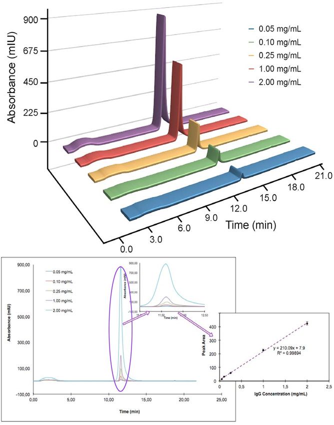
concentrations of IgG solutions were loaded to
where w0.5 is the peak height at the the column. Figure 1 shows the chromatogram of
corresponding peak height fraction, tR,1 and tR,2 are standart IgG solutions having concentrations in
the retention times of two adjacent peaks, w1 and range of 0.05 mg/mL and 2.0 mg/mL (flow rate: 1
w2 are the widths of the two adjacent peaks at the mL/min, T: 27°C). The 24 h papain digestion of IgG
baseline. results in two major fragments which are Fab and
Fc fragments. Since Protein A specifically interacts
The unbound and bound fragments were with the Fc region of IgG molecule, the first peak in
collected by the Frac 920 fraction unit of the the unbound region in Figure 2 represents the Fab
FPLC system and the solvent part of the collected fragment of digested IgG molecules.
fragments were removed by the freeze dryer.
As shown in Figure 2, a successful separation
SDS-PAGE analysis was observed and Fab and Fc fragments were
For the determination of molecular weights and separately collected. The retention times of the
purities of unbound and bound fractions, sodium fragments were achieved at 3.06 and 11.64 min,
dodecyl sulfate polyacrylamide gel electropho- respectively. The calculated chromatographic
resis (SDS-PAGE) was performed. Each sample parameters such as tR, N, k’, , Rs values are
was loaded at a volume of 5 μL and then separated summarized and given in Table 1. Rs value was136 G. Ertürk et al. / Hacettepe J. Biol. & Chem., 2011, 39 (2), 133–138
Figure 1. FPLC chromatogram of standart IgG solutions.
Flow-rate: 1.0 mL/min, T: 27°C, Detection was performed at 280 nm.
calculated as 4.70 for Fc fragment. Because the Rs SDS-PAGE analysis
value should be higher than 1.0 for a good resolution The purity of unbound and bound fractions of
of two peaks in such a chromatography system, the papain digested IgG was determined by SDS-PAGE
results for the resolution of Fab fragment/Fc fragment as described before. In Figure 3, Lane-1 represents
can be accepted as good resolution values. the Protein A unbound fraction of papain digestedG. Ertürk et al. / Hacettepe J. Biol. & Chem., 2011, 39 (2), 133–138 137
Figure 2. FPLC chromatogram of IgG after digestion with
papain. IgG concentration: 0.5 mg/mL, Flow-rate: 1.0 mL/
min, T: 27°C. Detection was performed at 280 nm.
Table 1. Chromatographic separation data.
Fragment tR N k’ Rs
Fab 3.06 52.39 1.19 6.14 -
Fc 11.64 1089.60 7.31 - 4.70
IgG, Lane-2 is the Protein A bound fraction of
papain digested IgG and Lane-3 contains proteins
as markers with known molecular weights; 60
kDa bovine serum albumin, 14.7 kDa lysozyme.
Molecular weight of immunoglobulin (IgG) is 150
kDa and papain digests IgG molecule from the
Figure 3. SDS-PAGE of IgG fragments from Protein A
hinge region. Therefore, polypeptide chains which column. The fractions were assayed by SDS-PAGE using
have 50 kDa molecular weight were formed as the 10% separating gel (9 cm x 7.5 cm) was stained with 0.25%
result of papain digestion in this study. In Figure 3, (w/v) Coomassie Brillant R 250 in acetic acid–methanol–
water (1:5:5, v/v/v) and destained in ethanol–acetic acid–
the bands having 50 kDa molecular weight were water (1:4:6, v/v/v). Lane 1, unbound Fab fragment; Lane
clearly seen. The results show that enzymatic 2, bound Fc fragment; Lane 3, marker proteins (BSA and
hydrolysis and fractionation of fragments with Lysozyme).
FPLC are achieved, respectively.
Fragments of antibodies are also widely
used as immunochemical tools, diagnostic and
ELISA results
therapeutic reagents because of the more rapid
The amount of IgG in 1:1 diluted plasma sample
pharmacokinetic answer and avoided potential
was found as 1280 mg/dL and the amount of IgG
non-specific binding of intact antibody molecules
in the unbound fraction of papain digested IgG
to constant region (Fc) receptors on lymphocytes.
was found as 100 mg/dL.
Since antibody fragments usually retain the antigen
binding activity of the original antibody, researchers
CONCLUSION
have attempted to use bioengineered antibody
fragments instead of intact antibody.
Antibodies have been used for a long time as
probes for research in biology and biopharmaceu-
In this study, we digested IgG molecule with
ticals. They are also used for immunodiagnosis
papain for gaining Fab and Fc fragments. For
and immunotherapy for several human diseases.
this purpose, we utilized Fast Protein Liquid
Some understandings of the structure of
Chromatography system that has been introduced
antibodies are obtained on degrading them
as one of the most preferred methods for purifi-
with proteolytic enzymes and examining the
cation of antibody fragments that are prepared
fragments which retain antibody activity.138 G. Ertürk et al. / Hacettepe J. Biol. & Chem., 2011, 39 (2), 133–138
by enzymatic digestion of whole Immunoglobulins. 8. M.L. Yarmush, A.M. Weiss, K.P. Antonsen, D.J. Odde,
For the separation of Fab and Fc fragments, we used D.M. Yarmush, Immunoaffinity purification basic
HiTrap_r Protein A column and we collected the principles and operational considerations, Biotechnol.
fragments by the fraction unit of the FPLC system. Adv., 10 (1992) 413.
9. S. Özkara, H. Yavuz, A. Denizli, Purification of
In the last part, we performed the SDS-PAGE and
Immunoglobulin G from human plasma by metal-
ELISA analysis of the fragments. As a result, we
chelate affinity chromatography, J. Appl. Polym. Sci.,
can digest human IgG molecule with papain and
89 (2003) 1567.
we can separate the IgG subfragments from each 10. N. Bereli, S. Akgöl, H. Yavuz, A. Denizli, Antibody
other by Fast Protein Liquid Chromatography. So purification by Concanavalin A affinity chromatogra-
that, we can obtain Fab and Fc fragments which phy, J. Appl. Polym. Sci., 97 (2005) 1202.
have several advantages over intact IgG for use in 11. Q. Luo, X. Mao, L. Kong, X. Huang, H. Zou, High-
immunochemical and therapeutic applications. performance affinity chromatography for characteri-
zation of human immunoglobulin G digestion with
papain, J. Chromatogr. B, 776 (2002) 139.
REFERENCES 12. D. Beale, Molecular fragmentations. Some applica-
tions in immunology., Dev Comp. Immunol., 11 (1987)
287.
1. T. Vo-Dinh, B. Cullum, Biosensors and biochips:
13. S.J. Boguslawski, Improved procedure for preparation
advances in biological and medical diagnostics,
of F(ab’)2 fragments of mouse IgGs by papain
Fresenius J. Anal. Chem., 366 (2000) 540.
digestion, J. Immunol. Meth., 120 (1989) 51.
2. L. Uzun, R. Say, A. Denizli, Porous poly(hydroxyethyl
14. K.M Wilson, A new enzymatic method to obtain high-
methacrylate) based monolith as a new adsorbent for
yield F(ab’)2 suitable for clinical use from mouse IgG1.,
affinity chromatography, React. Functl. Polym., 64
Mol. Immunol., 28 (1991) 69.
(2005) 93.
15. R.S. Kerbel, B.E. Elliot, Detection of Fc receptors.,
3. K. Huse, H. Bohme, G.H. Scholz, Purification of
Method. Enzymol., 93 (1983) 113.
antibodies by affinity chromatography, J. Biochem.
16. K. Haupt, K. Mosbach, Plastic antibodies: develop-
Biophys. Methods, 51 (2002) 217.
ments and applications, Tibtech November, Vol 16,
4. C.L. Morgan, D.J. Newman, C.P. Price, Immunosensors:
1998.
technology and opportunities in laboratory medicine,
17. R. Cortese, Identification of biologically active
Clin. Chem., 42 (1996) 193.
peptides using random libraries displayed on phage,
5. D.S. Hage, Immunoassays, Anal. Chem., 67 (1995)
Curr. Opin. Biotechnol., 6 (1995) 73.
455R.
18. L. Uzun, D. Türkmen, V. Karakoç, H. Yavuz, A. Denizli,
6. R.S Yalow, The Nobel lectures in immunology. The
Performance of protein A based affinity membranes
Nobel Prize for Physiology or Medicine, 1977, awarded
for antibody purification, J. Biomat. Sci. Polym. Ed.,
to Rosalyn S. Yalow Scand., J. Immunol., 35 (1992) 4.
2011, In Press, doi: 10.1163/092050610X538731.
7. T. Porstmann, S.T. Kiessig, Enzyme immunoassay
techniques: an overview., J. Immunol. Methods, 150
(1992) 5.You can also read