Case Report Myasthenia gravis and dyspnea associated with Castleman's disease
←
→
Page content transcription
If your browser does not render page correctly, please read the page content below
Int J Clin Exp Med 2018;11(7):7491-7494
www.ijcem.com /ISSN:1940-5901/IJCEM0064702

Case Report
Myasthenia gravis and dyspnea associated
with Castleman’s disease
Laiwei Cao, Zhentao Zhang, Zhaohui Zhang

Department of Neurology, Renmin Hospital of Wuhan University, Wuhan 430060, China
Received August 30, 2017; Accepted March 20, 2018; Epub July 15, 2018; Published July 30, 2018

Abstract: Castleman’s disease is a rare lymphoproliferative disorder. It is usually benign and the resection of the
tumor is curative. However, Castleman’s disease is occasionally associated with autoimmune diseases such as
paraneoplastic pemphigus, POEMS syndrome (polyneuropathy, organomegally, endocrinopathy, monoclonal gam-
mopathy, and skin changes), which usually leads to poor outcomes. Here we report a patient with Castleman’s
disease who developed progressive myasthenia gravis and dyspnea. Myasthenic symptoms were responsive to
acetylcholinesterase inhibitors and corticosteroids, but the patient’s respiratory functions continued to decline in
spite of intensive medical therapy. To our knowledge, this is the second case of Castleman’s disease associated
with simultaneous myasthenia gravis and dyspnea. Considering the immunostimulatory nature of the tumor, we
propose that the myasthenia gravis and bronchiolitis obliterans are both components of the aberrant systemic im-
munoreaction.

Keywords: Castleman’s disease, myasthenia gravis, bronchiolitis obliterans, respiratory insufficiency

Introduction                                                  Case report

Myasthenia gravis is an acquired autoimmune                   A 25-year old woman presented to our clinic
disorder characterized by skeletal muscle fa-                 with dyspnea of ten months’ duration. She had
tigue and weakness. The clinical feathers of                  no recent infections and her medical history
myasthenia gravis are mediated by antibodies                  was unremarkable. She experienced increasing
to the nicotinic acetylcholine receptor (nAChR)               inspiratory dyspnea on exertion. She also expe-
at the neuromuscular junction. Myasthenia gra-                rienced a nonproductive cough and oral muco-
vis is often associated with thymoma or thymic                sa ulcerations. A neck computed tomography
hyperplasia. Other diseases that are known to                 (CT) confirmed the presence of a well-defined
be associated with myasthenia gravis include                  and margined mass near the trachea (Figure
rheumatoid arthritis, pernicious anemia, sys-                 1). Excision of the mass was performed, and
temic lupus erythematosus, sarcoidosis, Sjo-                  a 3.3 × 2 × 5.3 cm uniformly solid lobulated
gren’s disease, and mitochondrial myopathy                    mass was removed. Histologic evaluation of
[1]. These associated conditions suggest an                   the mass showed immunoproliferative chan-
autoimmune basis for myasthenia gravis. Al-                   ges with abnormal follicular architecture, oblit-
though rare, a few myasthenia gravis cases                    eration of sinuses, hypervascular interfollicular
have been reported to be associated with Cas-                 tissue, and concentric perivascular arrange-
tleman’s disease, a lymphoproliferative disor-                ment of the mantle zone (Figure 2). The patient
der [2, 3]. We present the case of a patient with             was diagnosed with Castleman’s disease.
myasthenia gravis and progressive dyspnea
related to Castleman’s disease. Furthermore,                  Unfortunately, the dyspnea did not subside
the onset of the myasthenic symptoms occurr-                  postoperatively. Her respiratory symptoms gra-
ed 5 months after the resection of the tumor.                 dually progressed to the point that she requir-
This phenomenon suggests that the patient                     ed ventilatory support. Physical examination
suffered from a progressive systematic immune                 revealed three depressions sign and rhonchi.
disturbance.                                                  Pulmonary function test showed severe ob-
A case report of myasthenia gravis
trolled by these drugs, but the
pulmonary symptoms progres-
sively worsened. The condi-
tion of the patient deteriorated
in spite of intensive drug tre-
atment. She died four weeks
later from respiratory insuffi-
ciency.
Discussion
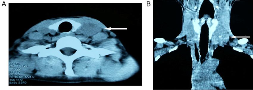
Figure 1. (A) Neck CT scan and (B) CT reconstruction image of neck. Neck
computed tomography (A) and the reconstructive imagine (B) showing a Castleman’s disease is a rare
well-defined and margined homogeneous mass at the right of the trachea
lymphoproliferative disease ch-
(arrow).
aracterized by non-clonal lym-
ph node hyperplasia. This tu-
mor may adopt a unicentric or multicentric pre-
sentation. The former is usually asymptomatic
and amenable to surgical treatment, and the
latter often exhibits complicated symptomatic
manifestations. The most common location of
these tumors is retroperitoneal or mediastinal.
Pathologically it is classified into the common
hyaline vascular type, the rarer plasma cell
type, and the mixed type. About 20-40% of
the tumors are discovered incidentally. Similar
to the present case, it usually manifests as a
solitary mass with marked enhancement on
computed tomography. The etiology and patho-
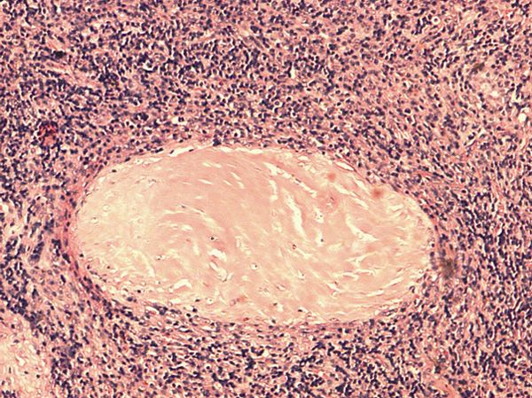
Figure 2. Histology of a patient with Castleman’s dis- genesis of Castleman’s disease are poorly
ease. Histopathologic sections (hematoxylin & eosin understood. Our current understanding of the
stain) show typical features of hyaline vascular Cas- pathogenesis of Castleman’s disease points
tleman disease with hyalinized vessels within germi- to reactive follicular hyperplasia in response to
nal follicles that are surrounded by onion-like lym-
phocytes. (hematoxylin and eosin staining, × 400). an unknown antigenic stimulus [4].
An association of multicentric Castleman’s dis-
structive ventilatory disturbances. There was ease with HIV infection has been demonstrat-
bronchiectasis, but no evidence of multicen- ed. In spite of the fact that unicentric
tric or residual tumors on computed tomogra- Castleman’s disease is frequently asymptom-
phy scan. No enlarged lymph nodes were found atic, there is a remarkable association of
around the neck, axillary fossa, or retroperito- Castleman’s disease with other diseases, such
neal under ultrasonic tomography. The comput- as lichen planus, pemphigus vulgaris, POEMS
ed tomography findings and clinical symptoms syndrome, myasthenia gravis, nephrotic syn-
were characteristic of bronchiolitis obliterans. drome, amyloidosis, plasmacytoma, and rheu-
Furthermore, she gradually developed gene- matoid arthritis [4-6]. The association of Cas-
ralized weakness and bilateral ptosis three tleman’s disease with various immune phe-
months after the operation. The neurologic nomena and immunological diseases presents
examination, electromyography, neostigmine a challenging pathophysiological model of anti-
test, and fatigability test were suggestive of bodies and variable immunodeficiencies.
myasthenia gravis.
We present this case because of the rare asso-
Because she was receiving ventilatory support ciation of myasthenia gravis, bronchiolitis oblit-
and could not easily swallow, she was treated erans, and Castleman’s disease. Myasthenia
with continuous intravenous administration of gravis is caused by autoantibodies against
neostigmine (4 mg/d) and prednisolone (40 nAChRs at neuromuscular junctions, resulting
mg) in the morning. The myasthenia was con- in symptoms of muscular weakness and fatiga-
7492 Int J Clin Exp Med 2018;11(7):7491-7494A case report of myasthenia gravis
bility. However, the mechanisms that initiate medication and lung transplantation is the
and maintain the autoimmune response are only potential treatment. It should be noted
not well understood. In addition to the antibo- that bronchiolitis obliterans is also a common
dy-mediated autoimmune response, T-cells are devastating complication of lung transplanta-
also thought to participate in the pathogenesis tion, which affects up to 50-60% of patients
of this disease. Several cases of Castleman’s who survive five years after surgery [11].
disease associated with myasthenia gravis Theoretically, the underlying autoimmune dis-
have been reported [2, 3, 5]. In one case the ease should be well controlled before the lung
myasthenic symptoms improved after removal transplantation to decreases the risk of devel-
of the tumor, but the condition deteriorated opment of bronchiolitis obliterans in the trans-
after several months [2]. In another case, the planted organ [6].
myasthenic symptoms were well controlled by
drugs. In our case, the myasthenic symptoms Acknowledgements
emerged five months after the surgery. These
This work was supported by the National Na-
cases suggest that the association between
tural Science Foundation of China [Grant no.
the two diseases is more than coincidental, but
81571249 to Zhentao Zhang, and Grant no.
they share common underlying autoimmune
81671051 to Zhaohui Zhang].
mechanisms.
Disclosure of conflict of interest
Bronchiolitis obliterans is particularly notewor-
thy because it is the most severe concomitant None.
disease state of Castleman’s disease [5-8].
Bronchiolitis obliterans is characterized by Address correspondence to: Dr. Zhaohui Zhang,
inflammation of the bronchioles with elabora- Department of Neurology, Renmin Hospital of
tion of fibrous granulation tissue and bronchial Wuhan University, Wuhan 430060, China. Tel:
exudates into the lumens. It is a lethal condi- +86-15172314891; E-mail: zzhan2@emory.edu;
tion with the common endpoint of functional zhangzhaohui18@hotmail.com
obstruction of the bronchioles. Indeed, all
reported cases of Castleman’s disease with References
bronchiolitis obliterans have been fatal, the
[1] Sthoeger Z, Neiman A, Elbirt D, Zinger H, Ma-
most serious pathological manifestation being
gen E, Burstein R, Eitan S, Abarbanel J, Mozes
rapidly progressive respiratory insufficiency
E. High prevalence of systemic lupus erythe-
[5-7]. Our patient died eleven months after matosus in 78 myasthenia gravis patients: a
the appearance of the first symptoms of bron- clinical and serologic study. Am J Med Sci
chiolitis. The fatal outcome was the result of 2006; 331: 4-9.
the bronchiolitis obliterans. The most common [2] Paşaoğlu I, Doğan R, Topçu M, Güngen Y. Mul-
mucocutaneous manifestation of Castleman’s ticentric angiofollicular lymph-node hyperpla-
disease is paraneoplastic pemphigus [9, 10]. sia associated with myasthenia gravis. Thorac
The mucous membrane of the mouth was Cardiovasc Surg 1994; 42: 253-6.
observed in our case, but we did not ob- [3] Day JR, Bew D, Ali M, Dina R, Smith PL. Castle-
serve polymorphous mucocutaneous erupti- man’s disease associated with myasthenia
ons, which are pathognomonic of paraneoplas- gravis. Ann Thorac Surg 2003; 75: 1648-50.
[4] Roca B. Castleman’s disease. A review. AIDS
tic pemphigus.
Rev 2009; 11: 3-7.
Castleman’s disease is an essentially benign [5] Chorzelski T, Hashimoto T, Maciejewska B, Am-
agai M, Anhalt GJ, Jablonska S. Paraneoplastic
condition. Surgical resection is the mainstay
pemphigus associated with Castleman tumor,
of treatment for uncomplicated cases. How
myasthenia gravis and bronchiolitis obliterans.
ever, complications may develop even after J Am Acad Dermatol 1999; 41: 393-400.
removal of the tumor. This may create a diag- [6] Chin AC, Stich D, White FV, Radhakrishnan J,
nostic and therapeutic dilemma for surgeons. Holterman MJ. Paraneoplastic pemphigus and
Castleman’s disease-associated myasthenia bronchiolitis obliterans associated with a me-
gravis may be controlled by acetylcholinester- diastinal mass: a rare case of Castleman’s dis-
ase inhibitors and corticosteroids. However, ease with respiratory failure requiring lung
bronchiolitis obliterans is not responsive to any transplantation. J Pediatr Surg 2001; 36: E22.
7493 Int J Clin Exp Med 2018;11(7):7491-7494A case report of myasthenia gravis
[7] Radzikowska E, Pawlowski J, Chabowski M, [10] Wang J, Zhu X, Li R, Tu P, Wang R, Zhang L, Li T,
Langfort R. Constrictive bronchiolitis obliterans Chen X, Wang A, Yang S, Wu Y, Yang H, Ji S.
in patient with Castelman’s disease. Monaldi Paraneoplastic pemphigus associated with
Arch Chest Dis 2005; 63: 226-9. Castleman tumor: a commonly reported sub-
[8] Hoffman MA, Qiao X, Anhalt GJ. CD8+ T type of paraneoplastic pemphigus in China.
lymphocytes in bronchiolitis obliterans, para- Arch Dermatol 2005; 141: 1285-93.
neoplastic pemphigus, and solitary Castle- [11] Nicod LP. Mechanisms of airway obliteration
man‘s disease. N Engl J Med 2003; 349: 407- after lung transplantation. Proc Am Thorac Soc
8. 2006; 3: 444-9.
[9] Alarcón-Torres I, Bastida-Iñarrea J, Rodríguez-
Salido MJ, Gómez-Duaso J, Rua-Figueroa I,
García-Aguilar GD, Martín-Alfaro R. Paraneo-
plastic pemphigus associated with Castle-
man’s disease: usefulness of the laboratory of
autoimmunity in the diagnosis of this disease.
Ann N Y Acad Sci 2007; 1107: 231-8.
7494 Int J Clin Exp Med 2018;11(7):7491-7494You can also read