Partial Obstruction of the Small Intestine by a Trichobezoar in a Dog - UFRGS
←
→
Page content transcription
If your browser does not render page correctly, please read the page content below
Acta Scientiae Veterinariae, 2017. 45(Suppl 1): 210.

                                                                     CASE REPORT                                                      ISSN 1679-9216
                                                                      Pub. 210

                          Partial Obstruction of the Small Intestine by a Trichobezoar in a Dog
                 Vinicius Gonzalez Peres Albernaz1, Renato Tavares Conceição1, Talita Caterine Eising2,
                   Isabella de Almeida Fabris1,Maria Jaqueline Mamprim2 & Sheila Canevese Rahal1

                                                                   ABSTRACT

Background: Bezoars are accumulations of foreign material and indigestible organic substances in the gastrointestinal
tract. There are different classifications for bezoars based on its primary composition. The trichobezoars are concretions
composed of hair or hair-like fibers and are often associated with trichophagia in humans. The obstruction by a trichobe-
zoar occurring in the stomach, with its tail extending to or beyond the ileocecal valve or jejunum is rare in humans. This
condition is called Rapunzel Syndrome. Obstruction by trichobezoar has been reported few times in cats and dogs. This
paper aims to describe an uncommon clinical presentation of a young dog with partial obstruction of the small intestine
by a trichobezoar.
Case: A 2-year-old, 5.5 kg, intact male poodle was referred due to kyphosis and a history of pain in the thoracolumbar
region for approximately 10 months. Physical examination revealed that the dog walked without any difficulty or ataxia,
but had pain on palpation of the lumbar vertebral column. Thoracolumbar spine radiographies failed to show any sign of
disease. Conservative therapy for intervertebral disk disease did not shown any improvement. In addition, the dog showed
signs of pain on abdominal palpation and 18-month history of hyporexia, apathy and dark colored diarrhea. Abdominal
ultrasonography detected a 5-cm intraluminal intestinal structure at the ileo-jejunal junction, forming an acoustic shadow,
with focal thickening of the intestinal wall. Exploratory celiotomy followed by jejunal enterotomy revealed a trichobezoar
consisting of undigested hair and textile fibers partially obstructing that segment. The intestinal wall in that region formed
a sacculation, so a 5 cm jejunal resection with end-to-end anastomosis was performed. Histopathology of this segment
did not show any neoplastic formation. After 20 days of surgical procedure, no clinical sign was reported by the owner,
the animal return to normal appetite and back pain was not present. Ultrasonography confirmed normal intestinal flow. At
the last follow-up 180 days after surgery, the dog was in excellent condition with no obvious clinical sign related to the
disease or surgical procedure.
Discussion: The mild chronic signs presented by the animal lead to an initial inaccurate diagnosis, since abdominal pain
may seem like a back pain. The ultrasonography was useful to identify the presence of an initially unknow foreign body.
However, definitive diagnosis was only possible after exploratory celiotomy, since trichophagia was not reported by the
owner. The trichobezoar found in this case cannot be classified as Rapunzel Syndrome, since it is not a gastric trichobezoar
with a tail extending up to the small intestine. The occurrence of trichobezoar is usually associated with overgrooming,
tumor or end-to-end anastomosis, but none of this conditions was present. The presence of omental adhesion on jejunum
wall is suggestive of previous damage, probably caused due to long-term permanence of the trichobezoar in this segment.
The intestinal perforation caused by trichobezoar is one of the most common life-threatening complication observed in
human patients. A sacculation observed during surgery may have contributed to its formation. The case presented may be
considered extremely uncommon, due to the partial obstruction of the intestinal lumen and long-term evolution.

Keywords: surgery, ultrasound, jejunum, obstruction.

Received: 15 March 2017                                         Accepted: 17 July 2017                                        Published: 8 August 2017
1
 Departamento de Cirurgia e Anestesiologia Veterinária & Departamento de Reprodução Animal e Radiologia Veterinária, Universidade Estadual Paulista
                                                        2

“Júlio de Mesquita Filho”. Botucatu, SP, Brazil. CORRESPONDENCE: S.C. Rahal [sheilacr@fmvz.unesp.br - Tel.: +55 (14) 3880-2041]. Departamento
de Cirurgia e Anestesiologia Veterinária, Universidade Estadual Paulista “Júlio de Mesquita Filho” (UNESP). Rua Prof. Dr. Walter Mauricio Correra, s/
nº. Bairro Rubião Júnior. CEP 18618-970 Botucatu, SP, Brazil.

                                                                         1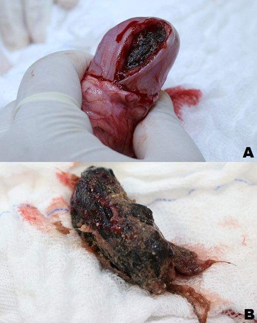
V.G.P. Albernaz, R.T. Conceição, T.C. Eising, I.A. Fabris, M.J. Mamprim & S.C. Rahal. 2017. Partial Obstruction of the
Small Intestine by a Trichobezoar in a Dog. Acta Scientiae Veterinariae. 45(Suppl 1): 210.
INTRODUCTION Abdominal ultrasonography showed focal
Bezoars are considered concretions of foreign intestinal wall thickening (0.75 mm) at the transition
material or indigestible organic substances that accumu- between the jejunum and the ileum (5 cm length),
late in the gastrointestinal tract [4,12,15]. The bezoar may with loss of layer stratification, and the presence of
be classified based on the composition of the primary con- an irregular hyperechoic content with strong acoustic
stituent as either trichobezoar, phytobezoar, lactobezoar, shadow. This intestine segment was distended. Mes-
pharmacobezoar or miscellaneous [12,14,15]. enteric and jejunal lymph nodes showed enlargement.
The trichobezoars are composed of hair or hair- The diagnosis was partial obstruction of the jejunum
like fibers [4,12,15]. In human patients, the trichobezoar attributable to a foreign body. Another proposed dif-
is related to hair accumulation mainly in the stomach, ferential diagnosis was intestinal neoplasm and severe
and Rapunzel syndrome is related to hair accumulation inflammatory bowel disease.
extending into the small intestine [4,5,14,15]. Both The dog was pre-medicated with morphine (0.5
diseases occur in humans, in general, with a history of mg/kg, IM) and meloxicam (0.2 mg/kg, IV). General
trichotillomania or trichophagia [5,10,14]. The Rapunzel anesthesia was induced with a combination of propofol
syndrome is considered rare in humans while the intesti- (4 mg/kg) and ketamine (1 mg/kg) administered intrave-
nal obstruction may occur when the gastric trichobezoar nously, and maintained with isoflurane in 100% oxygen
tail extends to or beyond the ileocecal valve or at least via an endotracheal tube. Intraoperative analgesia was
to the jejunum [10,12,14]. performed with infusion of fentanyl (10 mcg/kg/h),
A few reports have described intestinal obstruc- lidocaine (0.5 mg/kg/h) and ketamine (1.2 mg/kg/h),
tion by a trichobezoar in cats [1] and dogs [2,9,11]. plus intraperitoneal administration of bupivacaine (2
Therefore, the purpose of this report is to describe an mg/kg). Ceftriaxone was administered intravenously
uncommon clinical presentation of a dog with partial (30 mg/kg) at the time of induction of anesthesia.
obstruction of the small intestine by a trichobezoar. Exploratory celiotomy via a midline approach
revealed a dilated jejunal segment of approximately 5
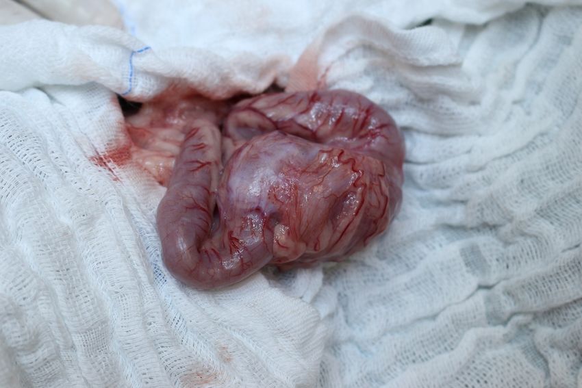
CASE cm with small areas of serosal hyperemia and larger
A 2-year-old, 5.5 kg intact male poodle was areas of omental adhesions (Figure 2). An unattached
referred due to a history of pain in the thoracolumbar firm intraluminal structure was palpated in the jejunum.
region and kyphosis for approximately 10 months. Be- An antimesenteric longitudinal enterotomy was per-
cause survey radiographs of the thoracolumbar spine had formed and a trichobezoar consisting of undigested hair
shown no signs of intervertebral disk disease, the dog and textile fibers (5 cm x 2.5 cm x 2 cm) was removed
had been treated conservatively without any improve- (Figure 3). Due to the presence of intestinal sacculation
ment for 10 days. Strict cage rest and therapy with tra-
madol hydrochloride, meloxicam, dipyrone, ranitidine
hydrochloride, and B complex had been prescribed. The
owner then reported that for approximately 18 months
the dog had been experiencing a loss of appetite, apathy,
and recurrent episodes of dark-colored foul-smelling
diarrhea. Overgrooming was not observed.
Physical examination revealed that the dog
walked without any difficulty or ataxia, but had pain
on palpation of the vertebral column especially in the
lumbar region. Spinal reflexes and proprioception were
normal. On abdominal palpation, the dog showed signs
of pain, but the presence of a mass was not detected.
Alopecia or other skin disorder was not detected. Figure 1. Intestinal Ultrasound image of a dog. Abdominal
Laboratory tests revealed anemia (Hematocrit 27%, ultrasonography demonstrated an irregular hyperechoic
interfacewith strong acoustic shadowing of the 0.44 cm foreign
Hemoglobin 7.5 g/dL, Red Blood Cells 4.44 million
body, in this segment of small bowel, was wall-thickening
cells/mm³), hypoproteinemia (5 g/dL), hypoalbumin- hypoechogenic, without preservation of layers. Enlargement of
emia (2.5 g/dL), and hypoglobulinemia (2.5 g/dL). mesenteric and jejunal lymph nodes was detected.
2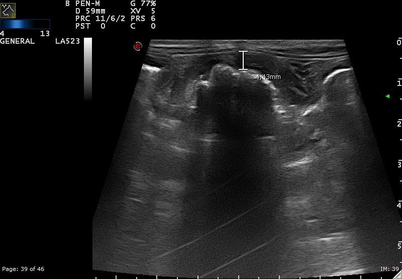
V.G.P. Albernaz, R.T. Conceição, T.C. Eising, I.A. Fabris, M.J. Mamprim & S.C. Rahal. 2017. Partial Obstruction of the
Small Intestine by a Trichobezoar in a Dog.                                  Acta Scientiae Veterinariae. 45(Suppl 1): 210.

after trichobezoar removal, a 5 cm jejunal resection with                 Necrosis and cell debris were also present. The diagno-
end-to-end anastomosis was performed with 4-0 nylon1                      sis was mucosal hyperplasia and chronic inflammation.
using a simple interrupted pattern. After abdomen lavage                          Cephalexin2 (30 mg/kg, q12 h), metronidazole3
with warm saline, the anastomotic site was covered with                   (25 mg/kg, q12 h), meloxicam4 (0.1 mg/kg, q24 h), di-
omentum. The abdomen was closed routinely.                                pyrone5 (25 mg/kg, q 8h), and ranitidine hydrochloride6
          Histopathological analysis (Hematoxylin &                       (2.2 mg/kg, q12 h), orally for 5 days were prescribed.
Eosin Staining) of the removed intestinal fragment                        Feeding was started 24 h after surgery, and gradually
showed hyperplasia of the mucosal layer, and infiltra-                    liquids, bland diet and normal diet were introduced
tion of inflammatory cells, mainly mononuclear cells.                     over 21 days. Ten days after surgery, the dog showed
                                                                          good recovery with no signs of pain on abdominal or
                                                                          vertebral column palpation. According to the owner,
                                                                          episodes of diarrhea or vomiting were not observed.
                                                                          An abdominal ultrasound performed 20 days postop-
                                                                          eratively demonstrated intestinal peristalsis and no sign
                                                                          of obstruction or free liquid.
                                                                                  At the last follow-up 180 days after surgery, the
                                                                          dog was in excellent condition with no obvious clinical
                                                                          sign related to the disease or surgical procedure.
                                                                                                 DISCUSSION

                                                                                   Clinical signs of weight loss, poor appetite,
Figure 2. Intra-operative view of small bowel dilated segment             intermittent vomiting, and reduced quantities of feces
with omental adherence.                                                   have been observed in dogs that developed intestinal
                                                                          trichobezoar [2,11]. The initial history of kyphosis and
                                                                          pain in the thoracolumbar region contributed to make an
                                                                          inappropriate diagnostic in the present case. Sometimes
                                                                          abdominal pain may seem like back pain, an effect
                                                                          called referred pain [8,17]. However, the obtainment of
                                                                          an accurate anamnesis helped to define the diagnostic
                                                                          process given that the owner reported a loss of appetite,
                                                                          apathy, and recurrent episodes of diarrhea. The diarrhea
                                                                          in cases of partial obstruction of the intestine is associ-
                                                                          ated with osmotic effects of unabsorbed substances and
                                                                          the secretory action of enterocytes [13].
                                                                                   Ultrasonography and computed tomography
                                                                          (CT) are considered reliable methods for diagnosing
                                                                          gastrointestinal bezoars in humans, but endoscopy is
                                                                          the method of choice for gastric bezoars [4,15]. In the
                                                                          present study, ultrasound was useful for identifying
                                                                          the presence of a foreign body obstructing the small
                                                                          bowel. Sonographic features of the trichobezoar in the
                                                                          small intestine in humans has been an arc-like echo
                                                                          casting a clear posterior acoustic shadow within the
                                                                          dilated lumen [12]. In a study of 11 dogs and five cats
                                                                          with clinical signs of gastrointestinal obstruction, the
Figure 2. A- Intra-operative view after anti-mesenteric enterotomy,       foreign bodies were identified by ultrasound due to dis-
visualization of a foreign body composed of non-digested fibers
and textile; B- Trychobezoar appearance after removal from the            tal acoustic shadowing and variable degrees of surface
intestinal segment in which it had been housed.                           reflection [18]. However, the definitive diagnosis in the
                                                                      3
V.G.P. Albernaz, R.T. Conceição, T.C. Eising, I.A. Fabris, M.J. Mamprim & S.C. Rahal. 2017. Partial Obstruction of the
Small Intestine by a Trichobezoar in a Dog. Acta Scientiae Veterinariae. 45(Suppl 1): 210.
present study was made during exploratory laparotomy, [3,16]. The presence of areas of omental adhesions in the
similar to other case reports of a gastrointestinal jejunum in the present report is suggestive of previous
trichobezoar in dogs and cats that used radiography damage to the intestinal wall, probably due to longer
or ultrasound in the abdominal evaluation [1,2,7,11]. permanence of the trichobezoar in this area. A study
The trichobezoar in the present report was showed that 63% of the obstruction of the gastrointes-
located exclusively in the small intestine, as observed tinal foreign bodies in dogs occur in the jejunum [6].
in two other reports on dogs [2,11]. Thus, these cases In summary, although intestinal obstruction
cannot be classified as Rapunzel syndrome, since it is secondary to ingestion of a foreign body is frequently
not a gastric trichobezoar with a tail extending up to observed in dogs, the intraluminal obstruction caused
the small intestine [10]. On the other hand, a report by a trichobezoar is considered uncommon. The case
described a trichobezoar located in the stomach and presented herein may be considered extremely uncom-
duodenum of a 12-year-old female Briard dog [7]. mon, due to a partial obstruction of the intestinal lumen
The development of trichobezoars in dogs have and long-term evolution.
been associated with overgrooming [7], stricture due to
tumor [11], and as a complication of circular end-to- MANUFACTURERS
1
Shalon Medical. Goiânia, GO, Brazil
end anastomosis stapling [2]. In the present report, the 2
Medley Indústria Farmacêutica Ltda. Campinas, SP, Brazil.
dog did not have alopecia or any other skin disorder at 3
LaboratórioTeutoBrasileiro S/A. Anápolis, GO, Brazil.
the moment of patient consultation, and no history of 4
Ourofino Saúde Animal Ltda. Cravinhos, SP, Brazil.
overgrooming. However, the area of intestinal saccula- 5
Sanofi-aventis Farmacêutica Ltda. São Paulo, SP, Brazil.
tion observed during surgery may have contributed to
6
EMS Pharma. Hortolândia, SP, Brazil.
trichobezoar formation. Declaration of interest. The authors report no conflicts of in-
Intestinal perforation caused by a trichobezoar terest. The authors alone are responsible for the contents and
is one of the complications reported in human patients writing of the paper.
REFERENCES
1 Barrs V.R., Beatty J.A., Tisdall P.L., Hunt G.B., Gunew M., Nicoll R.G. & Malik R. 1999. Intestinal obstruction
by trichobezoar in five cats. Journal of Feline Medicine and Surgery. 1(4): 199-207.
2 Carobbi B., Foale R.D. & White R.A.S. 2009. Trichobezoar obstruction after stapled jejunal anastomosis in a dog.
Veterinary Surgery. 38(3):417-420.
3 Diop B., Ngom G., Ndjaye A., Elmouhib R., Fall I. & Ndoye M. 2004. Trichobezoard revealed by intestinal perfora-
tion. A case report. Dakar Medical. 49(2): 83-85.
4 Eng K. & Kay M. 2012. Gastrointestinal bezoars: history and current treatment paradigms. Gastroenterology & Hepa-
tology. 8(11): 776-778.
5 Gonuguntla V. & Joshi D.D. 2009. Rapunzel syndrome: a comprehensive review of an unusual case of trichobezoar.
Clinical Medicine and Research. 7(3): 99-102.
6 Hayes G. 2009. Gastrointestinal foreign bodies in dogs and cats: a retrospective study of 208 cases. Journal of Small
Animal Practice. 50(11): 576-583.
7 Hettlich B.F. & Bahr A.M. 2000. What is your diagnosis? Journal of American Veterinary Medical Association.
217(4): 477-478.
8 McMahon S.B., Dmitrieva N. & Koltzenburg M. 1995. Visceral pain. British Journal of Anaesthesia. 75(2): 132-44.
9 Morgan A.F. & Miller E.R. 1980. A large trichobezoar in a Pekingese. Canine Practice. 7(1): 65.
10 Naik S., Gupta V., Naik S., Rangole A., Chaudhary A.K., Jain P. & Sharma A.K. 2007. Rapunzel syndrome reviewed
and redefined. Digestive Surgery. 24(3): 157-161.
11 O’Brien C.R. & Wong W.T. 2001. Intermittent vomiting and weight loss in an old dog. Australian Veterinary Journal.
79(4): 251-260.
12 O’Sullivan M.J., McGreal G., Walsh J.G. & Redmond H.P. 2001. Trichobezoar. Journal of the Royal Society of
Medicine. 94(2): 68-70.
13 Papazoglou L.G., Patsikas M.N. & Rallis T. 2003. Intestinal foreign bodies in dogs and cats. Compendium on Con-
tinuing Education for the Practicing Veterinarian. 25(11): 830-844.
4V.G.P. Albernaz, R.T. Conceição, T.C. Eising, I.A. Fabris, M.J. Mamprim & S.C. Rahal. 2017. Partial Obstruction of the
Small Intestine by a Trichobezoar in a Dog. Acta Scientiae Veterinariae. 45(Suppl 1): 210.
14 Parshad R., Prabhu S., Kumar G.V.R., Mukherjee D. & Bhramrah A. 2002. Trichobezoars: case reports and review
of literature. JK Science. 4(4): 202-205.
15 Sanders M. 2004. Bezoars: from mystical charms to medical and nutritional management. Practical Gastroenterology.
28(1): 37-50.
16 Sharma V. & Sharma I.D. 1992. Intestinal trichobezoar with perforation in a child. Journal of Pediatric Surgery.
27(4): 518-519.
17 Sikandar S. & Dickenson A.H. 2012. Visceral Pain – the Ins and Outs, the Ups and Downs. Current Opinion in Sup-
portive and Palliative Care. 6(1): 17-26.
18 Tyrell D. & Beck C. 2006. Survey of the use of radiography vs. ultrasonography in the investigation of gastrointestinal
foreign bodies in small animals. Veterinary Radiology & Ultrasound. 47(4): 404-408.
CR 210
www.ufrgs.br/actavet
5You can also read