Primary Retroperitoneal Teratoma with Predominant Neurogenic Elements Masquerading as Adrenal Tumor
←
→
Page content transcription
If your browser does not render page correctly, please read the page content below
Case Report                                                                                                          doi: 10.5146/tjpath.2016.01365

    Primary Retroperitoneal Teratoma with Predominant
    Neurogenic Elements Masquerading as Adrenal Tumor
                                Sonam SHARMA, Leelavathi DAWSON, Ashish Kumar MANDAL
                           Department of Pathology, Vardhman Mahavir Medical College and Safdarjung Hospital, NEW DELHI, INDIA

                                                                     ABSTRACT
Primary retroperitoneal teratomas are uncommon extragonadal nonseminomatous germ cell tumors that are composed of well differentiated
parenchymal tissues which are derived from more than one of the three embryonic germ cell layers. Here we report an unusual and first of its
kind, a case of primary mature cystic retroperitoneal teratoma mimicking as adrenal tumor in a 7-month-old female in which the tumor was
predominantly composed of neurogenic tissue histologically which is unlike the usual pattern seen in the teratomas.
Key Words: Retroperitoneal teratoma, Neurogenic elements, Adrenal neoplasm

                         INTRODUCTION                                          within normal range, with normal birth history. Her past
                                                                               history and medical history were non-contributory.
Teratomas also known as dysembryoma, teratoblastoma,
organoid tumor and teratoid tumor, are encapsulated                            On physical examination, a large intra-abdominal mass was
neoplasms, that are the most common form of all germ cell                      palpable in the left upper quadrant which was also extending
tumors (GCTs) and belong to nonseminomatous group                              into the left epigastric and left lumbar region. It measured
of GCTs (1). They arise from abnormal development of                           around 9 x 8 cm and was firm, non-tender, moving with
pluripotent cells: germ cells and embryonal cells, which in                    respiration and dull on percussion. The overlying skin was
turn greatly influences the age of presentation and involved                   unremarkable. All other systemic examinations were within
location. Teratomas of germ cell origin can be congenital                      normal limits. Routine haematological investigations were
or acquired and are usually gonadal. In contrast, teratomas                    unremarkable. Urine and blood cultures were negative.
of embryonic cell sources, which are always congenital                         Kidney, liver function tests, and X-ray of chest were normal.
and are usually found in extragonadal (15%) locations,                         Serum antibodies to human immunodeficiency virus and
such as sacrococcygeal , intracranial, cervical, mediastinal                   hepatitis B surface antigen were negative.
and retroperitoneal (2). Major differences in their clinical                   Abdominal ultrasonography (USG) showed a large,
behavior suggest that gonadal and extragonadal tumors                          multicystic mass located between the spleen and left kidney.
are biologically different, though histological, serological,                  There was no evidence of calcification in the tumor mass
and cytogenetic characteristics of all GCTs are similar (3).                   or ascitis. Contrast enhancement computed tomography
The present case study describes a child with an atypical                      (CECT) scan of abdomen and pelvis demonstrated a large
presentation of a rare case of primary retroperitoneal                         well circumscribed predominantly cystic retroperitoneal
teratoma which posed a diagnostic challenge and is first of                    mass occupying predominantly the left suprarenal region
its kind in terms of histology to be reported in the world                     (Figure 1A,B). It measured about 9.9 x 8.8 x 6.8 cm and
literature.                                                                    it showed multiple septae, a tiny fat and an enhancing
                                                                               soft tissue attenuation area. No calcification was seen.
                           CASE REPORT
                                                                               The mass displaced the aorta, celiac axis and superior
A 7-month-old female presented with a gradually                                mesenteric vessels to the contralateral side and the left
increasing lump in the left upper abdomen, which was                           kidney caudally with indentations to its contour. The left
first noticed 3 months back. There was no history of fever,                    renal vein was displaced antero-medially and draped along
weight loss, gastrointestinal, genito-urinary or respiratory                   the medial margin of the mass. The left adrenal gland could
disturbances. Developmental milestones of the child were                       not be detected separately from the mass. The body and

(Turk Patoloji Derg 2019, 35:69-73)                                            Correspondence: Sonam SHARMA
                                                                               Vardhman Mahavir Medical College and Safdarjung Hospital,
Received : 26.03.2016 Accepted : 10.06.2016                                    Department of Pathology, NEW DELHI, INDIA
                                                                               E-mail: drsonamsharma@gmail.com Phone: +99 998 413 93

                                                                                                                                                 69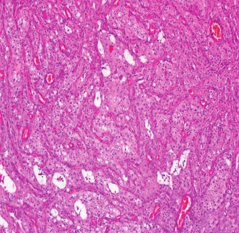
Turkish Journal of Pathology SHARMA S et al: Retroperitoneal Teratoma with Neurogenic Elements
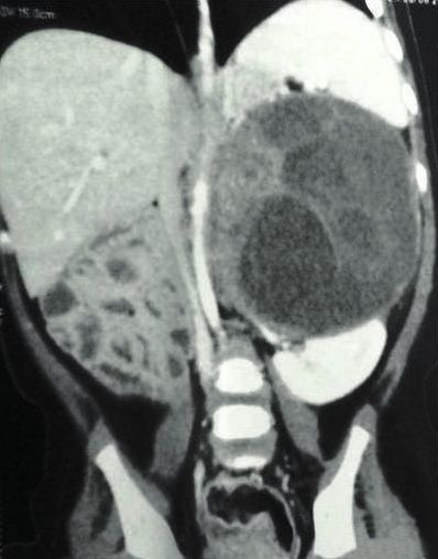
Figure 1: A,B) CECT abdomen
revealing a multicystic mass in the
A B retroperitoneum.
tail of pancreas were displaced anteriorly while descending Gross examination of the specimen received showed
colon and small bowel loops were displaced to right side. a well-circumscribed cystic mass measuring 9.8 x 9 x
There was no evidence of any significant abdominal and 8 cm with an intact and smooth surface. On incision,
pelvic lymphadenopathy or distant metastasis. Rest of the brownish fluid admixed with mucoid/jelly like material
abdominal and pelvic organs were unremarkable. Based on came out and thin walled cyst was left. On cut section,
these findings, a radiological suspicion of a cystic change multiloculated cysts were seen along with a very small solid
in a solid tumour originating from left adrenal gland (as grey yellow area measuring 0.8 x 0.8 x 0.5 cm (Figure 2A,B).
the normal left adrenal gland could not be recognized) or a Histopathological examination of cyst wall and small solid
retroperitoneal cystic teratoma was made. area showed predominantly mature neural tissue (Figure
Laboratory investigations indicating a functioning adrenal 3A). A glandular structure lined by ciliated columnar
tumour, consisting of plasma and urinary levels of cat- epithelium (Figure 3B) with occasional foci of adipose
echolamines, rennin, aldosterone, cortisol, adrenocortico- tissue and blood vessels could also be identified (Figure
trophic hormone levels were within normal limits. Tumor 4). Other mature or immature elements were not seen,
markers such as serum alpha-fetoprotein (AFP), lactate even on extensive sampling. On immunohistochemistry
dehydrogenase (LDH), neuron-specific enolase (NSE), car- (IHC), tumor cells were positive for synaptophysin and
cino embryonic antigen (CEA) and carbohydrate antigen glial fibrillary acidic protein. A final diagnosis of primary
19-9 (CA 19-9), were also estimated. Serum values of AFP retroperitoneal mature cystic teratoma with predominance
(18.9 μg/dL ), CEA (6.6 ng/ml) , CA19-9 (50.2 U/ml) were of neurogenic elements was made.
slightly higher whereas LDH and NSE were within normal The patient was discharged uneventfully in a stable
range , further ruling out the possibility of left adrenal condition. Post operative 1 year follow up failed to reveal
gland being the origin of this mass. any tumor recurrence.
On exploratory laparotomy, a large well defined cystic DISCUSSION
retroperitoneal mass occupying the left suprarenal area,
between the spleen and left kidney was seen. The left Primary retroperitoneal neoplasms are a rare but diverse
adrenal gland was compressed and adhered to the tumor group of benign and malignant tumors that arise within the
mass. The mass abutted the left kidney and displaced it retroperitoneal space but outside the major organs in this
inferiorly. The renal vessels were stretched and adherent space. They can be solid or cystic masses, each of which can
to the mass.The transverse and left colon were compressed be further subdivided into neoplastic and non-neoplastic
and displaced anteriorly whereas tail and body of the masses. Of the primary retroperitoneal neoplasms, 70%–
pancreas were displaced posteriorly. No invasion into the 80% are malignant in nature, and these account for 0.1%–
aorta or inferior vena cava was seen. No palpable regional 0.2% of all malignancies in the body (4). Among them,
nodes were identified. The tumor mass was completely primary retroperitoneal teratomas are extremely unusual
excised and sent for histopathological examination. neoplasms accounting for approximately 1–11% of all
70 Vol. 35, No. 1, 2019; Page 69-73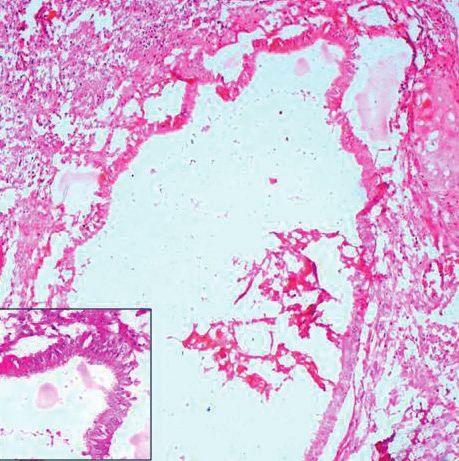
SHARMA S et al: Retroperitoneal Teratoma with Neurogenic Elements Turkish Journal of Pathology
Figure 2:
A) Gross specimen of
retroperitoneal mass.
B) Cut section revealing
multilocular cystic
A B component of the tumor.
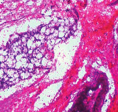
Figure 3:
A) Photomicrograph
showing predominantly
mature glial tissue (H&E;
x200). B) Glandular
structure lined by ciliated
columnar epithelium
(H&E; x200) [Inset:
Ciliated columnar
A B epithelium (H&E; x400)]
primary retroperitoneal neoplasms and typically occurs in
neonates, infants, and children (5).
Primary retroperitoneal teratomas involving adrenal
glands are exceedingly uncommon accounting for only
4% of all primary teratomas (6) and can be mistaken for
adrenal neoplasms (7). As in our case, based on radiology,
the left adrenal gland was inseparable from the mass, giving
an appearance that the tumor might have arisen from
the adrenal gland. Such an unusual radiological finding
may cause erroneous diagnosis. However, laboratory
investigations including serum tumor markers ruled out
the possibility of adrenal gland being the possible origin of
this tumor.
The diagnosis of this tumor is based on a combination of
high index of clinical suspicion, laboratory and radiological
investigations, though histopathology is the gold standard. Figure 4: Adipose tissue and blood vessels (H&E; x400).
Vol. 35, No. 1, 2019; Page 69-73 71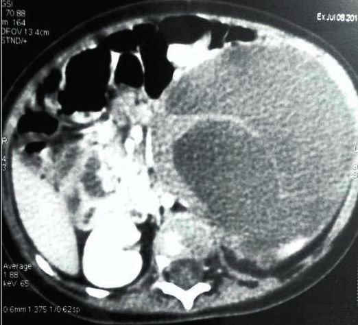
Turkish Journal of Pathology                                          SHARMA S et al: Retroperitoneal Teratoma with Neurogenic Elements

These tumors are usually asymptomatic but may manifest            case with similar predominance of neurogenic elements
as abdominal/back/flank pain, abdominal distention, or a          but that was in an ovarian mature cystic teratoma (14).
palpable abdominal mass, like in our case. Other symptoms         The pathogenesis of these predominant specific tissues in
can be of genito-urinary or gastrointestinal tract, limb/         teratoma is still obscure. In our case, teratoma components
genital swelling and secondary infections. Rarely malignant       were mature but approximately 90% of the tumor area
transformation and an acute syndrome can occur,                   showed glial tissue. Hence, the current case becomes a
involving peritonitis, intestinal obstruction, or renal colic     relevant value addition to the existing world literature.
(8). Laboratory investigations including serum tumor
                                                                  The prognosis of primary retroperitoneal teratomas is
markers played a pivotal role in our patient, in clinching
                                                                  generally good if the tumor is removed completely (15). In
the diagnosis. The retroperitoneal teratomas have a
                                                                  our case, the tumor was totally excised and the postoperative
property of expressing various serum tumor markers such
                                                                  as well as the follow up period was uneventful. Hence, it is
as elevated AFP, CEA and CA 19-9. These markers can also
                                                                  postulated that these tumors with predominant neurogenic
be used to monitor successful treatment or detect relapse in
                                                                  elements also behave in a benign manner as the primary
patients with specific tumor marker-secreting teratomas as
                                                                  retroperitoneal mature teratomas do. However, a close
suggested by few authors (9).
                                                                  follow up is mandatory as recommended by few researchers
Radiology has proved to be a valuable pre-operative               because of the possibility of its malignant transformation
diagnostic tool but has its own limitations (10,11). Plain        (12,16).
X-ray can demonstrate calcified material while USG can
                                                                  In conclusion, primary retroperitoneal teratoma, though a
differentiate between cystic and solid elements. CECT can
                                                                  rare entity, should always be considered among differentials
help to determine the size, extent of the tumor, relationship
                                                                  of adrenal masses, as it can masquerade a primary adrenal
to vessels and in differential diagnosis. Magnetic Resonance
                                                                  tumor, as seen in our case. Preoperatively laboratory and
Imaging (MRI) can offer better assessment of tumor staging
                                                                  radiological investigations do play an integral role, but it is
and distinction between benign and malignant neoplastic
                                                                  the histopathology which is confirmative. More insight is
features (12). In our case, no calcification was seen both
                                                                  required to understand the genesis and behaviour of these
on USG or CECT, and MRI was not done, owing to the
                                                                  tumors with one predominant element in teratomas.
unaffordability by the patient.
                                                                                   CONFLICT OF INTEREST
Various differential diagnosis of retroperitoneal cystic
lesions are cysts (mesenteric, omental, splenic, enteric dupli-   The authors declare no conflict of interest.
cations, mullerian, epidermoid, tailgut), solid neoplasms                                  REFERENCES
with cystic change (paraganglioma, neurilemmomas, leio-
                                                                  1. Mathur P, Lopez-Viego MA, Howell M. Giant primary
myosarcomas), lymphangiomas, lymphangioleiomyomas,
                                                                     retroperitoneal teratoma in an adult: A case report. Case Rep
mucinous/serous cystadenoma or cystadenocarcinoma,                   Med. 2010;2010. pii: 650424.
haematoma, urinoma, lymphocoele, pancreatic and non-              2. Bedri S, Erfanian K, Schwaitzberg S, Tischler AS. Mature cystic
pancreatic pseudocyst.                                               teratoma involving adrenal gland. Endocr Pathol. 2002;13:59-64.
Complete surgical excision, either by open surgery or by          3. Sharma S, Singh M, Bhuyan G, Mandal AK. Extragonadal
laparoscopy followed by histopathology evaluation is the             dysgerminoma presenting as neck metastasis and masquerading
                                                                     as a thyroid swelling. Clin Cancer Investig J. 2016;5:43-5.
mainstay for its definitive diagnosis as well as treatment
                                                                  4. Neville A, Herts BR. CT characteristics of primary retroperitoneal
(1,13). Usually teratomas, histopathologically consists of
                                                                     neoplasms. Crit Rev Comput Tomogr. 2004;45:247-70.
multiple parenchymal tissues that are derived from more
                                                                  5. Schmoll H. Extragonadal germ cell tumors. Ann Oncol.
than one germ cell layer (6). Our case was interesting as            2002;13:265-72.
numerous sections were taken to locate the different              6. Polo JL, Villarejo PJ, Molina M, Yuste P, Menendez JM, Babe
elements of teratoma microscopically, but we could only              J, Puente S. Giant mature cystic teratoma of the adrenal region.
find predominantly mature neurogenic element. However,               AJR Am J Roentgenol. 2004;183:837-8.
after extensive sampling, a glandular structure and a tiny        7. Hui JP, Luk WH, Siu CW, Chan JC. Teratoma in the region of
focus of adipose tissue could be identified. Therefore, this         an adrenal gland in a 77-year-old man. J Hong Kong Coll Radiol.
case cannot be considered as pure monodermal. Thus, we               2004;7:206-9.
designated this case as primary mature cystic retroperitoneal     8. Wolski Z, Jasinski Z. Retroperitoneum teratoma. Int Urol
teratoma with predominance of neurogenic elements. An                Nephrol. 1981;13:137-40.
extensive search of PubMed and Medline revealed one

72                                                                                                       Vol. 35, No. 1, 2019; Page 69-73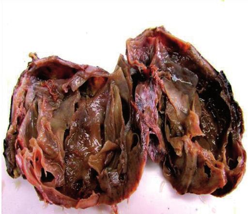
SHARMA S et al: Retroperitoneal Teratoma with Neurogenic Elements                                              Turkish Journal of Pathology

9. McKenney JK, Heerema-McKenney A, Rouse RV. Extragonadal                13. Ratkala JM, Shaikb NJ, Salia D, Choukimatha SM. Rare primary
   germ cell tumors: A review with emphasis on pathologic                     retroperitoneal teratoma masquerading as adrenal incidentaloma.
   features, clinical prognostic variables, and differential diagnostic       African Journal of Urology. 2015;21:96-9.
   considerations. Adv Anat Pathol. 2007;14:69-92.                        14. Akbulut M, Kelten EC, Ege CB. Mature cystic teratoma with
10. Shinagare AB, Jagannathan JP, Ramaiya NH, Hall MN, Van den                predominately neurogenic elements–case report. Aegean
    Abbeele AD. Adult extragonadal germ cell tumors. AJR Am J                 Pathology Journal. 2006;3:18-20.
    Roentgenol. 2010;195:W274‑80.                                         15. Aldhilan A, Alenezi K, Alamer A, Aldhilan S, Alghofaily
11. Barka M, Mallat F, Hmida W, Ahmed KB, Chavey SO, Abdallah                 K, Alotaibi M. Retroperitoneal teratoma in 4 months old
    AB, Tlili K. Giant primary retroperitoneal teratoma in an adult           girl: Radiology and pathology correlation. Curr Pediatr Res.
    male: A rare entity. Int J Case Rep Images. 2014;5:558-61.                2013;17:133-6.
12. Chaudhary A, Misra S, Wakhlu A, Tandon RK, Wakhlu                     16. Okulu E, Ener K, Aldemir M, Isik E, Irkkan C, Kayigil O. Primary
    AK. Retroperitoneal teratomas in children. Indian J Pediatr.              mature cystic teratoma mimicking an adrenal mass in an adult
    2006;73:221-3.                                                            male patient. Korean J Urol. 2014;55:148-51.

Vol. 35, No. 1, 2019; Page 69-73                                                                                                           73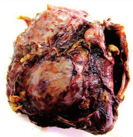
You can also read