Lily Poisoning in Domestic Cats - UFRGS
←
→
Page content transcription
If your browser does not render page correctly, please read the page content below
Acta Scientiae Veterinariae, 2019. 47(Suppl 1): 357.

                                                                     CASE REPORT                                                      ISSN 1679-9216
                                                                      Pub. 357

                                              Lily Poisoning in Domestic Cats

               Welden Panziera1, Claiton Ismael Schwertz1, Luan Cleber Henker1, Guilherme Konradt2,
               Daniele Mariath Bassuino2, Rochana Rodrigues Fett3, David Driemeier1 & Luciana Sonne1

                                                                   ABSTRACT

Background: Cases of plant intoxication in small animals are observed frequently in the domestic environment, mainly
because most dogs and cats live in households and occasionally have access to streets and rural areas. Among such toxic
agents, ornamental plants of the genus Lilium and Hemerocallis, which are potentially nephrotoxic to the feline species,
are highlighted. Affected cats start presenting clinical signs 1-6 h after plant ingestion. Renal failure takes place in 12-72
h, and death may occur in an interval ranging from three to seven days. The objective of this article is to describe the
epidemiological, clinical and pathological findings of lily (Lilium sp.) poisoning in two cats.
Case: The aspects of lily poisoning in two cats are described (cat #1 and cat #2). Cat #1 was a 3-year-old, mixed breed
female cat, which presented a clinical history of anorexia, apathy, drooling, vomiting and polydipsia. Serum biochemical
analysis revealed creatinine elevation (21.2 mg/dL), as well as hyperphosphatemia (19 mg/dL). Seventy-two h after the
onset of clinical signs, renal failure progressed to anuria, followed by death. The second animal of this report (cat #2) was
a 2-year-old, mixed-breed male cat. The animal was found dead by the owner without displaying any previous clinical
signs. Cats #1 and #2 ingested leaves of lily, which were present in their households as ornamental plants. At necropsy, the
kidneys of both cats presented mild enlargement. Moderate perirenal edema was also noted. Cat #1 showed morphologic
extrarenal uremic lesions, characterized by ulcers in the oral mucosa and in the margin of the tongue ventral surface. Mi-
croscopic lesions observed in both cases were similar and compatible with acute toxic nephropathy. Histologically, severe
epithelial cell degeneration and necrosis of proximal and distal convoluted tubules were noted. Other renal microscopic
findings included hyaline and granular casts, tubule regeneration and occasional birefringent oxalate crystals. Cat #1 also
presented moderate white matter vacuolation in the telencephalon and cerebellum.
Discussion: The epidemiologic, clinical and pathological findings reported in the present study are similar to previous
descriptions of lily poisoning in cats. Lily poisoning has been described in both males and females, without breed and age
predisposition, similarly to what has been found in the present study. Kidney metabolite excretion, including the elimination
of molecules such as creatinine, urea, and phosphorus is usually compromised in these cases, which was noted in cat #1.
The same animal showed extrarenal manifestations of renal failure, leading to a clinical presentation of uremic syndrome,
which is not frequent in these intoxications. Animals intoxicated by lily usually die from renal failure and anuria. In most
cases, lesions are restricted to the kidneys. In the reported cases, the microscopical lesions consisted of tubule epithelial
cells degenerative changes and necrosis. Acute lily intoxication in cats must be differentiated from other conditions, such
as intoxications due to aminoglycoside antibiotics, heavy metals, nonsteroidal anti-inflammatory drugs, antifungal agents,
chemotherapeutic drugs, and ethylene glycol. The knowledge regarding the toxic potential of ornamental plants is fun-
damental in order to prevent such events of intoxication, as well as to reach the final diagnosis. Epidemiological, clinical
and pathological findings were essential to conclude the final diagnosis.
Keywords: feline diseases, toxic plants, pathology, toxic nephropathy, uremia.
Descritores: doenças de felinos, plantas tóxicas, patologia, nefropatia tóxica, uremia.

                                                           DOI: 10.22456/1679-9216.89516
Received: 8 August 2018                                      Accepted: 16 December 2018                                     Published: 15 January 2019
1
 Setor de Patologia Veterinária (SPV), Faculdade de Veterinária (FaVet), Universidade Federal do Rio Grande do Sul (UFRGS), Porto Alegre, RS, Brazil.
2
 Universidade de Cruz Alta (Unicruz), Cruz Alta, RS. 3Chatterie Centro de Saúde do Gato, Porto Alegre. CORRESPONDENCE: L. Sonne [lusonne@
yahoo.com.br - Tel.: +55 (51) 3308-6107]. Faculdade de Veterinária - UFRGS. Av. Bento Gonçalves n. 9090. Bairro Agronomia. CEP 91540-000 Porto
Alegre, RS, Brazil.

                                                                         1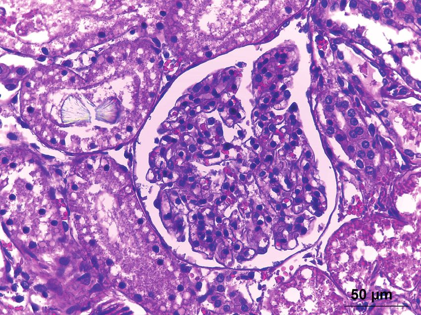
W. Panziera, C.I. Schwertz, L.C. Henker, et al. 2019. Lily Poisoning in Domestic Cats.
                                                                                    Acta Scientiae Veterinariae. 47(Suppl 1): 357.

                      INTRODUCTION                                  any previous clinical signs. The owner reported that
         Cases of plant intoxication in small animals               the cat was seen ingesting leaves of lily in the garden,
are observed frequently in the domestic environment,                corroborating to the clinical suspicion.
mainly because most dogs and cats live in househol-                          At the necropsy, cats #1 and #2 presented
ds, and occasionally have access to streets, parks                  moderate dehydration, characterized by ocular globe
and rural areas. Nonetheless, widespread ornamental                 retraction in the orbit. At necropsy, the kidneys of both
plants present in gardens and used as house decoration              cats presented mild enlargement as well as moderate
are responsible for the majority of intoxications [1].              perirenal edema. Cat #1 showed morphologic extra-
Among such toxic agents, ornamental plants of the                   renal uremic lesions, which characterized the clinical
genus Lilium and Hemerocallis, which are potentially                presentation of uremic syndrome. The lesions consisted
nephrotoxic to the feline species, are highlighted. Some            mainly of ulcerative stomatitis and glossitis, featured
species of the referred genus, which may lead to into-              as ulcers in the oral mucosa (Figure 1) as well as bila-
xication, include the Eastern lily (Lilium longiflorum),            teral and symmetrical ulcers and necrosis in the tongue
the Peace lily (Spathiphyllum wallisii), the Tiger lily             ventral surface (Figure 2). Additional gross findings in
(Lilium tigrinum), the Japanese lily (Lilium speciosum)             both cats included moderate hydrothorax, pulmonary
and Day lily (Hemerocallis spp.) [4,10].                            congestion and edema. During the necropsy, samples
         Lily intoxication is characterized by kidney               of several organs were collected, fixed in 10% buffered
tubular necrosis in the feline species, and death is                formalin, routinely processed for histology, and stained
caused by acute renal failure. Affected cats may start              by hematoxylin and eosin.
presenting clinical signs 1-6 h after plant ingestion,                       Microscopic lesions observed in both ca-
which may include anorexia, apathy, vomiting, diar-                 ses were similar, and compatible with acute toxic
rhea, and drooling. Signs of renal failure usually occur            nephropathy. Histologically, severe epithelial cell
from 12-72 h after lily ingestion, and are characterized            degeneration and necrosis of the proximal and distal
by polydipsia, polyuria, anuria, and azotemia [2-                   convoluted tubules were noted (Figure 3). Degene-
4,6,8,10]. The objective of this article is to describe             rated cells were enlarged, and presented irregular
the epidemiological, clinical and pathological findings             and vacuolated cytoplasm. The necrotic epithelium
of lily (Lilium sp.) poisoning in two cats.                         was characterized by cells presenting granular or
                                                                    hypereosinophilic cytoplasm, with nuclear pyknosis
                            CASE
                                                                    and karyorrhexis, and sometimes nuclear absence.
         The aspects of lily poisoning in two cats are              The lumen of numerous collecting ducts was filled
described (cat #1 and cat #2). Cat #1 was a 3-year-old,             with granular eosinophilic material (granular casts)
mixed breed female cat, which presented a clinical                  or amorphous eosinophilic material (hyaline casts).
history of anorexia, apathy, drooling, vomiting and                 Additionally, abundant amount of necrotic debris and
polydipsia. The cat owner informed that a few hours                 occasional birefringent oxalate crystal were observed
prior to the onset of clinical signs, the animal had
ingested some leaves of lily, which was present in the
house. Clinical examination revealed dehydration, hy-
pothermia, and hypotension. Intravenous fluid therapy
was performed. Abnormalities detected in serum bio-
chemical analysis included creatinine elevation (21.2
mg/dL {reference range from 0.8-1.8 mg/dL} [5]),
as well as hyperphosphatemia (19 mg/dL {reference
range from 4.5-8.1 mg/dL} [5]). Seventy-two h after
the onset of clinical signs, renal failure progressed to
anuria, followed by death. The second reported animal
(cat #2) was a 2-year-old, mixed-breed male cat that
had free access to the outside environment. The ani-                Figure 1. Spontaneous lily poisoning in domestic cats. Oral cavity. Multifo-
mal was found dead by the owner without displaying                  cal areas of ulcerative stomatitis are observed in cat #1.

                                                                2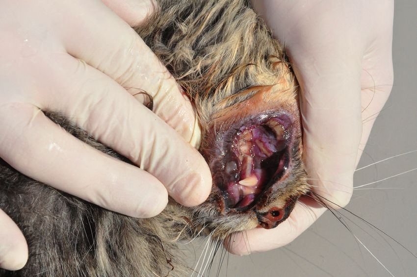
W. Panziera, C.I. Schwertz, L.C. Henker, et al. 2019. Lily Poisoning in Domestic Cats.
Acta Scientiae Veterinariae. 47(Suppl 1): 357.
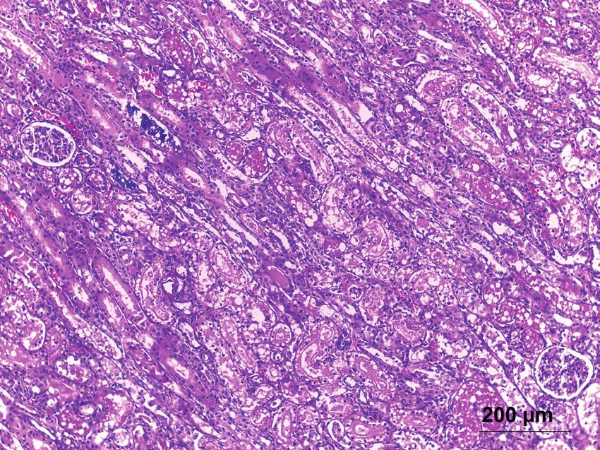
inside proximal and distal convoluted tubules (Figure
4). Frequently, different degrees of tubular epithelial
regeneration were also noted, as well as mild interstitial
inflammatory infiltrate of lymphocytes, plasma cells,
and macrophages.
Additional histological changes observed in
cat #1, which should be highlighted, included mode-
rate white matter vacuolation of the cerebellum and
telencephalon (occipital, parietal and frontal lobes);
as well as bilateral focally extensive areas of necrosis
and ulceration of the tongue epithelium, associated
with mild fibrin deposition and mild inflammatory
Figure 2. Spontaneous lily poisoning in domestic cats. Multifocal sym- infiltrate of lymphocytes, plasma cells and neutrophils.
metric bilateral areas of ulceration and necrosis are observed in the margins
of the tongue ventral surface in cat #2 (arrows). These gross changes,
DISCUSSION
along with the alterations noted in Figure 1, are consistent with extrarenal
uremic lesions.
The epidemiological, clinical and pathological
findings reported in the present study are similar to
previous descriptions of lily poisoning in the feline
species [2-4,6,8,10]. Cats are extremely susceptible
to the intoxication by Lilium and Hemerocallis. The
lack of knowledge regarding the toxic potential of such
plants by the pet owners is a major predisposing factor
[9]. Besides, lily intoxication risk is increased due to
the widespread utilization of such plant in households,
added to the fact that cats seem to present a peculiar
attraction towards lily species [10]. It is suggested that
some of these epidemiological factors may have pre-
disposed to the intoxication event in the reported cases.
Lily poisoning has been observed in both ma-
Figure 3. Spontaneous lily poisoning in domestic cats. Kidney. Severe les and females, without breed and age predisposition
epithelial cell degeneration and necrosis of the proximal and distal [4,10], similarly to what has been found in the present
convoluted tubules are noted. In addition, granular and hyaline casts are
seen in the lumen of numerous kidney tubules, associated with areas of study. All parts of the lily plant such as petals, stems,
mineralization [HE, x100].
leaves, and pollen present toxic potential [4,12]. It was
not possible to determine the ingested amount of lily
in the reported cases; however, the consumption of
small quantities, for instance, two leaves or a portion
of a single flower may lead to death [4]. An experi-
mental trial evaluating Oriental hybrid lily toxicity in
cats demonstrated that doses of 5 g/kg and 10 g/kg of
petals were lethal [12]. It is conjectured that cat #2 of
the present study may have ingested a greater amount
of the toxic plant, due to the sudden death with no
previous clinical signs.
Clinical signs are related to the nephrotoxic
effects of lilies; even though the toxic principle and
exact pathogenesis of renal toxicity are not known, the
acute clinical presentation indicates a rapid absorption
Figure 4. Spontaneous lily poisoning in domestic cats. Kidney. Birefrin-
gent oxalate crystal is observed in the lumen of a renal tubule [HE, x400]. rate of the involved toxic molecule. Conversely, the
3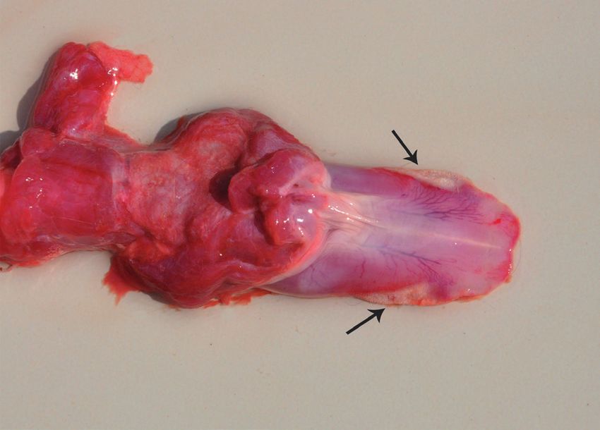
W. Panziera, C.I. Schwertz, L.C. Henker, et al. 2019. Lily Poisoning in Domestic Cats.
                                                                                    Acta Scientiae Veterinariae. 47(Suppl 1): 357.

nephrotoxic effects are not observed in dogs, even                  of individual mitochondria as well as mitochondrial
when great lily amounts are ingested [4]. The renal                 fusion. Additional ultrastructural changes described
excretion of metabolites, such as creatinine, urea and              include piknotic nuclei in the tubule epithelial cells and
phosphorus is compromised in such cases [2,4,8,10].                 fat infiltration [8]. Although not present in the reported
These changes were noted in cat #1 of this report,                  cases, occasionally pancreatic lesions are associated
which presented increased levels of creatinine and                  with kidney lesions [6].
phosphorus. The association of azotemia, hyperphos-                           Acute lily intoxication in cats must de diffe-
phatemia and cylindruria indicates primary renal                    rentiated from other conditions which lead to toxic
failure with tubular damage [2].                                    nephropathy and tubular necrosis. Among those, in-
         Affected cats may start presenting clinical                toxications due to aminoglycoside antibiotics, heavy
signs 1-6 h after plant ingestion. Renal failure is esta-           metals (for instance, lead, arsenic, and mercury), nons-
blished in 12-72 h, and death may occur in an interval              teroidal anti-inflammatory drugs, antifungal agents (for
ranging from three to seven days [2,4,8,10]. Similar                example, amphotericin B), chemotherapeutic drugs
aspects were noted in cat #1 of the present report. Cli-            (for instance, cisplatin), as well as ethylene glycol
nical signs of polydipsia, polyuria, and anuria indicate            poisoning are highlighted [3,7]. In the present reported
the acute renal failure. Additionally, the same animal              cases, the mentioned differential diagnoses were ruled
displayed extrarenal manifestations of renal damage,                out through the clinical history.
leading to a clinical presentation of uremic syndrome.                        Toxin absorption may be considerably redu-
Uremic extrarenal changes, identified at the clinical               ced, minimizing deleterious effects, when treatment is
examination or necropsy, are occasionally observed                  performed in the first hours of intoxication [11]. In the
in cats intoxicated by lily [4], and are related to the             reported case, intravenous fluid therapy was performed
longer survival time in the uremic phase of the disea-              in cat #1, aiming to avoid the anuric phase of renal
se. Therefore, the presence and severity of extrarenal              failure. Although the animal succumbed, fluid therapy
lesions are greater in cases with a prolonged course                plays a crucial role and must be conducted early in the
of renal failure [3,7], as probably observed in cat #1              disease progression, since dehydration, one of the main
of this study.                                                      consequences of intoxication, may lead to death [4,11].
         Animals intoxicated by lily usually die from                         The knowledge regarding the toxic potential
anuric renal failure and total compromise of renal                  of ornamental plants is fundamental in order to pre-
function [4]. In most cases, lesions are restricted to the          vent such intoxications, as well as to reach the final
kidneys [2-4,6,8,10]. In the reported cases, the renal              diagnosis and establish the adequate treatment [11].
microscopical lesions were compatible with acute                    Furthermore, small animal clinicians and pathologists
nephrotoxic tubular necrosis, consisting of tubule epi-             working with the feline species must consider lily
thelial cells degenerative changes and necrosis. Initial            intoxication as an important differential diagnosis in
kidney lesions affect the proximal convoluted tubules,              cats which present acute renal failure. Epidemiological,
and with disease progression, distal convoluted tubu-               clinical and pathological findings were essential to
les are also damaged. Ultrastructural changes include               conclude the final diagnosis.
mitochondrial swelling in the proximal convoluted                   Declaration of interest. The authors report no conflicts of
tubules epithelium, as well as megamitochondria for-                interest. All authors approved the manuscript and its submis-
mation, which may be the result of the enlargement                  sion to the journal.

                                                         REFERENCES

 1 Andrade S.F. 2011. Plantas tóxicas ornamentais. In: Nogueira R.M.B & Andrade S.F. (Eds). Manual de Toxicologia
   Veterinária. São Paulo: Roca, pp.34-58.
 2 Brady M.A & Janovitz E.B. 2000. Nephrotoxicosis in a cat following ingestion of Asiatic hybrid lily (Lilium sp.).
   Journal of Veterinary Diagnostic Investigation. 12(6): 566-568.
 3 Cianciolo R.E. & Mohr F.C. 2016. Urinary system. In: Maxie M.G. (Ed). Jubb, Kennedy, and Palmer’s Pathology of
   Domestic Animals. v.2. 6th edn. Philadelphia: Saunders Elsevier, pp.421-428.
 4 Fitzgerald K.T. 2010. Lily toxicity in the cat. Topics in Companion Animal Medicine. 25(4): 213-217.

                                                                4
W. Panziera, C.I. Schwertz, L.C. Henker, et al. 2019. Lily Poisoning in Domestic Cats.
Acta Scientiae Veterinariae. 47(Suppl 1): 357.
5 Kaneko J.J., Harvey J.W. & Bruss M.L. 1997. Clinical Biochemistry of Domestic Animals. 5th edn. San Diego:
Academic Press, 932 p.
6 Langston C.E. 2002. Acute renal failure caused by lily ingestion in six cats. Journal of the American Veterinary Medi-
cal Association. 220(1): 49-52.
7 Newman S.J. 2013. O Sistema urinário. In: Zachary J.F. & McGavin M.D. (Eds). Bases da Patologia em Veterinária.
5.ed. Rio de Janeiro: Mosby Elsevier, pp.592-662.
8 Rumbeiha W.K., Francis J.A., Fitzgerald S.D., Nair M.G., Holan K., Bugyei K.A. & Simmons H. 2004. A com-
prehensive study of easter lily poisoning in cats. Journal of Veterinary Diagnostic Investigation. 16(6): 527-541.
9 Slater M.R. & Gwaltney-Brant S. 2011. Exposure circumstances and outcomes of 48 households with 57 cats exposed
to toxic lily species. Journal of the American Animal Hospital Association. 47(6): 386-390.
10 Souza T.M., Fighera R.A., Kommers G.D. & Barros C.S.L. 2005. Poisoning by day lily (Hemerocallis sp.; Hemero-
callidaceae) in Brazilian cats. In: Panter K.E., Wierenga T.L. & Pfister J.A. (Eds). Poisonous plants: global research
and solutions. Wallingford: CABI, pp.46-49.
11 Stumpf A.R.L., Gaspari R., Bertoletti B., Amaral A.S. & Krause A. 2014. Intoxicação por lírio em um gato. Vet-
erinária e Zootecnia. 21(4): 527-532.
12 Xia Z., Wan J., Chen Y., He Y. & Yu J. 2013. Experimental oriental hybrid lilies (Lilium hybrids) poisoning in cats.
Journal of Clinical Toxicology. 3(1): 1-4.
CR357
http://seer.ufrgs.br/ActaScientiaeVeterinariae
5You can also read