First case of Phthirus pubis and Demodex co-infestation of the eyelids: a case report - BMC Ophthalmology
←
→
Page content transcription
If your browser does not render page correctly, please read the page content below
Huo et al. BMC Ophthalmology       (2021) 21:122
https://doi.org/10.1186/s12886-021-01875-w

 CASE REPORT                                                                                                                                      Open Access

First case of Phthirus pubis and Demodex
co-infestation of the eyelids: a case report
Yanan Huo1, Yanping Mo2, Xiuming Jin1, Xiaodan Huang1 and Wei Chen3*

  Abstract
  Background: Phthirus pubis is an obligate parasite of human beings. Demodex spp. is a much more common
  parasite of human beings. However, P. pubis infestation accompanied by Demodex mite infestation in eye has not
  been reported.
  Case presentation: We report the first case of Phthirus pubis and Demodex co-infestation on a 48-years-old woman.
  She presented to the hospital with itching and burning at her right eye for 2 weeks. Slit lamp examination revealed
  multiple nits and adults of P. pubis anchored to both upper and lower eyelashes. Eyelashes were trimmed,
  moxifloxacin eye ointment and fluorometholone eye drops were initiated daily. However, itching didn’t improve
  after 2 weeks of treatment. Light microscopy examination of eyelashes revealed infestation with Demodex. The
  patient was treated with lid scrubs with 25% tea tree oil daily for 4 weeks and was completely cured.
  Conclusion: Our report shows the importance of an early and comprehensive diagnosis, because both phthiriasis
  palpebrarum and demodicosis can be confused with blepharitis.
  Keywords: Phthirus pubis, Demodex, Co-infestation

Background                                                                           Demodex mite infestation in eye has not been reported.
Phthirus pubis is an obligate parasite of human beings. It                           Here, we describe the first case of co-infestation by these
is mainly detectable in the hair of pubic, rectal and in-                            two parasites.
guinal areas. The infestation of P. pubis in eyelid is rare,
unilateral infestation is extremely rare [1]. Transmission                           Case report
of P. pubis to eyelashes may be manual from the infested                             A 48-year-old-woman presented to our hospital with un-
body hair or during sexual contact. Indirect transmission                            bearable itching and stinging with black secretion on her
through clothes or towels contaminated with nits is less                             right eye for 2 weeks. She had no specific ocular, chronic
frequent.                                                                            or immune disease. Her best corrected visual acuity was
  Demodex spp., on the other hand, is a much more                                    20/20. Slit lamp examination revealed over three hun-
common parasite of human beings. Out of many Demo-                                   dred of translucent ovoid nits and over twenty live adult
dex species, only Demodex folliculorum and Demodex                                   P. pubis firmly attached to the base and shaft of eye-
brevis can parasitize in human eye. D. folliculorum is                               lashes. Dry blood and faeces appeared as granular and
most commonly found in eyelash follicles, whereas D.                                 dark dots on nearby eyelids, which the patient consid-
brevis colonizes in the sebaceous and meibomian glands                               ered as “black secretion” (Fig. 1). Lid margin vascular en-
[2]. However, P. pubis infestation accompanied by                                    gorgement and mild hyperemia were observed in the
                                                                                     conjunctiva. There was mild punctate defect on corneal
* Correspondence: wei_chen@zju.edu.cn                                                epithelium. No nit, lice or any abnormal of ocular sur-
3
 Department of Medical Oncology, Tongde hospital of Zhejiang Province,
NO, 234, Gucui Road, Hangzhou 310012, Zhejiang, China                                face were observed in the left eye. The left cornea was
Full list of author information is available at the end of the article               clear and no vision decline. The patient was referred to
                                      © The Author(s). 2021 Open Access This article is licensed under a Creative Commons Attribution 4.0 International License,
                                      which permits use, sharing, adaptation, distribution and reproduction in any medium or format, as long as you give
                                      appropriate credit to the original author(s) and the source, provide a link to the Creative Commons licence, and indicate if
                                      changes were made. The images or other third party material in this article are included in the article's Creative Commons
                                      licence, unless indicated otherwise in a credit line to the material. If material is not included in the article's Creative Commons
                                      licence and your intended use is not permitted by statutory regulation or exceeds the permitted use, you will need to obtain
                                      permission directly from the copyright holder. To view a copy of this licence, visit http://creativecommons.org/licenses/by/4.0/.
                                      The Creative Commons Public Domain Dedication waiver (http://creativecommons.org/publicdomain/zero/1.0/) applies to the
                                      data made available in this article, unless otherwise stated in a credit line to the data.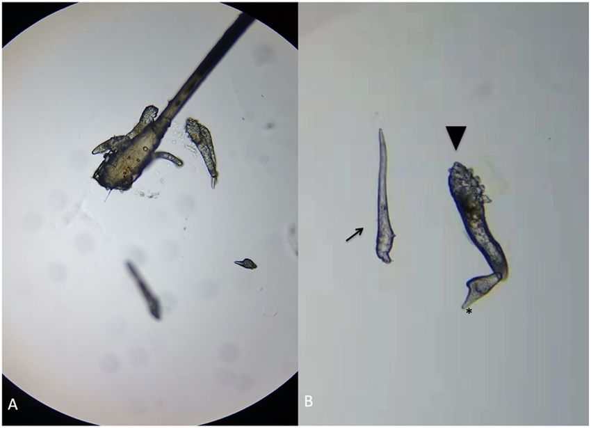
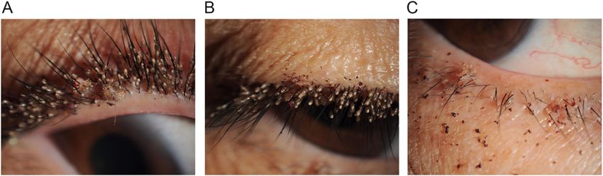
Huo et al. BMC Ophthalmology         (2021) 21:122                                                                                          Page 2 of 4

 Fig. 1 Photos of her right eye shows multiple nits and lice on eyelashes of upper lid (a) (b) and lower lid (c), with dried blood on skin. Under slit-
 lamp examination, adult lice adhere to eyelashes with crab-like bodies; the nits appear oval and translucent

dermatologist to detect potential phthiriasis lesions in                      prescribed daily moxifloxacin eye ointment and fluoro-
other body parts. On dermatologic examination, lice                           metholone eye drops four times a day for 2 weeks. The
were also detected in the pubic area. She had past his-                       pubic hair was shaved and hexachlorocyclohexane cream
tory of sexual contacts with her husband. The search of                       was applied daily. The patient was advised to avoid close
sexually transmitted diseases was negative. Initial diag-                     body contact and not to share clothing and towels.
nosis of unilateral phthiriasis palpebrarum was made.                           After two-week’s treatment, the patient returned to
  Mechanical removal of the lice and nits was attempted                       our hospital complaining with persistent itching of the
but failed as they were numerous. All eyelashes of the af-                    right eye. The corneal epithelial punctate defect
fected eye were trimmed with a scissors. Conjunctival                         remained unchanged. There were no lice or nits ob-
sac was irrigated and lids margin was disinfected by                          served on right eye. A co-infestation by Demodex was
compound iodine disinfectant cotton swab immediately                          suspected. Three eyelashes from each eyelid of both eyes
and followed at weekly intervals. The patient was                             were epilated and examined with light microscopy.

 Fig. 2 Microscopic features of Demodex in the patient’s right eye. a A group of Demodex and an egg were revealed with a lash follicle. b An
 egg (arrow), a larvae which had three pairs of poorly developed legs and a slender body (arrowhead), and an adult D. folliculorum with four pairs
 of well-developed legs (*) and a stumpier body
Huo et al. BMC Ophthalmology   (2021) 21:122                                                                                     Page 3 of 4

Demodex detection and counting in epilated lashes were          infestation on the left eye is too low to cause any
performed as proposed by Gao et al. [3]. A total of 14 D.       symptoms.
folliculorum was found in 6 lashes from the right eye              Because Demodex can be found in healthy asymptomatic
(Fig. 2) and a total of 5 D. folliculorum was found in 6        population, some authors have suggested that the relation-
lashes from the left eye. The diagnosis of demodicosis          ship between symptoms and the number of Demodex
was confirmed. The lid margin around the root of the            should be considered at the same time. Three mites per
eyelashes were scrubbed by a sterile cotton-tipped appli-       three lashes should consider as Demodex infestation posi-
cator saturated with 25% tea tree oil daily and eyelids         tive. If the related symptoms of Demodex infestation are
were heated with wet towel at about 40–45 °C twice per          serious, it can be diagnosed once the mite is detected [3, 8–
day for 2 months. Clothing, pillow cases, and towels            10]. Many methods, including tea tree oil, 1% yellow mer-
were recommended to be washed with hot water and                cury ointment, 2% topical metronidazole gel, 1% acaricide
then heat dried for up to 10 mins. The patient’s symp-          permethrin, and daily lid scrubbing and cleaning, can be
toms relived, and the affected corneal was clear after          used for eradicating ocular Demodex infestation [11–13].
two-week’s treatment. The number of mites during an             We used 25% tea tree oil daily and heated the eyelids with
examination 1 month later reduced to 2 in the right eye         wet towel at about 40–45 °C twice per day for 2 months.
and 0 in the left eye, and was reduced to 0 in both eye 2       The patient’s symptoms relived, and the affected corneal
months later. No recurrence was observed during 3               was clear after two-week’s treatment. No recurrence was
months of follow-up.                                            observed during 3 months of follow-up.
                                                                   Other authors have suggested that demodicosis may
                                                                be associated with leukemia or immunodeficient patients
Discussion and conclusions                                      with HIV-infection [14–16] or patients with end stage
This is the first report of co-infestation by two different     chronic renal failure [17]. However, the causal relation-
kinds of parasites in human eyelids, and also the most          ship between P. pubis infestation and Demodex infest-
severe case of phthiriasis palpebrarum that has been re-        ation has not yet been studied.
ported in literature.                                              In conclusion, here we report the first case of a severe
   The infestation of phthiriasis palpebrarum happens           infestation of P. pubis co-infested with Demodex in hu-
mainly in people who live in crowded places or poor hy-         man eye. This report shows the importance of an early
giene conditions. The infestation is usually transmitted        and comprehensive diagnosis, because both phthiriasis
by sexual activity because P. pubis is less mobile and          palpebrarum and demodicosis can be confused with
cannot fly or jump from the initial located area to the         blepharitis. In this case, the Demodex infestation of the
eyes. Therefore, sexual abuse should be considered in           right eye is much more severe than the left eye, and P.
children with P. pubis infestation [4]. There are multiple      pubis only infested in the right eye. Whether the host’s
treatment options available like mechanical removal with        immune reactions by Demodex make it susceptible for P.
forceps, pilocarpine hydrochloride, liquid Vaseline, mox-       pubis infestation is still unclear.
ifloxacin eye ointment, 1% mercury oxide, cryotherapy,
                                                                Acknowledgements
argon laser, topical botulinum toxin application, 50% tea
                                                                Not applicable.
tree oil, and 20% fluorescein eye drops [5–7]. In this
case, we trimmed the eyelashes and treated patient with         Authors’ contributions
moxifloxacin eye ointment and fluorometholone eye               HYN gathered and interpreted the patient data and was a major contributor
                                                                in writing the manuscript. MYP, JXM, and HXD gathered and interpreted the
drops. No lice or eggs were observed on the eyelashes.          patient imaging data and contributed in writing the manuscript. CW
   Eye infestation by Demodex is much more common than          provided clinical guidance and contributed in writing the manuscript. All
P. pubis. Among a wide range of species, only D. follicu-       authors read and approved the final manuscript.
lorum and D. brevis can parasitize the human eye. Demodex       Funding
folliculorum lives in the lash follicle, whereas Demodex bre-   This work is supported by Zhejiang Natural Science Foundation
vis lives solitarily and deeply in the sebaceous gland of the   (LY20H120009) and National Natural Science Foundation of China
                                                                (31751003).
eyelash and the meibomian gland [2]. Hence, when eye-
lashes are sampled, the detection rate of D. folliculorum is    Availability of data and materials
much higher than that of D. brevis. Patients has cylindrical    All data generated or analyzed during this study are included in this
dandruff in eyelash roots should highly considered Demo-        published article.

dex infestation [3, 8, 9]. However, in the presenting case,     Declarations
there was no typical cylindrical dandruff observed in both
eyes. The previous treatment of topical eye ointment and        Ethics approval and consent to participate
                                                                This research has obtained human research ethic approval from the Ethic
weekly lid cleaning might covered the tracks of Demodex         Committee of the second Affiliated Hospital, School of medicine, Zhejiang
and its secretion on right eye. And the number of Demodex       university (2019–395).
Huo et al. BMC Ophthalmology (2021) 21:122 Page 4 of 4
Consent for publication
Written consent was obtained from the patient for permission for
publication of her personal and clinical details along with identifying images
to be published in this study.
Competing interests
The authors declare that they have no competing interests.
Author details
1
Department of Ophthalmology, The Second Affiliated Hospital of Zhejiang
University School of Medicine, Hangzhou 310009, China. 2Department of
Ophthalmology, Huzhou Third Municipal Hospital, Huzhou 313000, China.
3
Department of Medical Oncology, Tongde hospital of Zhejiang Province,
NO, 234, Gucui Road, Hangzhou 310012, Zhejiang, China.
Received: 13 April 2020 Accepted: 22 February 2021
References
1. Panadero-Fontan R, Otranto D. Arthropods affecting the human eye. Vet
Parasitol. 2015;208(1–2):84–93.
2. English FP, Nutting WB. Demodicosis of ophthalmic concern. Am J
Ophthalmol. 1981;91(3):362–72.
3. Gao YY, Di Pascuale MA, Li W, Liu DT, Baradaran-Rafii A, Elizondo A, et al.
High prevalence of Demodex in eyelashes with cylindrical dandruff. Invest
Ophthalmol Vis Sci. 2005;46(9):3089–94.
4. Ryan MF. Phthiriasis palpebrarum infection: a concern for child abuse. J
Emerg Med. 2014;46(6):e159–62.
5. Rundle PA, Hughes DS. Phthirus pubis infestation of the eyelids. Br J
Ophthalmol. 1993;77(12):815–6.
6. Pinckney J 2nd, Cole P, Vadapalli SP, Rosen T. Phthiriasis palpebrarum: a
common culprit with uncommon presentation. Dermatol Online J. 2008;
14(4):7.
7. Couch JM, Green WR, Hirst LW, de la Cruz ZC. Diagnosing and treating
Phthirus pubis palpebrarum. Surv Ophthalmol. 1982;26(4):219–25.
8. Coston TO. Demodex folliculorum blepharitis. Trans Am Ophthalmol Soc.
1967;65:361–92.
9. Norn MS. Demodex folliculorum. Incidence and possible pathogenic role in
the human eyelid. Acta Ophthalmol Suppl. 1970;108:7–85.
10. English FP. Demodex folliculorum and oedema of the eyelash. Br J
Ophthalmol. 1971;55(11):742–6.
11. Rodriguez AE, Ferrer C, Alio JL. Chronic blepharitis and Demodex. Arch Soc
Esp Oftalmol. 2005;80(11):635–42.
12. Jansen T, Kastner U, Kreuter A, Altmeyer P. Rosacea-like demodicidosis
associated with acquired immunodeficiency syndrome. Br J Dermatol. 2001;
144(1):139–42.
13. Murube J. Demodex hominis. Ocul Surf. 2015;13(3):181–6.
14. Damian D, Rogers M. Demodex infestation in a child with leukaemia:
treatment with ivermectin and permethrin. Int J Dermatol. 2003;42(9):724–6.
15. Clyti E, Sayavong K, Chanthavisouk K. Demodecidosis in a patient infected
by HIV: successful treatment with ivermectin. Ann Dermatol Venereol. 2005;
132(5):459–61.
16. Seyhan ME, Karincaoglu Y, Bayram N, Aycan O, Kuku I. Density of Demodex
folliculorum in haematological malignancies. J Int Med Res. 2004;32(4):411–
5.
17. Karincaoglu Y, Esrefoglu Seyhan M, Bayram N, Aycan O, Taskapan H.
Incidence of Demodex folliculorum in patients with end stage chronic renal
failure. Ren Fail. 2005;27(5):495–9.
Publisher’s Note
Springer Nature remains neutral with regard to jurisdictional claims in
published maps and institutional affiliations.You can also read