Does subepineural injection damage the nerve integrity? A technical report from four amputated limbs
←
→
Page content transcription
If your browser does not render page correctly, please read the page content below
Korean J Pain 2021;34(1):132-136
https://doi.org/10.3344/kjp.2021.34.1.132
pISSN 2005-9159 eISSN 2093-0569

Case Report

Does subepineural injection damage the nerve integrity?
A technical report from four amputated limbs
Sandeep Diwan1, Abhijit Nair2, Parag Sancheti3, and André Van Zundert4
1
 Department of Anesthesiology, Sancheti Hospital, Pune, India
2
  Department of Anesthesiology, Basavatarakam Indo-American Cancer Hospital and Research Institute, Hyderabad, India
3
 Department of Orthopedics, Sancheti Hospital, Pune, India
4
  Royal Brisbane and Women’s Hospital and The University of Queensland and Queensland University of Technology, Brisbane, Australia

Received July 30, 2020
Revised September 15, 2020                  Local anesthetic (LA) injection outside the sheath in epineural or paraneural con-
Accepted September 16, 2020                 nective tissue is considered safe practice among regional anesthesiologists. There
                                            is limited evidence as to whether neurological complications occur if LA is injected
Handling Editor: Jong Yeon Park             inside the sheath (subepineural - intraneural). We performed ultrasound guided in-
                                            jections at the level of undivided sciatic nerve in four amputated lower limbs. In two
Correspondence                              specimens, LA was injected in epineural connective tissue (paraneural tissue) and
Abhijit Nair                                in another two specimens by penetrating the outer nerve sheath (hyperechoic epi-
Department of Anesthesiology,
                                            neurium). Ultrasonography demonstrated an increase in the size of nerve and mac-
Basavatarakam Indo-American Cancer
Hospital and Research Institute, Road No.
                                            roscopic findings revealed fascicular tracings with sub-epineural injections. Limbs
10, Banjara Hills, Hyderabad-500034,        were sent for histological analysis in formalin containers. Pathologist performed the
Telangana State, India                      analysis which demonstrated an intact perineurium and a breach in the epineurium.
Tel: +91-9963180495                         We conclude that sub-epineural injections are unsafe and injection should be done
Fax: +91-040-2354-2120                      in paraneural tissue to ensure safety and avoid unwanted neurological sequelae af-
E-mail: abhijitnair95@gmail.com             ter the block.

                                            Key Words: Anesthetics, Local; Injections; Nerve Block; Neuralgia; Pathology; Pe-
                                            ripheral Nerves; Sciatic Nerve; Ultrasonography, Interventional.

It is universally agreed that the epineurium should not                      this practice should be discontinued in clinical settings.
be breached during an isolated nerve block. Breach in                          To understand the histological changes in a nerve speci-
the epineurium, leads to intraneural placement of needle                     men after a sub-epineural injection, we conducted a US-
tip, which is not desirable [1,2]. Improved imaging tech-                    guided needle tip placement in two popliteal sciatic nerves
niques with ultrasound (US) has increased the possibility                    4-5 cm above the level of division, following an above-
of narrowing the needle tip-nerve distance, although the                     knee amputation. A pathologist independently evaluated
optimal needle tip-nerve distance remains elusive and                        the histological changes in the nerves of the amputated
undefined. The concept of intraneural placement of lo-                       limbs.
cal anesthetic (LA) has been erroneously described as a                        Our aim was to understand the dynamics of spread in
safe practice [3]. Furthermore, there is limited evidence of                 sciatic nerve specimens under US with extra-neural and
absence of neurological complications if LAs are injected                    sub-epineural injections in recently amputated limbs.
inside the sheath (sub-epineural-intraneural). Therefore,

   This is an open-access article distributed under the terms of the         Author contributions: Sandeep Diwan: Writing/manuscript prepara-
Creative Commons Attribution Non-Commercial License (http://cre-             tion; Abhijit Nair: Methodology; Parag Sancheti: Supervision; André Van
ativecommons.org/licenses/by-nc/4.0/), which permits unrestricted            Zundert: Writing/manuscript preparation.
non-commercial use, distribution, and reproduction in any medium,
provided the original work is properly cited.
© The Korean Pain Society, 2021

www.epain.org                                                          132                                             Korean J Pain 2021;34(1):132-136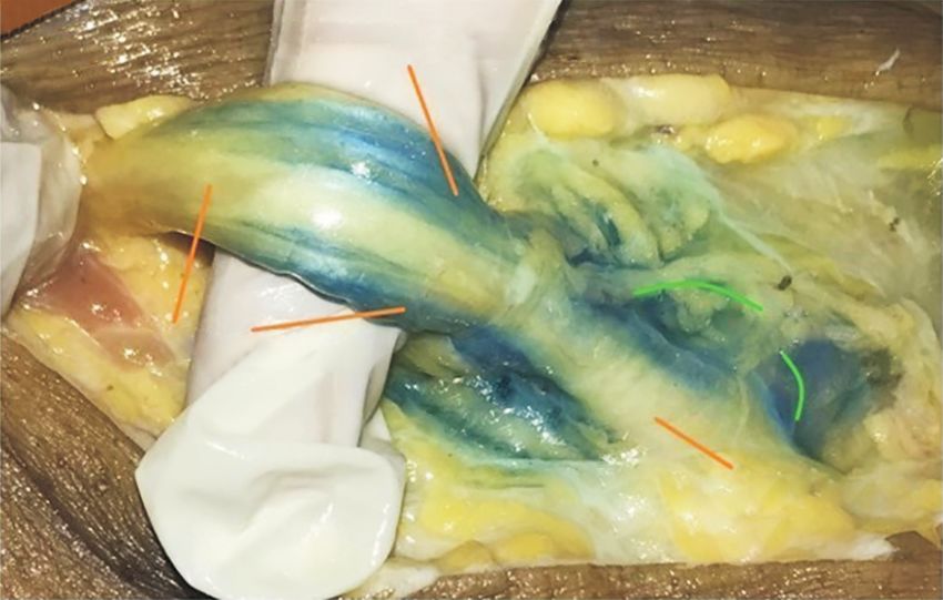
Nerve integrity after subepineural injection 133
CASE REPORT the epineurium – which was appreciated as a ‘pop’ – in 2
other specimens (L3 and L4). An in-plane technique was
This study was approved by the Institutional Ethics Com- chosen to place a 50 mm needle (Stimuplex ®; B. Braun,
mittee, Sancheti Institute for Orthopedics and Rehabilita- Melsungen, Germany) from the lateral to medial aspect of
tion, Pune (EC-SIOR/Agenda 060). Written informed con- the sciatic nerve (Fig. 1). In L1 and L2, the needle tip was
sent was obtained from four patients and their next of kin. tangential to the nerve at a 6-7 o’clock position, and was
The lower limbs of these four patients suffered from severe positioned in the EPI-ct. Three mL sterile water with MBD
superficial femoral arterial thrombosis, and the surgeon was injected without any resistance. In L3 and L4, with
deemed amputation the best option. The sensations were the needle tip close to the nerve at a 9 o’clock position, the
attenuated below the mid-calf level and minimal motor tip was advanced until a ‘pop’ was appreciated, and 3 mL
movements in the form of plantarflexion and dorsiflexion sterile water with MBD was injected, below the epineu-
of the foot and toe were possible. The time to surgery after rium in the ‘sub-epineural area’.
the injury was more than 8 hours in each case. After above Within 20 minutes after injection, with all specimens in
knee amputations the limbs were placed prone on a sterile prone position (L1, L2, L3, and L4), gentle dissection was
sheet (Fig. 1). We performed injection of methylene blue performed by the surgeon and anesthesiologist by peeling
dye (MBD) with sterile water under US guidance (linear- layer by layer to identify the superficial dye spread. Tissue
array probe, 3-12 Hz, Sonosite M-Turbo; FUJIFILM Son- was handled carefully to avoid unwanted spread of the
osite Inc., Bothell, WA) outside the epineurium in the epi- dye in different tissue planes. The findings were noted and
neural connective tissue (EPI-ct) in 2 specimens (labelled the specimens were immersed in the chamber filled with
as L1 and L2) and below the epineurium after penetrating 10% formalin and sent for histopathological analysis. The
histological findings were reported independently by a pa-
thologist. The tissues were fixed in formalin and subjected
Leg end Linear probe to increasing concentration of alcohol. The dehydrated
tissues were dipped in molten wax. The tissue blocks were
cut into thin ribbons through a microtome. After passing
the ribbons through a decreasing concentration of alcohol,
the tissue slides were stained with hematoxylin and eosin.
In L1 and L2, the spread was observed and images were
Insulated needle Midthigh
downloaded. The spread was in the EPI-ct (outside the
sheath), and no spread occurred in the sub-epineural area
(inside the sheath). There was no swelling of the nerve. (Fig.
2). In L3 and L4, upon sub-epineural injection, the nerve
Methylene blue dye
size briefly increased in all planes, evident from the dis-
sipation of the solution in several directions (Fig. 3). The
Fig. 1. L1 specimen in prone position, with ultrasound guided injection nerve returned to near normal size within a few seconds.
at the popliteal sciatic nerve through a sterile insulated needle under a In L3 and L4, upon sub-epineural injection, the nerve size
linear probe. also briefly increased in all planes, evident from the dis-
Epineurium
Fascicle
Needle in the
extra epineurial space LA spread
extra-epineurial
A space B
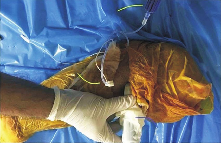
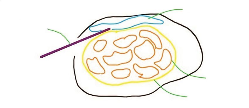
Fig. 2. L1 specimen in prone position. (A) The needle in-plane (white arrow) inserted from lateral to medial. The marker – dark blue – is on lateral side
depicts the orientation marker. Green line denotes paraneural covering i.e. , epineural connective tissue. The blue cross is the paraneural spread of the
solution. Orange dots are fascicles. (B) Schematic diagram of Fig. 2A. LA: local anesthetic.
www.epain.org Korean J Pain 2021;34(1):132-136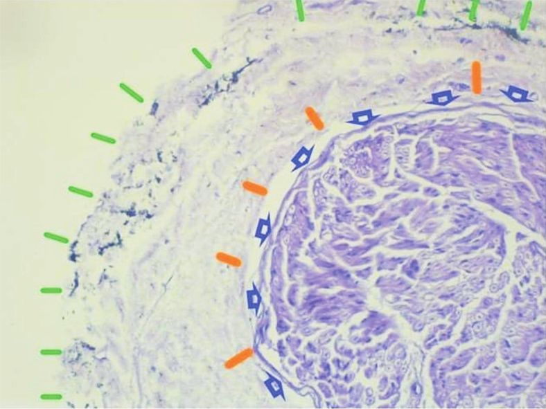
134 Diwan, et al
Epineurium
LA injected below
epineurium
Increase in
Subepineurial cross-sectional
needle area of nerve
placement
Paraneural
tissue
A B
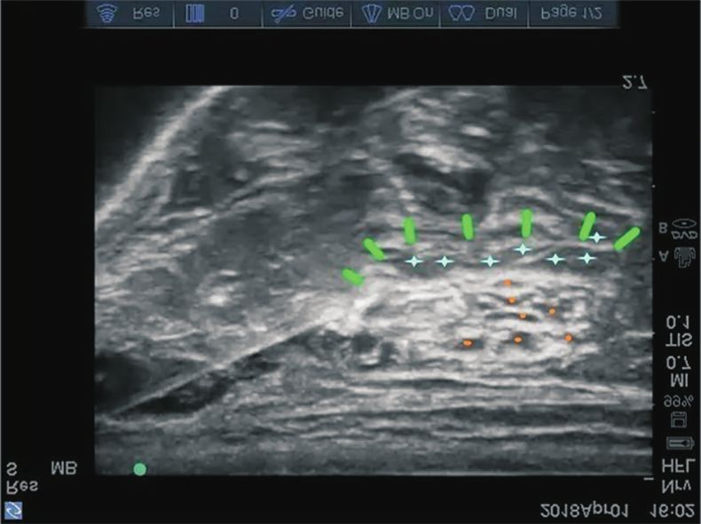
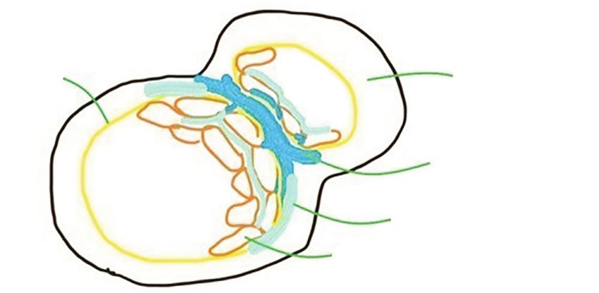
Fig. 3. L3 specimen in prone position. (A) The needle in-plane (white arrow) inserted from lateral to medial in L3. The marker – dark blue – is on lateral
side depicts the orientation marker. Green line denotes paraneural covering i.e. , epineural connective tissue. Blue asterisk is the intraneural solution
spread. Orange dots are fascicles. (B) Schematic diagram of Fig. 3A. LA: local anesthetic.
Tibial nerve
Popliteal sciatic nerve
EPI EPI-ct
Fascicles below EPI EPI-ct Fig. 4. L1 and L3 specimen in prone posi-
tion. (A) Macroscopic findings of solution
dispersal in L1. In the epineural connec-
EPI-ct tive tissue (EPI-ct) (paraneural) needle
placement and injection of methylene
blue dye (MBD) revealed an EPI-ct (para-
CPN A Fascicles below EPI C
neural) spread of MBD between the two
nerves at the level of division. The EPI-ct
Fascicle
Dilute MBD EPI-ct (paraneural) and the epineural coverings
Tibial
nerve in EPI-ct Sub-EPI = of the nerves are well delineated. (B)
EPI-ct
Concentrated intraneural Schematic diagram of Fig. 4A. (C) Macro-
MBD in EPI-ct scopic findings of solution dispersal in L3
CPN EPI-ct
EPI Sub-EPI = specimen. (D) Schematic diagram of Fig.
B D
Popliteal sciatic nerve intraneural 4C. CPN: common peroneal nerve.
sipation of the solution in several directions (Fig. 3). The The perineurium appeared to be intact, but the individual
nerve returned to near normal size within a few seconds. fascicles close to the ragged epineurium were darker
Macroscopic examination of L1 and L2 revealed a uniform, stained (Fig. 5B) than the rest of individual fascicles in the
dense, circumferential spread of MBD in the specimens in same nerve bundle. The nerve appeared to be edematous.
which injection was performed EPI-ct (outside the sheath), The other individual nerve bundles were intact. The US
and a distal cross-section of the nerve at the point of divi- and histological findings are summarized in Table 1.
sion revealed a spread in the EPI-ct between the 2 nerves
(Fig. 4A, B), and partially around it. In the specimens of
L3 and L4 with injections performed in sub-epineural DISCUSSION
area (below the sheath), the spread was not dense, and
was non-uniform around the nerve (Fig. 4C, D). Fascicular Our study demonstrated that in 2 specimens, at L3 and
tracings of MBD were observed with sub-epineural injec- L4, a US-guided injection below the epineurium, which is
tions. a sub-perineural injection of the undivided sciatic nerve,
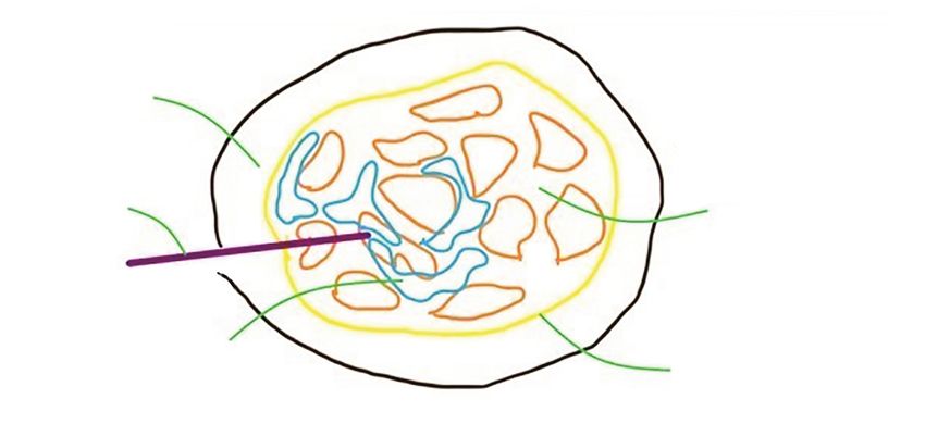
A histological transverse section of a thin nerve (Fig. disturbed the epineurium and increased the size of the
5A) showed the epineurium with MBD in the EPI-ct. The nerve with 3 mL of solution. Close to the ragged appear-
structures displaced included collagen fibers, elastin fi- ance of the epineurium (Fig. 3A) the fascicles were stained
bers, and fibroblasts. The transverse section of the thin with MBD, suggesting a possible breach in the perineu-
nerve (Fig. 5B) shows the injection site with a probable rium. In the other 2 specimens, at L1 and L2, the injection
puncture of the epineurium, at the ragged end of the epi- in the EPI-ct did not cause damage to any structures. In
neurium (Fig. 5B). MBD is imbedded in the epineurium. clinical practice at least 20 mL of LA is injected for an ad-
Korean J Pain 2021;34(1):132-136 https://doi.org/10.3344/kjp.2021.34.1.132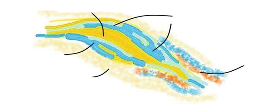
Nerve integrity after subepineural injection                                                                                                           135

                                                                         A                                                      B

Fig. 5. Histological information about L1 and L3 specimen (hematoxylin and eosin stain, low power [×10]). (A) Histological findings of specimen L1.
Epineural connective tissue (paraneural tissue-green lines) injection in L1, revealed localization of methylene blue dye in the epineural connective tissue
(paraneural). The intact inner epineurium (gold) and the perineurium (dark blue arrows) and internal architecture of the nerve are well preserved. (B) His-
tological findings of specimen L3. Sub-epineural needle placement (inside the sheath) revealed the breach of inner epineurium (gold).

Table 1. Ultrasound and macroscopic findings of 4 specimens

 Specimen                             Ultrasound                                                         Macroscopic findings
    no.                    EPI-ct                       Sub-EPI                                 EPI-ct                                 Sub-EPI

    L1          Epineurium connective tissue     NA                          Uniform and dense circumferential spread –      NA
                 spread                                                       MBD between TN and CPN
    L2          Epineurium connective tissue     NA                          Uniform and dense circumferential spread        NA
                 spread
    L3          NA                               Increased diameter of       NA                                              Fascicular tracings observed
                                                   nerve in all planes                                                        below sheath
    L4          NA                               Increased diameter of       NA                                              Fascicular tracings observed
                                                   nerve in all planes                                                        below sheath
EPI-ct: epineural connective tissue, Sub-EPI: sub-epineural, NA: not applicable, MBD: methylene blue dye, TN: tibial nerve, CPN: common peroneal nerve.

equate block, so one can imagine the nerve damage this                               Through this study we demonstrate that a pop or a click
would produce, with a sub-epineural injection.                                    in an US-guided popliteal sciatic nerve block could be
  Tran’s study group [4] concluded that in an US-guided                           detrimental regarding the nerve integrity, and recom-
popliteal sciatic nerve, a sub-epineural injection provides                       mend injections in the EPI-ct, better termed as paraneural
a higher success rate with a shorter performance time.                            tissue. Furthermore, the needle placement should be tan-
Well-defined experimental animal models have concluded                            gential (Fig. 2A) and not perpendicular to the nerve [10].
that a sub-epineural injection (intraneural) produces sig-                           Some unanswered questions were the disturbed (ragged)
nificant axonal damage and disruption of the blood-nerve                          epineurium, which appeared at the 6-7 o’clock position,
barrier [5-7].                                                                    the probable site of needle tip puncture (Fig. 5B), while
  In clinical practice, a ‘pop’ is appreciated as the needle                      the rest of the neural architecture demonstrated a nor-
penetrates what we visualize as a sheath around the undi-                         mal pattern (Fig. 3A). Perhaps a larger sample size would
vided sciatic nerve. This sheath is the epineurium, which                         have been more suited to answering the above-mentioned
should not be violated. This sub-neural injection is an                           questions. A comparative study between high and low
intraneural injection [8]. This is an unsafe practice and                         volumes in the paraneural and sub-epineural structure,
should be avoided in view of fascicular injury. Histologi-                        as well as subsequent histological analysis would answer
cally, there was no spread of MBD below the epineurium                            several queries. Recent practice patterns concerning iso-
after injecting into the EPI-ct. This EPI-ct is mentioned in                      lated peripheral nerve injections suggest that paraneural
the literature as the paraneural tissue [9]. The paraneural                       injections are considered safe [9,11]. Our histological study
injections are not accompanied by any damage of the ax-                           demonstrates that an injection in the EPI-ct (paraneural
ons. Clinically, in peripheral nerves, neither the pop, nor                       tissue) is a safe practice. A large volume sub-epineural
the current US resolution, differentiate between the epi-                         injection would be detrimental, with consequent nerve
neurium and perineurium.                                                          damage, considering the smaller volumes in our study

www.epain.org                                                                                                               Korean J Pain 2021;34(1):132-136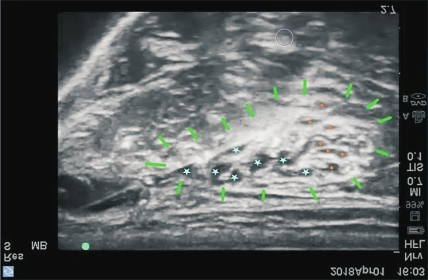
136                                                                                                                       Diwan, et al

producing histological changes suggestive of intraneural             105: 647-8.
injection.                                                        2. Al-Nasser B. Intraneural injection of local anesthetics during
  Though limited damage was evident on macroscopic                   ultrasound-guided peripheral nerve block may lead to nerve
and histological analysis of subepineural injections in our          injury. Anesthesiology 2007; 106: 1245-6.
study, we strongly recommend paraneural injections in             3. Bigeleisen PE. Nerve puncture and apparent intraneural
the isolated sciatic nerve.                                          injection during ultrasound-guided axillary block does not
                                                                     invariably result in neurologic injury. Anesthesiology 2006;
                                                                     105: 779-83.
ACKNOWLEDGEMENTS                                                  4. Tran DQ, Dugani S, Pham K, Al-Shaafi A, Finlayson RJ. A
                                                                     randomized comparison between subepineural and conven-
We acknowledge Dr. Asawari Manjarekar (MD), Patholo-                 tional ultrasound-guided popliteal sciatic nerve block. Reg
gist from CareFirst Diagnostics Pvt Ltd Pune, Maharashtra            Anesth Pain Med 2011; 36: 548-52.
State, India for histopathological analysis of the slides in-     5. Gentili F, Hudson AR, Hunter D, Kline DG. Nerve injection
volved in this manuscript.                                           injury with local anesthetic agents: a light and electron mi-
                                                                     croscopic, fluorescent microscopic, and horseradish peroxi-
                                                                     dase study. Neurosurgery 1980; 6: 263-72.
CONFLICT OF INTEREST                                              6. Westerlund T, Vuorinen V, Kirvelä O, Röyttä M. The endo-
                                                                     neurial response to neurolytic agents is highly dependent on
No potential conflict of interest relevant to this article was       the mode of application. Reg Anesth Pain Med 1999; 24: 294-
reported.                                                            302.
                                                                  7. Lupu CM, Kiehl TR, Chan VW, El-Beheiry H, Madden M,
                                                                     Brull R. Nerve expansion seen on ultrasound predicts histo-
FUNDING                                                              logic but not functional nerve injury after intraneural injec-
                                                                     tion in pigs. Reg Anesth Pain Med 2010; 35: 132-9.
No funding to declare.                                            8. Brull R, Chan VW, McCartney CJ, Perlas A, Xu D. Ultrasound
                                                                     detects intraneural injection. Anesthesiology 2007; 106:
                                                                     1244.
ORCID                                                             9. Andersen HL, Andersen SL, Tranum-Jensen J. Injection in-
                                                                     side the paraneural sheath of the sciatic nerve: direct com-
Sandeep Diwan, https://orcid.org/0000-0001-7950-070X                 parison among ultrasound imaging, macroscopic anatomy,
Abhijit Nair, https://orcid.org/0000-0003-2506-0301                  and histologic analysis. Reg Anesth Pain Med 2012; 37: 410-4.
Parag Sancheti, https://orcid.org/0000-0002-8903-1430            10. Sermeus LA, Sala-Blanch X, McDonnell JG, Lobo CA, Nich-
André Van Zundert, https://orcid.org/0000-0002-1836-6831             olls BJ, van Geffen GJ, et al. Ultrasound-guided approach to
                                                                     nerves (direct vs. tangential) and the incidence of intraneu-
                                                                     ral injection: a cadaveric study. Anaesthesia 2017; 72: 461-9.
REFERENCES                                                       11. Franco CD, Sala-Blanch X. Functional anatomy of the nerve
                                                                     and optimal placement of the needle for successful (and)
 1. Borgeat A. Regional anesthesia, intraneural injection, and       safe nerve blocks. Curr Opin Anaesthesiol 2019; 32: 638-42.
    nerve injury: beyond the epineurium. Anesthesiology 2006;

Korean J Pain 2021;34(1):132-136                                                                https://doi.org/10.3344/kjp.2021.34.1.132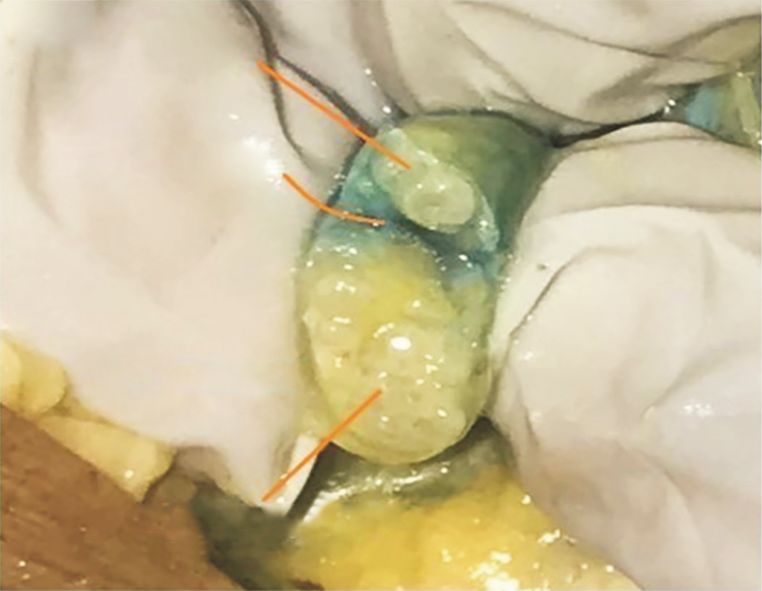
You can also read