Automatic X-ray Crack Inspection for Aircraft Wing Fastener Holes
←
→
Page content transcription
If your browser does not render page correctly, please read the page content below
2nd International Symposium on

  NDT in Aerospace 2010 - Mo.5.A.4

             Automatic X-ray Crack Inspection for
                Aircraft Wing Fastener Holes
               J. XU*, T. LIU*, X.M. YIN* , B. S. WONG**, Syahrom B. HASSAN**
      *
        Precision Measurement group, Singapore Institute of Manufacturing Technology, 71
                                Nanyang Drive, Singapore 638075;
               **
                  School of Mechanical and Aerospace Engineering, Nanyang Technological
                                      University Singapore 639798

                 Abstract. This paper presents an automatic X-ray image processing system for
                 fastener hole radial and circular crack detection. A robot guided X-ray imaging
                 system equipped with a digital detector is employed to take high resolution reference
                 images of fastener holes of various shapes, thicknesses and in different locations of
                 an aircraft wing. Learning module of the system is activated to train the system with
                 knowledge of hole shapes, segmentation thresholds, grey level distributions of holes
                 and backgrounds as well as hole pitches. Based on this knowledge, the system is
                 able to segment fastener holes of unknown incoming images with localized
                 thresholds, maximizing the shape similarity of the detected holes. Blob analysis is
                 then applied to derive hole gravity and boundary chain code. From the gravity point
                 of each hole, radial and circular scanning lines (arcs) are calculated to find subpixel
                 transitions (edges, which are potentially crack pixels) in the neighborhood of the
                 hole. Transition (edge) polarity and width are used to filter out unwanted
                 disturbances. Finally crack filtering helps removing noise and highlighting the
                 genuine cracks. The effectiveness of the system is demonstrated by processing real
                 aircraft wing images. We are able to achieve a processing speed of 300ms per 1024 x
                 1024 X-ray image ( Intel Dual Core PC 2.0GHz).

 1. Introduction

 Aircrafts are fabricated from a lightweight aluminum alloy that allows for an optimum
 balance of flight properties and cargo capacity. Aircraft wings, when subjected to the
 stresses of normal operation, fatigue damage can occur in high stress locations around
 fasteners. Crack growth, if left undetected, can lead to catastrophic failure. The inspection
 of aging aircraft for fastener hole cracks has been a time-consuming but essential task for
 the military services and commercial airlines alike.
         In order to identify the presence of cracks before they reach critical lengths, aircraft
 operators have updated their inspection techniques. While visual inspection is an important
 part of the detection process, many cracks in their initiation are too small to see visually,
 may be hidden beneath the head of the rivet, or may initiate in the inner layer of the lap
 joint and cannot be seen from outside of the aircraft. To assist with the detection of these
 small and hidden cracks, non-destructive inspection (NDI) methods are used. Some of the
 more common NDI methods used in aircraft crack detection are eddy current, ultrasound
 and digital radiography.
         Recent advances in magnetic sensor technology made the electromagnetic non-
 destructive evaluation method able to address crack detection issue. To detect deeply
 buried flaws, a low frequency electromagnetic field must be induced in the specimen under
 test. Traditional eddy current testing methods based on excitation-detection coils is

                                                           1
License: http://creativecommons.org/licenses/by/3.0/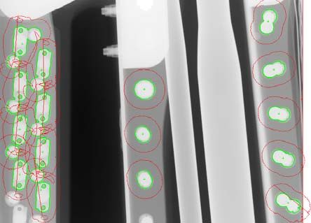
fundamentally limited by the poor sensitivity of the detection coils at low frequencies. Both
anisotropic magnetoresistive sensors (AMR) and giant magnetoresistive sensors (GMR)
have been successfully used in prior work for detecting deep cracks under installed
fasteners [1][2].
        The use of phased array ultrasonic [3][4] crack detection is another option.
Ultrasonic phased array transducers are made of multiple crystals that are electronically
pulsed in a sequence using a specified delay between pulses to create electronic scanning,
beam steering, and focusing capability. Ultrasonic phased array inspection is gaining wide
acceptance and has many advantages over conventional A-scan inspection. These
advantages include the ability to perform scanning with no mechanical movement, the
ability to perform angular (sectorial) scans and dynamic focusing without setup alteration,
two-dimensional visualization of results, and similar portability to A-scan equipment.
Ultrasonic phased array inspection results can be difficult to understand due to the
complexity of angular ultrasound compounded by multiple transducer element excitation.
Computational simulation can be used to predict the feasibility [5].
        Digital radiography has emerged as a leading technology for aircraft crack
inspection. It offers many advantages over conventional film-based radiography [6][7].
Digital Radiography enhances productivity as it records a ready to process image in short
time and eliminates the need for any chemical processing required as in the case of using a
film. In our previous work [8], we proposed methods and procedures to identify suitable x-
ray sources and digital detectors to meet the high sensitivity required to inspect an aircraft.
Different sensors and x-ray tubes were evaluated by using a set of a wire IQI penetrameter,
aluminium specimens with 6mm thickness and a 2T hole penetrameter. We recommended
amorphous silicon flat panels to be the most suitable detector for the aircraft inspection
applications.
        In this paper we propose an automatic X-ray image processing system for fastener
hole radial and circular crack detection. Learning is performed to acquire from multiple
aircraft wing X-ray images a sequence of fastener hole segmentation thresholds, hole sizes
and shapes. Then, a so called “smart threshold” is applied based on learnt knowledge to
optimize the finding of fasteners for any incoming wing image. Radial and circular
scanning around fastener holes is performed to find subpixel crack locations. For post
processing, three crack filters are applied to remove noise in crack images.
        The rest of the paper is organized as follows: in section 2, the hardware used for the
on- wing X-ray inspection, a robot guided X-ray imaging system equipped with a digital
detector, is described. In section 3, the proposed fastener hole crack detection software is
described in detail. Experimental results with real field trial images are given in section 4.
Finally in section 5 conclusions and future work are provided.

2. A Robot Guided X-Ray System for Aircraft Wing Imaging

Currently, the detection of deeply buried flaws is carried out by using either eddy current
techniques or ultrasound methods. The drawback of ultrasound and Eddy current methods
is that they are difficult to apply to areas hidden within aircraft wing structure. For
automatic fastener hole crack detection, we proposed a robot guided digital X-ray imaging
system (Fig. 1). A large raster scanning manipulator system has been developed to
automatically move an x-ray tube and a digital sensor along the wing of the aircraft.
Radiographs are taken as the system moves and a large montage of the whole wing area can
be built up gradually. The movement is programmed from a database drawing of the wing.

                                              2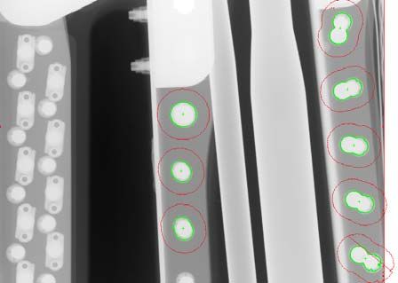
Fig. 1 A robot guided scanning manipulator positioned over an aircraft wing and digital radiographs

3. Automatic Image Processing for Fastener Hole Crack Detection

Fig. 2 illustrates the process flow of the proposed automatic image processing for fastener
hole crack detection system. To equip the system with learning capability for various
fasteners and imaging contrasts, the system has a learning module which allows user to
learn manually a sequence of X-ray images. Knowledge about the segmentation thresholds,
hole sizes and shapes are acquired and stored. For any unknown incoming wing image,
fastener hole segmentation module will apply a localized “smart threshold” for each
individual hole to determine its location and boundary. After that, radial and circular
scanning lines (arcs) are calculated to find subpixel transitions (edges, which are potentially
crack pixels) in the neighborhood of the hole. Transition polarity and width are used to
filter out unwanted disturbances. Finally crack filtering helps removing noise and
highlighting the genuine cracks.

     Fig. 2 Process flow of the proposed automatic image processing for fastener hole crack detection

                                                    3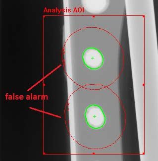
3.1 Knowledge Acquisition for Different Fastener Holes
As shown in Fig. 3, X-ray 2D scanning of an aircraft wing can generate images containing
various objects in the background, such as wires, piping, screws and other structures. It is
essential for an automatic algorithm to distinguish fastener holes and locate them from
background disturbances. To do this, a learning module is incorporated to train the software
with knowledge of what a fastener hole is and what is not.
The learning module allows user to load multiple images with different types of
fasteners. User can either select manually a number of thresholds to evaluate the
binarization results and identify the best one, or activate a semi-automatic process which
uses Otzu’s auto-threshold within a predefined adjustment percentage, say +/-25% to
perform auto binarization. Note that the global threshold selected for each image is for a
rough search of fastener holes. A localized threshold will be determined in segmentation
module to fine tune the binarization result for each hole.
The following knowledge about fastener holes will be acquired for segmentation
module:
A set of possible thresholds t , i 1. . N
Max hole number which is used to reject binarizations with too many objects
Hole shape in Fourier Descriptor for shape matching later on
Background pixel grey-level distribution outside holes; this will be used for filtering
out the false alarm pixels. The distribution is represented by an average value of all
pixels surrounding the hole and a statistical deviation.
Max and min hole areas and max and min hole extends in X and Y directions
A learning dada structure is used to record up to 100 different thresholds, 100 fastener
shapes, and other attributes.
Fig. 3 X-ray 2D scanning of an aircraft wing: images contain many objects and structures which make
fastener hole detection difficult
3.2 Localized Smart Threshold for Fastener Hole Localization
Let g x, y be an X-ray image pixel at location x, y with intensity within [0, 255],
binarization using a learnt threshold t can be formulated as
1 g x, y t
b x, y (1)
0 otherwise
where i 1,2, … N.
Applying object analysis algorithm to a binary image, a number of blobs ω can be
detected
O Ω ∑ ω (2)
whereby blob ω meets the following requirements
4 Its shape has been defined in learning result, e.g. there exists a learnt shape a such
that ∑ |a b |4th picture of Fig. 5) can be used. Again, the scan line length and incremental angle can be
defined by users.
Fig. 5 Radial (1st picture from the left) and circular cracks (2nd picture) and their detection using scan
lines perpendicular to the radial direction (3rd picture) and using radial scans (4th picture)
Subpixel Crack Detection Algorithm: When a crack detection scan line cuts across a crack
perpendicular to the crack propogation direction, its grey level profile typically has the
form displayed in Fig. 6a. Subpixel edge detection alorithm can be deployed to determine
the crack position and width. We use an energy balanced approach to calculate subpixel
edge location in a digital curve, a proprietary high speed subpixel edge detection method.
As shown in Fig. 6a, for a rising edge, edge start and end points can be determined from
each scan line using a minimum contrast constraint. Then, a model based double resolution
is performed to interpolate edge region (Fig. 6b) before an energy balanced subpixel edge
detection function is applied to derive subpixel edge position (Fig. 6c):
t s (3)
where A , A are areas of edge profile defined in Fig. 6c. For rising edge, we define the
edge polarity as minus. Similarly we can calculate a descending edge (plus edge).
(a) (b) (c)
Fig. 6 Subpixel crack detection by determining transitions in a rising and falling edge: (a) edge start and end
point and subpixel transition t; (b) double resolution to gain more accuracy; (c) calculate A1 and A2 for
subpixel position t calculation
Filtering for Noise Reduction: Many a time, when X-ray images are noisy, not only the
cracks, but also noise are detected by the edge detection algorithm mentioned earlier. To
reduce the noise, a few constraints are introduced:
Edge pair polarity check: crack should process a edge polarity from plus to minus
(e.g. profile transitions are from black pixels to white and then back to black)
Background consistency check: this option allows user to compare the background
grey level deviation with the learnt background. Only backgrounds with similar
grey level distributions are potential cracks
Max crack width check: crack width detected should be less than a predefined value
derived during learning
Another technique used in increasing the SNR of edge profile is a so called adaptive
linesum function which traces the edge propogation and performs edge directional
6integration to enhance SNR before applying subpixel edge detection algorithm. In this way
an improvement in SNR by a factor of 10 or above can be achieved.
4. Experimental Results
We now describe the details of our method used in real X-ray images to verify the validity
of the proposed approach. We use two groups of images: the first group is on-wing
acquired images. Since there are no cracks detected even with human eyes for the images in
the first group, we use a second group of real crack images from aluminum samples. Fig. 7
shows two smart threshold processing results using different learnt knowledge. The result
in Fig. 7a is achieved by learning three hole patterns while Fig. 7b is achieved by learning
only two patterns.
(a) (b)
Fig. 7 (a) Fastener hole detection by learning three fastener patterns; (b) Fastener hole detection by learning
two fastener patterns. The red circle covering each hole indicates the crack search area, which can be defined
by user
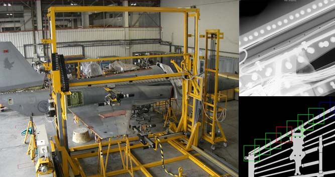
Fig. 8 shows the crack detection results with and without noise filtering. As can be
seen from Fig. 8a, noise pixels are falsely detected as cracks around hole boundary and
other areas, especially when user selects a big crack search area. After applying background
consistency and edge polarity check, the noise is removed (Fig. 8b).
(a) (b)
Fig. 8 (a) Crack detection without filtering: some red dots indicating detected cracks are found near hole
boundary; (b) Crack detection with edge polarity and background grey level consistency filters
7In Fig. 9, we apply the crack detection to a low SNR crack image. Linesum function
with 35 integrations is used to reduce the noise level. As can be seen from the image, one
real crack is detected, but there are some noise displayed in the crack map.
(a) (b)
Fig. 9 (a) original crack image; (b) Crack detection with only one crack detected; Using a lower contrast
constraint to get the other two cracks will produce very noisy result
5. Conclusions and Future Works
A crack detection software system developed for aircraft wing fastener hole inspection
from digitized radiographic image has been presented. We used a learning module to train
the system on fastener shapes and segmentation threshold and then used a smart threshold
to locate the fastener holes. Subpixel edge detection algorithm is applied subsequently to
detect all potential cracks both in radial direction as well as in circular direction. Finally a
few crack filtering mechanisms are implemented to remove the noise and output the crack
map. The future works include the enhancement of crack detection sensitivity and the
development of a crack model based detection algorithm.
References
[1] Lebrun, B., Jayet, Y. and Baboux, J.C., 1995, “Pulsed eddy current application to the detection of deep
cracks”, Materials Evaluation, Vol. 53, No. 11, pp. 1296-1300.
[2] Avrin, W.F., , “Eddy-current measurements with magnetoresistive sensors: third-layer flaw detection in a
wingsplice structure 25 mm thick”, Proceedings of SPIE, Vol. 3994, 2000, pp. 29-36.
[3] Roth, D.J., Tokars, R.P., Martin, R.E., Rauser, R.W., Aldrin, J.C., and Schumacher, E.J., “Ultrasonic
Phased Array Inspection Simulations of Welded Components at NASA”, Materials Evaluation, vol. 67,
no. 1, January 2009.
[4] Mahaut S., Chatillon S., Kerbrat E., Porré J., Calmon P., and Roy O., “New features for phased array
techniques inspections: simulation and experiments,” 16th World Conf on Non Destructive Testing,
Montréal, 2004.
[5] Aldrin, J.C. and Knopp, J.S., “Modeling and Simulation for Nondestructive Testing With Applications to
Aerospace Structures,” Materials Evaluation vol. 66. no. 1 pp. 53–59 (2008).
[6] Halmshaw R., Industrial radiology: theory and practice, second edition, Chapman and Hall, UK, 1995.
[7] Ewert, U., Zscherpel, U., Bavendiek, K., “Replacement of Film Radiography by Digital Techniques and
Enhancement of Image Quality” Online NDT Journal, vol 12. No.6, 2007.
[8] Xin Wang, B. Stephen Wong, Chen Guan Tui, Kai Peng Khoo, Frederic Foo, “Real-time Radiographic
Non-destructive Inspection for Aircraft Maintenance”, 17th World Conference on Nondestructive
Testing, 25-28 Oct 2008, Shanghai, China
8You can also read