Pediatric Multisystem Inflammatory Syndrome in Children as a Challenging Problem for Pediatric Surgeons in the COVID 19 Pandemic-A Case Report
←
→
Page content transcription
If your browser does not render page correctly, please read the page content below
CASE REPORT
                                                                                                                                                 published: 11 June 2021
                                                                                                                                         doi: 10.3389/fped.2021.677822

                                                Pediatric Multisystem Inflammatory
                                                Syndrome in Children as a
                                                Challenging Problem for Pediatric
                                                Surgeons in the COVID 19
                                                Pandemic—A Case Report
                                                Beata Jurkiewicz 1 , Magdalena Szymanek-Szwed 1 , Piotr Hartmann 2 , Joanna Samotyjek 1*,
                                                                2
                                                Eliza Bredowska
                                                        ˛         , Joanna Kaczorowska 2 , Ewa Wajszczuk 1 ,
                                                Martyna Twardowska-Merecka 1 and Joanna Cybulska 1
                                                1
                                                 Department of Pediatric Surgery and Pediatric Urology, Medical Center of Postgraduate Education, Dziekanów Leśny,
                                                Poland, 2 Department of Pediatrics, Children’s Hospital, Dziekanów Leśny, Poland

                           Edited by:
                        Juan A. Tovar,          The first cases of severe acute respiratory syndrome coronavirus-2 (SARS-CoV-2)
     University Hospital La Paz, Spain
                                                infection were identified at the end of 2019 and, in the next few months, coronavirus
                         Reviewed by:
                                                disease (COVID-19) spread throughout the world. Initially, it was believed that this disease
                    Leopoldo Martinez,
         Hospital Infantil La Paz, Spain        mainly affected elderly individuals with comorbidities, in whom respiratory failure often
                Ana Catarina Fragoso,           occurs. It was believed that children fell ill from the infection more often, although the
          University of Porto, Portugal
                                                course of infection in the vast majority of pediatric cases has been asymptomatic or
                  *Correspondence:
                   Joanna Samotyjek
                                                mildly symptomatic. In April and May 2020, the first report of a rapidly progressing
              asiamed@poczta.onet.pl            disease, similar to Kawasaki syndrome, was found in children who had been infected
                                                with SARS-CoV-2. Shortly thereafter, children with symptoms of pediatric inflammatory
                    Specialty section:
          This article was submitted to
                                                multisystem syndrome (PIMS-ST [temporally associated with SARS-CoV-2 infection])
                       Pediatric Surgery,       began presenting to pediatric hospitals around the world. The syndrome has a mortality
                a section of the journal        rate of up to 2%. Symptoms of PIMS-TS include those that may suggest the need for
                  Frontiers in Pediatrics
                                                surgical treatment (severe abdominal pain with the presence of peritoneal symptoms,
            Received: 08 March 2021
             Accepted: 17 May 2021              ascites, high levels of inflammatory markers, intestinal inflammation, and appendages
            Published: 11 June 2021             revealed on ultrasound examination). However, there are few reports addressing surgical
                               Citation:        cases associated with this condition. The authors present a case involving an 11-year-old
    Jurkiewicz B, Szymanek-Szwed M,
                                                boy who was admitted to hospital with severe abdominal pain and underwent surgery for
             Hartmann P, Samotyjek J,
        Bredowska
           ˛         E, Kaczorowska J,          symptoms of peritonitis and was diagnosed with PIMS in the post-operative period. Due
Wajszczuk E, Twardowska-Merecka M               to the large number of illnesses caused by SARS-CoV-2 infection in recent months, the
       and Cybulska J (2021) Pediatric
  Multisystem Inflammatory Syndrome             diagnosis of PIMS-TS/MISC should be considered in the differential diagnosis of acute
 in Children as a Challenging Problem           abdominal symptoms, especially in atypical courses and interviews indicating exposure
  for Pediatric Surgeons in the COVID
                                                to SARS-CoV-2.
        19 Pandemic—A Case Report.
              Front. Pediatr. 9:677822.         Keywords: pediatric inflammatory multisystem syndrome, appendectomy, coronavirus disease-19, children, case
      doi: 10.3389/fped.2021.677822             report

Frontiers in Pediatrics | www.frontiersin.org                                         1                                            June 2021 | Volume 9 | Article 677822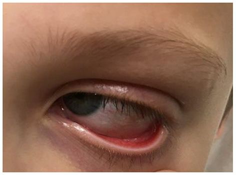
Jurkiewicz et al. PIMS in Pediatric Surgeon Practice
INTRODUCTION fraction, the appearance of coronary aneurysms, and embolic
complications (7, 8). The pathogenesis of the syndrome is
The first cases of coronavirus disease (COVID-19), caused by not fully understood. Only ∼45% of patients exhibit positive
infection with severe acute respiratory syndrome coronavirus-2 polymerase chain reaction (PCR) results for SARS-CoV-2,
(SARS-CoV-2), were reported at the end of 2019 in the city of although the presence of antibodies is found in 75%. Symptoms
Wuhan, Hubei Province, China. Within a few months, COVID- of the syndrome often appear only several weeks after contact
19 spread around the world (1). Initially, it was believed that with a sick person or an infection transmitted from an
COVID-19 mainly affects the elderly and those with specific asymptomatic or slightly symptomatic individual. Pre-disposing
comorbidities (2, 3). factors include age (∼≥9 years), male sex, obesity, and African–
According to data from the American Pediatric Society, American ethnicity (7, 8). The syndrome has a mortality rate
childhood cases constitute ∼12% of the total. Initially, it of up to 2%. Symptoms of PIMS-TS include those that may
was reported that, apart from the fact that children are less suggest the need for surgical treatment (severe abdominal pain
affected, the course of infection in the pediatric population is with the presence of peritoneal symptoms, ascites, high levels of
asymptomatic or mildly symptomatic in the vast majority of inflammatory markers, intestinal inflammation, and appendages
cases. Hospitalization rates for COVID-19 in pediatric patients revealed on ultrasound examination). However, there are few
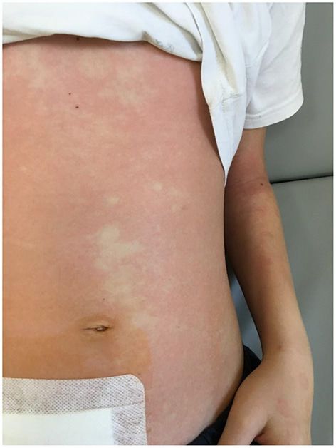
wereJurkiewicz et al. PIMS in Pediatric Surgeon Practice negative for SARS-CoV-2. Due to the characteristic history and physical examination, acute appendicitis was suspected and the patient was referred for emergency laparotomy. Intraoperatively, an unchanged appendix and a significant amount of serous fluid were found. Classic appendectomy was performed, the peritoneal cavity was drained, and no other source of peritoneal inflammation was found. The intestines were normal, and the mesenteric lymph nodes were not enlarged. The patient’s early post-operative course was uneventful. Perioperatively, the patient received antibiotic prophylaxis with cefazolin (5 doses in total) due to large amount of fluid in abdominal cavity. Good tolerance of an easily digestible diet was observed. After the surgery, the boy experienced watery stools (a total of up to 4 days) without pathological impurities. Upon suspecting bacterial infection of the gastrointestinal tract, antibiotic therapy was modified on day 2 of hospitalization, and cefazolin was discontinued and FIGURE 1 | Skin lesions. intravenous cefotaxime was empirically administered. Before treatment modification, serology for yersiniosis was performed, and feces were sent for culture. The boy complained of persistent pain and low-grade fever. In the following days, the results of stool culture and serological tests for yersiniosis were negative. On day 4 of hospitalization, a single ring-shaped skin lesion was observed. Suspected allergic background was treated with a second-generation antihistamine (levocetirizine). The next day, a fever of >39◦ C was observed. The boy’s condition deteriorated, and he reported feeling unwell and weak. Control laboratory investigations revealed an increase in inflammatory marker FIGURE 2 | Conjunctivitis. levels (CRP, 137.6 mg/dL; procalcitonin, 1.96 ng/mL), with normal peripheral blood counts and leukocytes count, neutrophil smear of 86.2%, signs of normocytic anemia (hemoglobin, 10.4 g/dL; mean corpuscular volume, 79.5 fL), and normal platelet concentration (5 g/dL). To determine the cause of the boy’s count. Antibiotic therapy was extended to include metronidazole poor general condition, diagnostics were directed toward a and intravenous amikacin. A control ultrasound examination proliferative etiology. Normal tumor marker (carcinoembryonic of the abdominal cavity 5 days after appendectomy revealed antigen and alpha-fetoprotein) levels were also determined. On an increased amount of fluid with increased echogenicity in day 1 after the repeat laparotomy, fever persisted. Additionally, the bladder area, thickened walls of the cecum and terminal a reddened appearance was observed on the skin of the trunk. intestine, and enlarged mesenteric nodes (up to 20 × 14 mm) The annular lesions intensified and fused to form a “garland” in the area of the removed appendix. Owing to the ultrasound (Figure 1). image suggesting the possibility of purulent lesions, computed Additionally, the patient developed bilateral non-pyrogenic tomography (CT) examination of the abdominal cavity was conjunctivitis, as well as chapped and reddened lips (Figure 2). performed and revealed traces of fluid in the pleural cavities, the He exhibited a temperature of 40◦ C despite a constant presence of fluid in the peritoneal cavity, interloop and bladder supply of paracetamol. During consultation with pediatrics, fluid (∼150 mL), thickened walls of the cecum (up to 19 mm), information regarding exposure to SARS-CoV-2/COVID-19 ∼1 thickened wall of the end intestine (up to 8 mm), without the month before the onset of disease symptoms was obtained. This presence of free gas in the peritoneal cavity, and numerous explained the high positivity for SARS-CoV-2 antibodies [cut- lymph nodes at the cecum and on the iliopsoas muscle (up off index (COI), 102 (normal,
Jurkiewicz et al. PIMS in Pediatric Surgeon Practice
TABLE 2 | Patient’s hospitalization timeline.
Day of Patient condition and Laboratory tests Imaging tests Treatment
hospitalization sympthoms
1 Symptoms of appendicitis. CRP 66.8 mg/L Presence of vesicular fluid and Emergency laparotomy was
Leukocyte count 4.04 × 10³ /µL thickening of the cecum wall performed: intraoperatively, an
Neutrophilic smear 70.7% unchanged appendix and a significant
Lymphopenia (0.77 × 10³ /µL) amount of serous fluid were found.
Cefazolin as antibiotic prophylaxis
was administered
2 Watery stools, low-grade Serology for yersiniosis feces for Antibiotic modification: cefotaxime
fever, persistent pain culture was administered
abdominal complaints
4 Single ring-shaped skin Second-generation antihistamine
lesion was observed (levocetirizine)
5 A fever of >39◦ C An increase in inflammatory An increased amount of fluid with Metronidazole and intravenous
marker levels (CRP, 137.6 increased echogenicity in the amikacin was additionally
mg/dL; procalcitonin, bladder area, thickened walls of administered
1.96 ng/mL), with normal the cecum and terminal intestine,
peripheral blood counts and and enlarged mesenteric nodes
leukocytes count, neutrophil (up to 20 × 14 mm) in the area of
smear of 86.2%, signs of the removed appendix.
normocytic anemia (hemoglobin,
10.4 g/dL; mean corpuscular
volume, 79.5 fL), and normal
platelet count
6 CT traces of fluid in the pleural Relaparotomy: a large amount of
cavities, the presence of fluid in slightly cloudy serous fluid was
the peritoneal cavity, interloop aspirated from the peritoneal cavity,
and bladder fluid (∼150 mL), which was sent for culture (negative),
thickened walls of the cecum (up and a drain was inserted into the
to 19 mm), thickened wall of the bladder area. Intraoperative images of
end intestine (up to 8 mm), the intestines did not concur with the
without the presence of free gas ultrasound and CT descriptions
in the peritoneal cavity, and
numerous lymph nodes at the
cecum and on the iliopsoas
muscle (up to 28 mm)
7 Fever persisted, a reddened Carcinoembryonic antigen and PIMS-TS was suspected human
on the skin of the trunk. The alpha-fetoprotein) levels = N, immunoglobulin [2 g/kg (i.e., 80 g of
annular lesions intensified high positivity for SARS-CoV-2 the preparation)] and ASA were
and fused to form a antibodies [cut-off index (COI), administered intravenous antibiotic
“garland,” patient developed 102 (normal, < 0.99) therapy with cefotaxime and
bilateral non-pyrogenic CRP, 176.1 mg/L; procalcitonin, metronidazole was continued, and
conjunctivitis, as well as 2.97 ng/mL; ferritin, 301 ng/mL amikacin was discontinued oral
chapped and reddened lips (normal, < 124 ng/mL) D-dimer, supplementation and drip infusion
transferred to the 4,460 µg/dL (normal, < 250 with potassium
Department of Pediatrics µg/dL)] hypokalemia,
hypoalbuminia, INR 1.34
(normal, 0.85–1.25); D-dimer,
4,460 µg/dL hypokalemia
11 Drain from the abdominal Less fluid in the abdominal cavity Ursodeoxycholic acid was included
cavity was removed and thinner walls of the
large intestine. Lung—presence
of fluid in the right pleural cavity
up to 10 mm in the left up to
5 mm, and fluid in the pericardial
sac up to 5 mm at the
widest point
13 No fever, no skin lesions, Normalization of inflammatory Due to the necessity of performing
good appetite markers echocardiography and further
cardiological treatment on day 13 of
hospitalization, the patient was
transferred to the Department of
Cardiology of the Medical University
of Warsaw (Warsaw, Poland) for
further treatment.
Frontiers in Pediatrics | www.frontiersin.org 4 June 2021 | Volume 9 | Article 677822Jurkiewicz et al. PIMS in Pediatric Surgeon Practice
human immunoglobulin [2 g/kg (i.e., 80 g of the preparation)] presented gastrointestinal symptoms (11). In presented case
and ASA were administered. The course of the immunoglobulin report the first symptoms were worsening abdominal pain
infusion was uneventful. Due to the inserted drain and increased and low-grade fever, what is very similar to other cases. Also
risk for bleeding, the recommended dose of ASA was reduced. Periyakaruppan presents a case with a boy who manifested
Considering the boy’s condition after two surgeries, intravenous gastrointestinal symptoms and fever (12). However, laparotomy
antibiotic therapy with cefotaxime and metronidazole was was not performed because CT abdomen revealed normal
continued, and amikacin was discontinued. While in the appendix. Similar to the authors’ practice, the use of intravenous
Department of Pediatrics, the patient experienced gradual immunoglobulins as a treatment method quickly improved the
improvement in his clinical condition. His vital signs, including patient’s condition.
blood pressure, were normal. The boy experienced fever during Our experience and the experiences of other authors should
the first 2 days of hospitalization. An albumin infusion was increase the awareness of surgeons who treat children with
performed in response to persistent hypoalbuminemia. Due abdominal pain and suspected appendicitis during the Sars CoV
to severe hypokalemia, simultaneous oral supplementation and 2 pandemic (13). In multisystem inflammatory syndrome the
drip infusion with potassium was performed, which achieved patients condition can rapidly deteriorate. Valitutti et al. based on
normokalemia on day 4 of hospitalization in the pediatric their own experience, encourages the assessment of the activity of
ward. In the days following the hospital stay, the boy’s the heart muscle before the operation of exploring the abdominal
skin lesions gradually disappeared and his appetite improved cavity. A preliminary assessment of troponin, BNP, D-Dimer,
significantly. In addition, the boy was receiving high-calorie ferritin and echocardiography can help to establish a precise
“nutri-drink” preparations, which he tolerated well. Four days differential diagnosis in children with acute abdomen, especially
after laparotomy, the drain from the abdominal cavity was when in MIS-C evolution toward cardiogenic shock should
removed. Follow-up abdominal ultrasound revealed less fluid in not be neglected (14). Another recomendation is presented by
the abdominal cavity and thinner walls of the large intestine. Khesrani et al. They advice that in atypical abdominal pain
At the same time, gallbladder examination revealed thickened syndrome during this pandemic an abdominal CT angio-scan
bile, with a slightly thickened wall and discreetly increased should be performer to look for vascular damages in order to
echogenicity in both kidneys. Lung ultrasound revealed the establish an appropriate medical treatment (immunoglobulins,
presence of fluid in the right pleural cavity up to 10 mm, in the corticoids) (15). In presented case none of above were performed
left up to 5 mm, and fluid in the pericardial sac up to 5 mm at before patients surgery. However, after analyzing the literature
the widest point. Electrocardiography revealed sinus rhythm and and acquiring our own experience, we apply the presented
non-specific ST segment abnormalities. Ursodeoxycholic acid recommendations in our center.
was included in the treatment regimen due to characteristics
suggestive of non-calculus cholecystitis. In subsequent laboratory CONCLUSION
investigations, a trend toward normalization of CRP level
was observed. Peripheral blood counts revealed leukopenia Due to the large number of illnesses caused by SARS-
(3.47 × 10³ /µL), with slight neutropenia (1.28 × 10³ /µL), CoV-2 infection in recent months, the diagnosis of
and normalization of the lymphocyte count (1.54 × 10³ /µL). PIMS-TS/MISC should be considered in the differential
An increase in hemoglobin concentration (10.9 g/dL) was diagnosis of acute abdominal symptoms, especially in
observed. Creatine phosphokinase concentration was normal, atypical courses and interviews indicating exposure
and albumin concentration normalized. Troponin T and CK-MB to SARS-CoV-2.
concentrations were normal, as was urinalysis. Two days after
the fever resolved, the dose of ASA was reduced to 300 mg daily DATA AVAILABILITY STATEMENT
(6.5 mg/kg body weight). Due to the necessity of performing
echocardiography and further cardiological treatment on day 13 The raw data supporting the conclusions of this
of hospitalization, the patient was transferred to the Department article will be made available by the authors, without
of Cardiology of the Medical University of Warsaw (Warsaw, undue reservation.
Poland) for further treatment (Table 2).
ETHICS STATEMENT
DISCUSSION
Written informed consent was obtained from the minor(s)’
Multisystem inflammatory syndrome (MIS-C) is a life- legal guardian/next of kin for the publication of any potentially
threatening condition occurring in children. It is most frequently identifiable images or data included in this article.
post-infectious, rather than related to acute SARS-CoV-2
infection. Gastrointestinal symptoms are the most common AUTHOR CONTRIBUTIONS
clinical manifestations of MIS-C (87% of children), followed by
muco-cutaneous (73%), cardiovascular (71%), respiratory (47%) BJ, MS-S, PH, and JS contributed to conception and design
and neurologic symptoms in 22% (9). In the first published paper of the study. MS-S organized the database. BJ wrote the first
reporting MIS-C 100% of presented patients had gastrointestinal draft of the manuscript. JS, EB, JK, EW, MT-M, and JC
symptoms (10). Similar results were presented in the material wrote sections of the manuscript. All authors contributed to
published by the group from Italy where 6 out of 10 patients manuscript revision, read, and approved the submitted version.
Frontiers in Pediatrics | www.frontiersin.org 5 June 2021 | Volume 9 | Article 677822Jurkiewicz et al. PIMS in Pediatric Surgeon Practice
REFERENCES 10. Riphagen S, Gomez X, Gonzalez-Martinez C, Wilkinson N,
Theocharis hyperinflammatory shock in children during COVID-
1. Wang C, Horby PW, Hayden FG, Gao GF. A novel coronavirus 19 pandemic. Lancet. (2020) 395:1607–8. doi: 10.1016/S0140-6736(20)
outbreak of global health concern, Lancet. (2020) 395:15–21, Pages 31094-1
497–506. doi: 10.1016/S0140-6736(20)30185-9 11. Verdoni l, Mazza A, Gervasoni A, Martelli L, Riggeri M, Ciuffreda M,
2. Paules CI, Marston HD, Fauci AS. Coronavirus infections-more than just the et al. An outbreak of severe Kawasaki-like disease at the Italian epicentre
common cold. JAMA. (2020) 323:707–8. doi: 10.1001/jama.2020.0757 of the SARS-CoV-2 epidemic: an observational cohort study. Lancet. (2020)
3. Siordia JA Jr. Epidemiology and clinical features of COVID- 395:1771–8. doi: 10.1016/S0140-6736(20)31103-X
19: a review of current literature. J Clin Virol. (2020) 12. Periyakaruppan M, Kumar S, Kandasamy S, Sangaralingam T, Srinivasan
127:104357 doi: 10.1016/j.jcv.2020.104357 S, Thiagarajan A, et al. COVID abdomen: SARS-CoV-2 infection
4. Kim L, Whitaker M, O’Halloran A, Kambhampati A, Chai SJ, presenting as ‘acute abdomen’ in a Child. Indian J Pediatr. (2020) 88:1–2.
Reingold A. Hospitalization rates and characteristics of children doi: 10.1007/s12098-020-03508-4
agedYou can also read