Clinical course of COVID 19 patients needing supplemental oxygen outside the intensive care unit - Nature
←
→
Page content transcription
If your browser does not render page correctly, please read the page content below
www.nature.com/scientificreports
OPEN Clinical course of COVID‑19
patients needing supplemental
oxygen outside the intensive care
unit
Ayham Daher1,3, Paul Balfanz2,3, Maria Aetou1, Bojan Hartmann2, Dirk Müller‑Wieland2,
Tobias Müller1, Nikolaus Marx2, Michael Dreher1 & Christian G. Cornelissen1*
Patients suffering from CVOID-19 mostly experience a benign course of the disease. Approximately
14% of SARS-CoV2 infected patients are admitted to a hospital. Cohorts exhibiting severe lung failure
in the form of acute respiratory distress syndrome (ARDS) have been well characterized. Patients
without ARDS but in need of supplementary oxygen have received much less attention. This study
describes the diagnosis, symptoms, treatment and outcomes of hospitalized patients with COVID-19
needing oxygen support during their stay on regular ward. All 133 patients admitted to the RWTH
Aachen university hospital with the diagnosis of COVID-19 were included in an observational registry.
Clinical data sets were extracted from the hospital information system. This analysis includes all 57
patients requiring supplemental oxygen not admitted to the ICU. 57 patients needing supplemental
oxygen and being treated outside the ICU were analyzed. Patients exhibited the typical set of
symptoms for COVID-19. Of note, hypoxic patients mostly did not suffer from clinically relevant
dyspnea despite oxygen saturations below 92%. Patients had fever for 7 [2–11] days and needed
supplemental oxygen for 8 [5–13] days resulting in an overall hospitalization time of 12 [7–20] days.
In addition, patients had persisting systemic inflammation with CRP levels remaining elevated until
discharge or death. This description of COVID-19 patients requiring oxygen therapy should be taken
into account when planning treatment capacity. Patients on oxygen need long-term inpatient care.
Since December 2019, the novel coronavirus “severe acute respiratory syndrome coronavirus 2 (SARS-CoV-2)”,
has been causing a rapidly spreading international outbreak1. Due to the different related clinical scenarios which
vary from asymptomatic infection to multiorgan involvement and failure, to even death in worse cases2–5, this
disease has been causing a substantial burden on healthcare systems worldwide at every level and especially on
available resources. However, the epidemiological studies from different countries have shown that the majority
of infected patients (> 80%) are asymptomatic or have mild symptoms, whereas about 14% of infected patients
have a severe disease and need to be h ospitalized2–7. Nevertheless, among hospitalized patients, the presence
and severity of respiratory failure are usually the most important clues in making the decision about admitting
to the intensive care unit (ICU) in order to provide ventilatory support (non-invasive or invasive ventilation) or
to treat on the regular ward. The group of ICU admitted patients has been very well characterized, and thera-
peutic approaches regarding ventilatory support have been well e stablished8–11. On the other hand, there is a
group of patients which have hypoxemic respiratory failure, but still could be managed on regular ward with
supplemental oxygen therapy. The characteristics of these patients have—to the best of our knowledge—not
been well described so far.
We therefore describe the diagnosis, symptoms, treatment and outcomes of hospitalized patients with
COVID-19 needing oxygen support during their stay on regular ward.
1
Department of Pneumology and Internal Intensive Care Medicine, University Hospital RWTH Aachen,
Pauwelsstrasse 30, 52074 Aachen, Germany. 2Department of Cardiology, Angiology and Internal Intensive Care
Medicine, University Hospital RWTH Aachen, Aachen, Germany. 3These authors contributed equally: Ayham Daher
and Paul Balfanz. *email: ccornelissen@ukaachen.de
Scientific Reports | (2021) 11:2256 | https://doi.org/10.1038/s41598-021-81444-9 1
Vol.:(0123456789)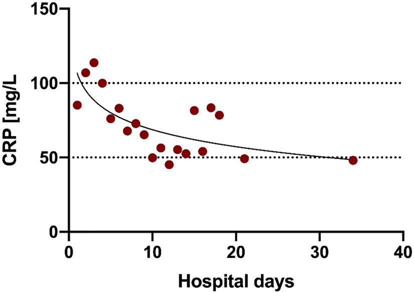
www.nature.com/scientificreports/
Total (N = 57) Non-Survivors (N = 13) Survivors (N = 42)
Characteristics
Age, years 72 [60–81] 81 [76–86] 65 [56–78]
Female sex 13 (23%) 2 (15%) 10 (24%)
Initial symptoms
Fever 39 (68%) 9 (69%) 29 (69%)
Cough 34 (60%) 10 (77%) 23 (55%)
Dyspnea 25 (44%) 4 (31%) 21 (50%)
Fatigue 21 (37%) 3 (23%) 18 (43%)
Gastrointestinal Symptoms 17 (30%) 1 (8%) 16 (38%)
Diarrhea 13 (23%) 0 (0%) 13 (31%)
Emesis 3 (5%) 1 (8%) 2 (5%)
Nausea 9 (16%) 1 (8%) 8 (19%)
Tiredness 16 (28%) 1 (8%) 15 (36%)
Myalgia 12 (21%) 1 (8%) 11 (26%)
Loss of Taste 10 (18%) 1 (8%) 9 (21%)
Loss of Smell 9 (16%) 1 (8%) 8 (19%)
Headache 7 (12%) 0 (0%) 7 (17%)
Sore throat 4 (7%) 0 (0%) 4 (7%)
Angina pectoris 4 (7%) 0 (0%) 4 (10%)
Pharyngalgia 3 (5%) 1 (8%) 2 (5%)
Rhinorrhoea 2 (4%) 0 (0%) 2 (5%)
Symptom onset to Hospitalization, days 4 [0–7] 0 [0–2] 6 [1–7]
Inpatient treatment
Patients with antibiotic therapy1 22 (39%) 9 (69%) 11 (26%)
Duration of antibiotic therapy, days 5 [4–6] 4 [4, 5] 5 [4–7]
COVID-19 specific treatment – – –
Periods, days
Fever days 7 [2–11] 8 [4–11] 8 [2–11]
Hospitalization 12 [7–20] 9 [6–15] 13 [8–20]
Oxygen supplementation 8 [5–13] 7 [4–10] 9 [5–13]
Outcome
Survivor 42 (74%) – –
Non-Survivor 13 (23%) – –
Ongoing hospitalization 2 (4%) – –
Discharge location
Home 39 (68%) – 39 (93%)
Rehabilitation 0 (0%) – –
Hospice 0 (0%) – –
Nursing facility 3 (5%) – 3 (7%)
Discharge with oxygen therapy 3 (5%) – 3 (5%)
Table 1. Baseline characteristics. Data in N (%) or Median [IQR]. IQR, Interquartile range. a Antibiotic classes
most commonly used: aminopenicillines, cephalosporines.
Methods
The protocol for this study was approved by the ethics committee of the University Hospital Aachen, Germany
(EK 080/20). All investigations were performed in accordance with the ethical standards laid down in the Decla-
ration of Helsinki in its latest revision and all patients provided written informed consent; in case patients could
not provide consent, written consultant advice and next of kin permission was obtained.
Previously we compared patients with and without ARDS regarding differences and outcome7. The current
analysis primarily focusses on patients with hypoxemic respiratory failure (defined as peripheral oxygen satu-
ration on pulse oximetry (SpO2) < 92% on ambient air), which were admitted to a regular ward. Demographic
data, disease history, coexisting medical conditions, presence of chronic respiratory failure, smoking history,
and medication history were recorded for all patients. Symptoms at admission and a detailed history of present
symptoms were also documented. Patients were assessed for eligibility on the basis of a positive reverse-tran-
scriptase-polymerase-chain-reaction (RT-PCR) assay for SARS-CoV-2 in a respiratory tract sample tested by
the local diagnostic laboratory. Viral load was also determined using RT-PCR. The threshold value Ct represents
the time point, at which the exponential phase of amplification begins, which therefore is inverse proportional
to virus concentration in the material investigated and reflects the relative difference on a logarithmic scale. The
Scientific Reports | (2021) 11:2256 | https://doi.org/10.1038/s41598-021-81444-9 2
Vol:.(1234567890)www.nature.com/scientificreports/
Total (N = 57) Non-Survivors (N = 13) Survivors (N = 42)
Comorbidities
Total 56 (98) 13 (100) 41 (98)
Arterial hypertension 33 (58) 10 (77) 21 (50)
Pre-existing heart diseases 22 (39) 5 (38) 15 (36)
Cardiovascular disease 15 (26) 4 (31) 11 (26)
Atrial fibrillation 11 (19) 4 (31) 6 (14)
Heart failure 12 (21) 2 (15) 8 (19)
Pre-existing respiratory disease 20 (35) 5 (38) 15 (36)
COPD 10 (18) 1 (8) 9 (21)
Obstructive sleep apnea syndrome 5 (9) 2 (15) 3 (7)
Bronchial asthma 5 (9) 2 (15) 3 (7)
Other pulmonary diseases 8 (14) 3 (23) 5 (12)
Smoking 19 (33) 2 (15) 17 (40)
Former smoking 8 (14) 2 (15) 6 (14)
Continued smoking 7 (12) 0 (0) 7 (17)
Overweight (BMI ≥ 25 kg/m2, < 30 kg/m2) 17 (30) 2 (15) 14 (33)
Obesity (BMI ≥ 30 kg/m2) 12 (21) 1 (8) 11 (26)
Diabetes mellitus 17 (30) 5 (38) 10 (24)
Prediabetes 10 (18) 3 (23) 7 (17)
Chronic kidney disease 10 (18) 3 (23) 6 (14)
Malignancy 10 (18) 3 (23) 6 (14)
Cerebrovascular disease 6 (11) 1 (8) 4 (10)
Chronic hepatitis 4 (7) 1 (8) 3 (7)
Peripheral arterial occlusive disease 3 (5) 1 (8) 2 (5)
Chronic liver failure 3 (5) 1 (8) 2 (5)
Premedication
ACE-Inhibitors 20 (35) 6 (46) 14 (33)
Angiotensin-receptor blockers 14 (25) 2 (15) 11 (26)
Beta blocker 20 (35) 7 (54) 11 (26)
Calcium antagonists 15 (26) 2 (15) 12 (29)
Diuretics 29 (51) 10 (77) 17 (40)
Antidiabetics 13 (23) 4 (31) 8 (19)
Lipid-lowering agents 16 (28) 6 (46) 10 (24)
Antiplatelet therapy 19 (33) 5 (38) 14 (33)
Anticoagulants 11 (19) 4 (31) 6 (14)
Inhalation therapy 16 (28) 2 (15) 13 (31)
Inhaled Glucocorticoids 5 (9) 1 (8) 4 (10)
Systemic Glucocorticoids 5 (9) 1 (8) 4 (10)
Immunosuppressive therapy 4 (7) 0 (0) 3 (7)
NSAIDs 11 (19) 0 (0) 11 (26)
Antibiotics 11 (19) 5 (38) 5 (12)
Antiviral therapy 1 (2) 0 (0) 1 (2)
Table 2. Comorbidities and premedication. Data in N (%) COPD, chronic obstructive pulmonary disease;
BMI, body mass index; ACE, Angiotensin-converting enzyme; NSAIDs, nonsteroidal anti-inflammatory drugs.
threshold value of the sample gene < 20, was classified as high. Values > 30 were classified as low virus load, and
values between ≥ 20 and < 30 as medium virus load.
Overweight was defined as BMI > 25 kg/m2 and obesity as BMI > 30 kg/m2. Diabetes or prediabetes was
defined by clinical history, medication and H
bA1c values ≥ 6.5%, or ≥ 5.7 to < 6.5%, respectively.
Vital signs including SpO2 were measured at least two times per day and if clinically indicated and docu-
mented in the hospital electronic medical record system. The worst values in 24 h were depicted for analysis.
Febrile days were defined as the time from fever onset until the last documented value above 38.5 ℃.
Generally, supplemental oxygen was given to target SpO2 values of > 94% and clinical relief in patients with-
out risk of hypercapnia, and S pO2 values of 88–92% with clinical relief if there was a risk of hypercapnia. The
flow rates of supplemental oxygen where documented two times per day and adjusted when needed. An oxygen
saturation below 92% led to an increase by 1 L/min while a sO2 above 96% triggered a decrease in oxygen flow
by 1 L/min. Compliance to this process was checked daily by the attending physicians and is part of the hospital’s
quality management program.
Scientific Reports | (2021) 11:2256 | https://doi.org/10.1038/s41598-021-81444-9 3
Vol.:(0123456789)www.nature.com/scientificreports/
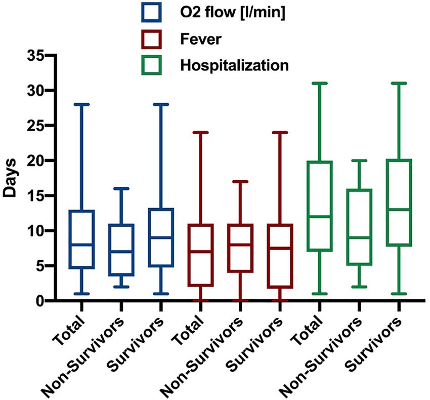
Figure 1. Clinical course of COVID-19 pneumonia. Box plots (Median with IQR and Min. to Max.) of
oxygen supplementation (blue), fever (red) and hospitalization (green) each for total cohort, non-survivors and
survivors.
Figure 2. Example for the long-lasting elevation of inflammatory parameters in COVID-19 pneumonia. Mean
of CRP (C reactive protein, red) in mg/L for complete cohort by hospital days.
Serum, plasma, and whole blood samples were obtained routinely at the time of admission in all patients as
per standard of care. Further blood tests were analyzed regularly as indicated, therefore patient numbers vary
between different time points in the figures. Radiological and further microbiological tests were performed
based on clinical decision making.
Values are displayed as median with interquartile ranges or mean ± standard error of the mean.
Results
57 consecutive patients being hospitalized outside the ICU on an isolation ward for SARS-CoV-2 pneumonia
between February and April 2020 were included into the analysis. All of them needed supplemental oxygen. At
the time of this analysis, 13 of the 57 patients were deceased (23%), while 42 (74%) had been discharged from the
hospital and 2 (4%) were still hospitalized. 12 of the 13 non-survivors (92%) expressed their will for a limitation
of therapy during hospitalization and therefore did not wish to be resuscitated, intubated or treated on the ICU.
Patient characteristics. Baseline characteristics of all patients as well as the subgroups of non-survivors
and survivors are summarized in Table 1. The median age (IQR) of the overall cohort was 72 (60–81) years, and
23% were women. Non-survivors were older compared to survivors (Table 1). All but one patient had comor-
bidities, which are, as well as concomitant medication, displayed in Table 2. Survivors and non-survivors exhib-
ited a similar prevalence of arterial hypertension, pre-existing respiratory diseases or pre-existing heart diseases
(Table 2).
Scientific Reports | (2021) 11:2256 | https://doi.org/10.1038/s41598-021-81444-9 4
Vol:.(1234567890)www.nature.com/scientificreports/
Reference values Total (N = 57) Non-Survivors (N = 13) Survivors (N = 42)
Vital parameters
Height, cm 172 [166–178] 173 [164–180] 173 [167–178]
Weight, kg 75 [66–80] 75 [66–80] 84 [72–91]
BMI, kg/m2 24.8 [23.5–27.2] 24.8 [23.5–27.2] 27.7 [24.2–30.3]
Respiratory rate, bpm 22 [18–26] 24 [17.5–28.3] 21 [18–25.8]
Oxygen saturation, % 94 [91–96] 94 [92–95] 95 [90.3–97]
Oxygen flow, l/min 2 [0–4] 3 [2–7] 2 [0–2]
Temperature, °C 37.9 [36.9–38.6] 37.8 [37–38.5] 37.9 [36.9–38.6]
sBP, mmHg 125 [101–140] 127 [110–140] 126 [103–140]
dBP, mmHg 71 [60–80] 67 [57–78] 74 [60–80]
Heart rate, bpm 88 [80–100] 88 [71–103] 90 [80–100]
Laboratory tests at admission
Leukocytes, 1/nl 4.0–10.0 6.9 [5.1–9.5] 8.7 [5.9–9.3] 6.7 [4.5–9.4]
m: 14.0–18.0
Hb, g/dl 13.4 [10.3–14.5] 10.4 [10.1–13.5] 13.9 [12.1–14.6]
w: 12.0–16.0
Thrombocytes, 1/nl 150–400 180 [144–232] 148 [134–315] 187.5 [148.5–225.8]
Lymphocytes, % 22.0–53.0 10.4 [7–15.2] 7 [4.4–12.3] 11.6 [9.1–20.5]
INR 1.2 [1.1–1.3] 1.2 [1.1–1.4] 1.2 [1.1–1.3]
aPTT, sec 25.1 – 36.5 27.8 [26–30.7] 28.6 [26.1–30.9] 27.8 [26.2–30.4]
D-Dimer, ng/ml < 500 855 [622–1086] 16,401 [11158–21644] 738.5 [595.8–881.3]
HbA1c, % < 5.7 6.1 [5.5–6.7] 6.1 [5.8–6.8] 5.8 [5.4–6.6]
Sodium, mmol/l 136–145 139 [136–141.3] 141 [138–143] 139 [136–141]
Potassium, mmol/l 3.6–5.5 4.2 [3.8–4.6] 4.2 [3.6–4.7] 4.2 [3.8–4.6]
Albumin, g/dL 3.5–5.2 3.2 [2.7–3.8] 3.8 [3.6–3.9] 2.9 [2.7–3.8]
Total Bilirubin, mg/dL < 1.2 0.6 [0.5–0.9] 0.9 [0.4–1.1] 0.6 [0.5–0.7]
AST, U/l < 35 41.5 [30–55.3] 42 [27–72] 41 [33–55]
ALT, U/l < 35 27 [20–37] 25.5 [20.8–29.5] 30 [20–41]
Gamma-GT, U/l < 40 31 [23–68] 57 [32.5–110.8] 31 [23–65]
AP, U/l 35–105 65 [47–82.5] 98 [84–158.5] 61.5 [44.3–80.3]
m: 135–225
LDH, U/l 339 [277–442] 416 [332–452] 339.5 [273.3–436]
w: 135–214
m: < 174
CK, U/l 132 [82.8–285.5] 96 [62–400] 150.5 [98–285.5]
w: < 140
CK-MB-Activity, U/l < 26 U/l 16 [11–18] 18 [14–18.5] 16 [12–18]
hsTroponin T, pg/ml < 14.0 83.5 [27.3–131] 81 [68.5–93.5] 93 [28–179]
NTproBNP, pg/ml < 220 726 [198.2–1393] 1730.5 [663.7–3028.3] 323.9 [71.5–1237]
Urea, mg/dl 16.6–48.5 40 [29.5–65.5] 48.5 [41–69.3] 38 [27–58]
Creatinine, mg/dl 0.5–1.2 1 [0.9–1.4] 1 [0.9–2] 1 [0.9–1.2]
CRP, mg/lwww.nature.com/scientificreports/
Total (N = 57) Non-Survivors (N = 13) Survivors (N = 42)
Chest radiography
No infiltrates 7/53 (13%) 3/13 (23%) 3/38 (8%)
Unilateral infiltrates 5/53 (9%) 2/13 (15%) 3/38 (8%)
Bilateral infiltrates 41/53 (77%) 8/13 (62%) 32/38 (84%)
Viral load
High 11/52 (21%) 4/12 (33%) 7/38 (18%)
Medium 29/52 (56%) 7/12 (58%) 21/38 (55%)
Low 12/52 (23%) 1/12 (8%) 10/38 (26%)
Ct S-Gen 24,8 [2–29] 22,8 [2, 9–28] 24,1 [8–28]
Viral detection
Respiratory detection 52/56 (93%) 12/13 (92%) 38/41 (93%)
positive out of hospital 7/57 (12%) 1/13 (8%) 6/42 (14%)
Extra-respiratory detection 11/34 (32%) 3/4 (75%) 8/28 (29%)
Serum 6/28 (21%) 2/4 (50%) 4/22 (18%)
Stool 5/15 (33%) 1/3 (33%) 4/12 (33%)
Urine 4/24 (17%) 2/4 (50%) 2/18 (11%)
Bacterial detection
Blood culture 3/44 (7%) 1/12 (8%) 2/30 (7%)
Urine culture 16/38 (42%) 3/9 (33%) 11/27 (41%)
Table 4. Radiological and microbiological findings. Data in N/total available N (%), or Median [IQR] IQR,
Interquartile range.
Vital signs as well as laboratory parameters, radiological and microbiological findings assessed at baseline
are displayed in Tables 3 and 4. The median oxygen supply at admission was at 2 (0–4) L/min. 3 patients (5%)
had already been on long-term oxygen therapy (LTOT). The median oxygen flow during the whole period of
hospitalization was 2 (0.4–2.5) L/min. The majority of patients (77%) had bilateral infiltrates. Patients who died
had higher values of inflammatory parameters compared to survivors (Table 3). In addition, D-dimers were
elevated in all patients with higher values in those who died compared to survivors (Table 3).
Discussion
This study characterizes patients suffering from COVID-19 that require supplemental oxygen therapy but do
not exhibit severe Acute Respiratory Distress Syndrome (ARDS) and can be treated outside the ICU. Several
studies have focused on COVID-19 patients needing intensive care medicine8–11, but to our knowledge, patients
requiring supplemental oxygen on a general ward have not been described in detail.
The patients included in this study presented with a typical set of symptoms2: fever, cough or fatigue were
usually present. Of note, less than half (44%) of the patients exhibited dyspnea despite their hypoxemic respira-
tory failure, which might easily result in underestimation of the clinical severity of the disease. Peripheral oxygen
saturation should thus be measured in all patients with COVID-19 at admission and routinely on regular basis
during the hospital stay. All but one patient had at least one comorbidity; with hypertension, heart diseases and
overweight being the most common ones.
Survivors and non-survivors in this study should be regarded as two different patient groups. While survivors
were on average 16 years younger, non-survivors declined intensive care treatment. Respiratory failure led to
death in the latter patients. Marked systemic inflammation reflected by IL-6 levels highlights the difference in
disease severity between these groups. Also, an extra pulmonary manifestation of COVID-19 was detected in
75% of non-survivors, comparable to patients with mild A RDS11.
The single most outstanding finding of this study is the length of hospitalization and the need of supplemental
oxygen: patients were treated for 12 days and needed oxygen therapy for 8 days on average. Hospital duration
exceeded oxygen therapy whenever the overall patient status did not yet permit discharge or when home quar-
antine requirements could not be met. Importantly, comparing to patients being hospitalized because of severe
influenza12, patients with COVID-19 seem to need a significantly longer hospital stay and are longer on oxygen
therapy.
The severity and the prolonged course of COVID-19 in these patients might be caused by persisting systemic
inflammation reflected in fever and elevated C-reactive protein (CRP) as well as interleukin-6 (IL-6). In fact,
CRP levels remained elevated until discharge or death (ref. Figure 2).
In Conclusion patients with COVID-19 requiring oxygen therapy need long-term inpatient care with a
median of 12 days in hospital including 8 days on supplemental oxygen, which should be taken into account
when planning treatment capacity. This result could be partially explained by the prolonged inflammatory course
of the disease.
Scientific Reports | (2021) 11:2256 | https://doi.org/10.1038/s41598-021-81444-9 6
Vol:.(1234567890)www.nature.com/scientificreports/
Received: 27 September 2020; Accepted: 7 January 2021
References
1. World Health Organization. Director-General’s remarks at the media briefing on 2019-nCoV on 11 February 2020. (Accessed 15
September 2020); https: //www.who.int/dg/speech es/detail /who-direct or-genera l-s-remark s-at-the-media- briefi ng-on-2019-ncov-
on-11-february-2020.
2. Huang, C. et al. Clinical features of patients infected with 2019 novel coronavirus in Wuhan, China. Lancet 395, 497–506 (2020).
3. Chen, N. et al. Epidemiological and clinical characteristics of 99 cases of 2019 novel coronavirus pneumonia in Wuhan, China: a
descriptive study. Lancet 395, 507–513 (2020).
4. Wang, D. et al. Clinical characteristics of 138 hospitalized patients with 2019 novel coronavirus-infected pneumonia in Wuhan,
China. JAMA 323(11), 1061–1069 (2020).
5. Liu, K. et al. Clinical characteristics of novel coronavirus cases in tertiary hospitals in Hubei Province. Chin. Med. J. (Engl) 133(9),
1025–1031 (2020).
6. Wu, Z. & McGoogan, J. M. Characteristics of and important lessons from the coronavirus disease 2019 (COVID-19) Outbreak
in China: summary of a report of 72 314 cases from the Chinese Center for Disease Control and Prevention. JAMA 323(13),
1239–1242 (2020).
7. dre Leber, B. et al. Charakteristik von 50 hospitalisierten COVID-19-Patienten mit und ohne ARDS. Dtsch Arztebl Int. 117,
271–278. https://doi.org/10.3238/arztebl.2020.0271 (2020).
8. Richardson, S. et al. Presenting characteristics, comorbidities, and outcomes among 5700 patients hospitalized with COVID-19
in the New York City Area. JAMA https://doi.org/10.1001/jama.2020.6775 (2020).
9. Myers, et al. Characteristics of hospitalized adults with COVID-19 in an integrated health care system in California. JAMA 23(21),
2195–2198 (2020).
10. Phua, J. et al. Intensive care management of coronavirus disease 2019 (COVID-19): challenges and recommendations. Lancet
Respir. Med. 8, 506 (2020).
11. Gattinoni, L. et al. Covid-19 does not lead to a “typical” acute respiratory distress syndrome. Am. J. Respir. Crit. Care Med. 201,
1299–1300 (2020).
12. Lee, N. et al. Outcomes of adults hospitalised with severe influenza. Thorax 65(6), 510–515 (2010).
Author contributions
M.D., N.M. and D.M.W. designed the study. A.D., M.A., T.M. and C.G.C. acquired the data. A.D., P.B., B.H., M.D.,
N.M., D.M.W .and C.G.C. analyzed the data. A.D., P.B., C.G.C. and M.D. wrote the manuscript and prepared
figures & tables. All authors reviewed the manuscript.
Funding
Open Access funding enabled and organized by Projekt DEAL.
Competing interests
The authors declare no competing interests.
Additional information
Correspondence and requests for materials should be addressed to C.G.C.
Reprints and permissions information is available at www.nature.com/reprints.
Publisher’s note Springer Nature remains neutral with regard to jurisdictional claims in published maps and
institutional affiliations.
Open Access This article is licensed under a Creative Commons Attribution 4.0 International
License, which permits use, sharing, adaptation, distribution and reproduction in any medium or
format, as long as you give appropriate credit to the original author(s) and the source, provide a link to the
Creative Commons licence, and indicate if changes were made. The images or other third party material in this
article are included in the article’s Creative Commons licence, unless indicated otherwise in a credit line to the
material. If material is not included in the article’s Creative Commons licence and your intended use is not
permitted by statutory regulation or exceeds the permitted use, you will need to obtain permission directly from
the copyright holder. To view a copy of this licence, visit http://creativecommons.org/licenses/by/4.0/.
© The Author(s) 2021
Scientific Reports | (2021) 11:2256 | https://doi.org/10.1038/s41598-021-81444-9 7
Vol.:(0123456789)You can also read