Growth Assessment Protocol (GAP) Guidance - November 2020 Perinatal Institute
←
→
Page content transcription
If your browser does not render page correctly, please read the page content below
Growth Assessment Protocol (GAP)
                Guidance
    https://perinatal.org.uk/GAPguidance.pdf

             November 2020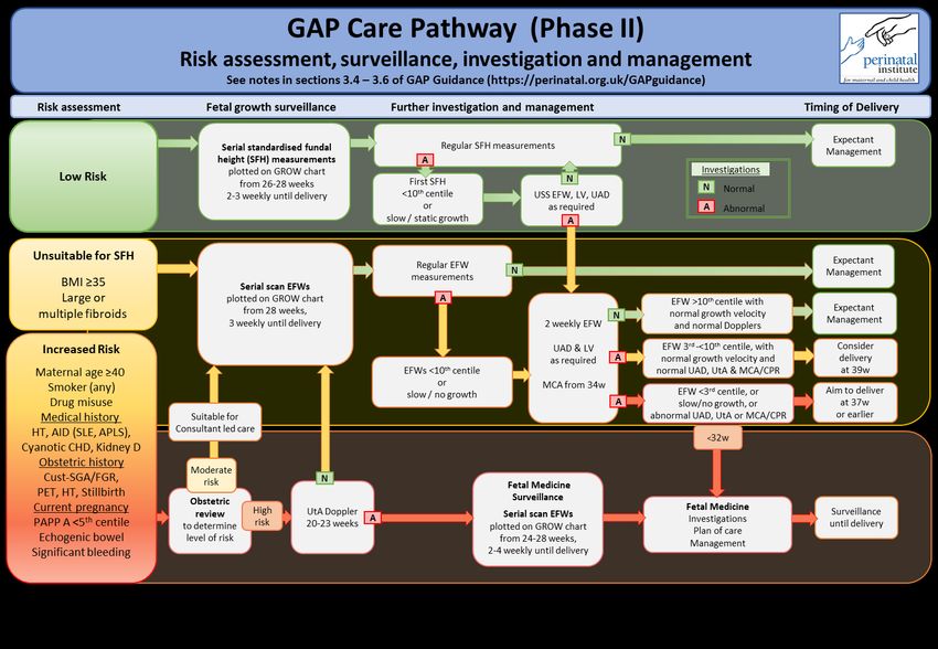
CONTENTS

1. Introduction

2. Elements of GAP
   2.1 Training and accreditation
   2.2 Protocols and Guidelines
   2.3 GROW charts
   2.4 Recording outcome and benchmarking
   2.5 Missed case audit
   2.6 Links to GAP service level agreement

3. GAP Care Pathway
   3.1 Introduction
   3.2 Phase I pathway
   3.3 Phase II pathway
   3.4 Risk Assessment
   3.5 Fetal growth surveillance
   3.6 Further investigation and management
   3.7 Audit of detection rates and missed cases

   3.8 Conclusion

   3.9 Next Steps

   3.10 References

                                                   2
1 | INTRODUCTION
The Perinatal Institute provides the Growth Assessment Protocol as a licensed and supported service to assist
clinicians and their health organisations with the assessment of fetal growth. This priority has arisen from
evidence that many adverse outcomes in maternal and perinatal care are associated with unrecognised fetal
growth problems, and can be prevented by improved awareness and detection of the pregnancies affected.
This document should be read in conjunction with the GAP service level agreement which sets out the
proposed partnership between the PI’s GAP team and each organisation’s ‘champions’ / leads tasked with
implementating and running the programme. A recent, award winning analysis of Office of National Statistics
(ONS) data has emphasised the benefits of thorough implementation and adherence to GAP protocol on
stillbirth prevention [1].
Here, we summarise the main components of GAP, and present the Care Pathway with evidence based
guidelines which can be adapted into local protocols.
2 | IMPLEMENTATION OF THE GAP PROGRAMME
The main components of GAP are training, protocols, growth charts, benchmarking and missed case audit.
2.1 Training and accreditation of all staff involved in maternity care
Face to face training and remote workshops on all aspects of theory as well as practice including standardised
fundal height measurements, ultrasound and Doppler parameters, generation and plotting on customised
charts, early pregnancy risk assessment and referral pathways, and data collection and audit. This is
supported by e-learning with theoretical and practical modules, which include a short theoretical assessment
and provision of an e-certificate. A designated e-learning lead/administrator can be given access to local e-
learning accounts and compliance reports.
2.2 Adoption and local adaptation of growth assessment protocols and guidelines
Includes the GAP Care Pathway, with early pregnancy ‘booking’ risk assessment, triage for low risk care
through serial fundal height measurement, and indications for referral; and increased risk care, obstetric
review, serial growth scans with assessment of size as well as growth rate, and appropriate additional
Doppler investigations and implications for management.
32.3 Implementation and training in use of customised GROW software
Provision of GROW-App, (stand-alone or integrated with hospital electronic record), for plotting
measurements of standardised fundal height (SFH) and estimated fetal weight (EFW). The growth chart is
customised for constitutional variation (ethnic origin, maternal size, parity) and optimised by excluding
pathological factors (e.g. high BMI, smoking), thus improving the ability to identify abnormal growth while
reducing false positive diagnoses.
2.4 Recording pregnancy outcome and generating reports on progress
The software prompts the recording of key indicators following delivery of the baby, including the
birthweight centile and, if SGA, whether it was detected antenatally. The false positive rate is also calculated.
Standardised unit-specific reports are generated for local feedback and benchmarking against a baseline
(established before GAP was implemented) to assess progress and compare with national GAP user average.
2.5 Missed case audit
Software and training is also provided to allow clinicians to undertake a ‘standardised clinical outcome review
and evaluation’ (SCORE) of their SGA deliveries that were not recognised antenatally. GAP leads are
encouraged to undertake this regularly on a proportion of cases, to check for avoidable factors such as failure
to follow risk assessment protocol, inaccurate measurement or plotting, lack of referral for investigation, etc.
2.6 Further information is available in the GAP service level agreement:
Annex A – Key responsibilities - sets out the respective roles and responsibilities for the Perinatal Institute
and the local health service to ensure effective implementation and running of the GAP programme.
Annex B – Help Desk Services - outlines the support that hospitals and individual users can expect when
assistance is required from the Perinatal Institute, including clinical and technical helpdesks.
Annex C – GROW Software, data Items and user accounts - provides details of the various software and
user accounts available to support the programme, including growth charts with API links to information
systems, audit tools for benchmarking and missed case review, and individual (clinical) and bulk (research)
centile calculators.
43 | THE GAP CARE PATHWAY
3.1 Introduction
Following publication in 2019 of NHS England’s Saving Babies’ Lives Care Bundle version 2 (CBv2) [2], the
Perinatal Institute has undertaken consultations on aligning it for NHS Trusts in the GAP programme, while
maintaining the benefits of a tried and tested protocol that has been credited as a main contributor to the
reduction of stillbirth rates in England [1, 3, 4, 5].
Our care pathway seeks to balance the need to identify, investigate and appropriately manage at-risk
pregnancies with the aim to avoid unnecessary intervention. It continues the emphasis on surveillance
according to risk assessment, by standardised fundal height (SFH) and estimated fetal weight (EFW)
measurements, serially plotted on customised charts to improve the distinction between constitutional and
pathological smallness [6, 7, 8, 9]. A focus of the revised pathway is a new, evidence-based definition of slow
growth (see Section B).
Concerns about the ultrasound and Doppler capacity demands of CBv2, expressed at the 2019 National GAP
User Symposium, have been confirmed by a survey we conducted in association with the British Medical
Ultrasound Society (BMUS), which showed that most ultrasound services in the NHS cannot currently meet
the requirements [10]. The Society of Radiographers have also expressed concern [11].
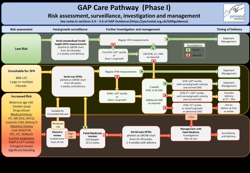
We have therefore developed a Care Pathway which can be implemented in two phases, according to
maternity unit capacity. Phase I should be achievable with current resources, while sonographic services seek
to acquire the additional training and resources required for Phase II. These Explanatory Notes are designed
to be read in conjunction with Care Pathway algorithms I and II.
Disclaimer: We have made this guidance as evidence based as possible. It is intended to supplement, not
substitute maternity care, which needs to be personalised and based on good clinical judgement.
The Perinatal Institute and its staff cannot be held responsible for any adverse outcome.
57
3.4 Risk Assessment

1. Early pregnancy risk assessment and triage into the correct care pathway is essential. As the
    significance of risk factors is often determined by the severity of current and previous conditions and
    various other circumstances, moderate and high risk categories are amalgamated into an ‘Increased
    risk’ group, which should undergo obstetric review to help determine the appropriate pathway and
    level required for investigations and surveillance.

2. Pre-existing diabetes, diabetes arising during pregnancy, or twins and other multifetal pregnancy are
    covered by their respective NICE guidelines and are out of scope of this document.

3. Previous ‘SGA’, if determined by customised centiles, is more likely to be due to fetal growth
    restriction (FGR) than if SGA is determined by population centiles (on which CBv2 is based). It is
    therefore represents increased risk and is often the only available indicator of the likelihood that
    there was indeed previous FGR, as it is often missed antenatally. Obstetric review should consider
    severity, gestational age at onset (if known), gestational age at delivery and associated factors such
    as preeclampsia.

4. Previous stillbirth is considered an increased risk, regardless of size and stated cause, unless placental
    insufficiency has been reliably excluded by histopathological examination.

5. Calculating an estimated fetal weight (EFW) at the time of the anomaly scan is not recommended, as
    good evidence is lacking about its efficacy to predict adverse outcome. Preliminary results presented
    at the 2019 Fetal Growth conference [12] suggest that the narrow normal range at 20-23 weeks can
    frequently lead to a false positive suspicion of SGA. The error is substantially increased with
    population based fetal weight standards. If units nevertheless wish to assess EFW at this time, we
    recommend using the customised centile calculator which works from 20 weeks gestation and is
    available from the GAP Team.

6. For women with a history of, or significant risk factor(s) for, placental dysfunction (including history
    of pre-eclampsia or fetal growth restriction), Aspirin 75-150mg nocte is recommended from 12 weeks
    to birth according to latest NICE guidelines [13].

7. The recommended initial obstetric review of pregnancies considered at increased risk of early or late
    onset FGR should determine next steps according to severity of risk and unit policy.

                                                     8
Phase 1: Pregnancies considered high risk should be referred to maternal-fetal medicine (MFM)
services, where available, for investigation and review:
- if uterine artery (UtA) Doppler is normal ➔ moderate risk pathway;
- if UtA Doppler is abnormal ➔ continue under MFM surveillance
Where MFM services are not available, frequent assessment from 24 weeks throughout
the third trimester is recommended, as per Care Pathway.
Phase 2: Pregnancies considered high risk can have UtA Doppler performed in the ultrasound
department, with referral to MFM if abnormal.
3.5 Fetal growth surveillance
1. The low risk pathway follows the existing algorithm, with 2-3 weekly clinical assessment and fundal
height measurement from 26-28 weeks until delivery, according to unit policy, using a standardised
technique [14] and with measurements plotted on the customised GROW chart. If suboptimal
growth is suspected (first fundal height measurement5. Slow or ‘restricted’ growth by serial EFWs is defined as a growth rate between scan measurements
which is slower than the slope of the 3rd centile line on the customised chart at the same gestational
age. A growth rate less than the slope of the customised 3rd centile line predicts adverse perinatal
outcome [15]. For precision and reduced effect of scan error, routine serial EFW measurements to
assess growth velocity should be at least 2 weeks apart [8]. While third trimester EFW measurements
in routine NHS practice have been shown to be accurate to within +/-10% in 70% of cases [16],
consideration should be given to the clinical implication of potential scan error, as well as the overall
need for quality assurance. The Perinatal Institute has developed a free audit tool for EFW error,
available from the GAP Team.
6. In 2020/21, we will be introducing auto-plotting and digital assessment in the new GROW App, with
alerts if the growth rate is outside the normal range at the respective stage in pregnancy. In the
interim, visual assessment is required to compare serial SFH and EFW measurements with the 10 th
and 3rd centile reference lines. Plotting can be assisted by set-squares, available from the GAP Team.
The figures below illustrate an example of manual assessment of growth rate:
Fig 1 (left) has two sample EFW measurements plotted at 36 and 39 weeks. The two measurement each
lie between the 50th and 90th centile but taken in sequence, they suggest slow growth. This is confirmed
in Fig 2 (right), where a line is drawn through the two plots to delineate the slope. Using a set square, a
parallel line is drawn through the 3rd centile line over the same gestational age interval; this shows that
the growth rate is slower than the lowest accepted rate in this pregnancy, over this gestation interval.
The use of a set square to draw parallel lines is illustrated in a short video clip.
103.6 Further Investigation and Management
1. The Care Pathway outlines the suggested level of surveillance when placental insufficiency is
suspected - by an EFWOptional data fields will be added in the GROW App in 2021 to enable units to undertake a more thorough assessment of service and outcome. These will include: • Proportion of pregnancies identified as increased risk in early pregnancy, and care offered • Aspirin use and outcome • Third trimester scan regime and outcome, stratified according to risk factors In addition, the new GROW-App will integrate with the GAP-SCORE tool which will facilitate ongoing case- note review of ‘missed’ SGA cases (neonates with birthweight
3.10 References

1.    Hugh O, Williams M, Turner S, Gardosi J. Reduction of stillbirths in England according to uptake of the Growth
      Assessment Protocol, 2008-2017: 10 year population based cohort study. Ultrasound in Obstetrics &
      Gynecology 2020 https://doi.org/10.1002/uog.22187

2.    Saving Babies’ Lives v.2: A care bundle for reducing perinatal mortality. NHS England, 2019
      www.england.nhs.uk/publication/saving-babies-lives-version-two-a-care-bundle-for-reducing-perinatal-mortality
3.    Gardosi J, Giddings S, Clifford S, Wood L, Francis A. Association between reduced stillbirth rates in England and
      regional uptake of accreditation training in customised fetal growth assessment. BMJ Open 2013;3(12).
      https://bmjopen.bmj.com/content/bmjopen/3/12/e003942.full.pdf
4.    Widdows K, Roberts S, Camacho E, Heazell A. Evaluation of the implementation of the Saving Babies’ Lives Care
      Bundle in early adopter NHS Trusts in England (SPIRE) Manchester, UK: Maternal and Fetal Health Research
      Centre, University of Manchester; 2018 https://bit.ly/2LZFbBv
5.    Williams M, Turner S, Butler E, Gardosi J. Fetal growth surveillance – current guidelines, practices and
      challenges. Ultrasound 2018 https://bit.ly/2XKypZB
6.    Gardosi J, Francis A. Controlled trial of fundal height measurement plotted on customised antenatal growth
      charts. Br J Obstet Gynaecol. 1999;106(4):309–317
      https://obgyn.onlinelibrary.wiley.com/doi/full/10.1111/j.1471-0528.1999.tb08267.x
7.    Clifford S, Giddings S, Southam M, Williams M, Gardosi J. The Growth Assessment Protocol: a national
      programme to improve patient safety in maternity care. MIDIRS Digest. 2013;23:516–23 https://bit.ly/2Do0tGw
8.    RCOG. The investigation and management of the small-for-gestational-age fetus. Green Top Guideline 31. RCOG
      2013 https://www.rcog.org.uk/globalassets/documents/guidelines/gtg_31.pdf
9.    Jayawardena L, Sheehan P. Introduction of a customised growth chart protocol increased detection of small for
      gestational age pregnancies in a tertiary Melbourne hospital. Aust N Z J Obstet Gynaecol. 2018
       https://obgyn.onlinelibrary.wiley.com/doi/abs/10.1111/ajo.12902
10.   Survey of ultrasound and Doppler services. Perinatal Institute and BMUS, 2019 https://bit.ly/2OpZHPt
11.   Society of Radiographers. SoR calls for review of the implementation of Saving Babies’ Lives Care Bundle V2
      2019 https://www.sor.org/news/scor-calls-review-implementation-saving-babies-lives-care-bundle-v2
12.   Hugh O, Francis A, Gardosi J. Customised vs population based fetal weight standards to define small for
      gestational age as a risk factor at 22 weeks gestation. 8th International Conference of Fetal Growth, Berlin 2019
      www.fetalgrowth.org Abstract PO8 https://bit.ly/2XQuiZH
13.   National Institute for Health and Care Excellence. Hypertension in pregnancy: diagnosis and management
      (Clinical Guideline 133). NICE 2019 www.nice.org.uk/guidance/ng133
14.   Morse K, Williams A, Gardosi J. Fetal growth screening by fundal height measurement.
      Best Practice Research Clin Obstet Gynaecol. 2009;23(6):809–18. https://bit.ly/327QJLM
15.   Gardosi J, Francis A, Hugh O, Turner S, Williams M. Customised limits for normal, restricted and accelerated
      fetal growth rates and their ability to predict perinatal outcome. Ultrasound Obstet Gynecol 2000;56:S1 –
      OC07.06 https://obgyn.onlinelibrary.wiley.com/doi/full/10.1002/uog.22247
16.   Francis A, Tonks A, Gardosi J. Accuracy of ultrasound estimation of fetal weight at term. ADC Fetal and Neonatal
      https://fn.bmj.com/content/96/Suppl_1/Fa61.1
17.   MacKay DF, Smith GCS, Dobbie R, Pell JP. Gestational Age at Delivery and Special Educational Need:
      Retrospective Cohort Study of 407,503 Schoolchildren. PLoS Med. 2010 Jun 8;7(6):e1000289
      https://journals.plos.org/plosmedicine/article?id=10.1371/journal.pmed.1000289
18.   Jacobsson B, Ahlin K, Francis A, Hagberg G, Hagberg H, Gardosi J. Cerebral palsy and restricted growth status at
      birth: population-based case-control study. Br J Obstet Gynaecol. 2008 Sep;115(10):1250–5
      https://obgyn.onlinelibrary.wiley.com/doi/10.1111/j.1471-0528.2008.01827.x
19.   Williams M, Turner S, Hugh O, Gardosi J. Reduction of small-for-gestational-age stillbirths at term in the GAP
      programme 2015-2018. Br J Obstet Gynaecol. 2019;126(S2):53. https://bit.ly/2YxH7a5
20.   Figueras F, Gratacos E. An integrated approach to fetal growth restriction. Best Practice Research Clin Obstet
      Gynaecol 2017;38:48-58. https://bit.ly/2XevtA5

                                                         13
You can also read