DETECTION OF FECAL SARS-COV-2 RNA IN A PROSPECTIVE COHORT OF CHILDREN WITH MULTISYSTEM INFLAMMATORY SYNDROME (MIS-C) - PREPRINTS.ORG
←
→
Page content transcription
If your browser does not render page correctly, please read the page content below
Preprints (www.preprints.org) | NOT PEER-REVIEWED | Posted: 10 May 2021                                   doi:10.20944/preprints202105.0187.v1

          Brief Report

          Detection of fecal SARS-CoV-2 RNA in a prospective cohort of
          children with multisystem inflammatory syndrome (MIS-C)
          Emilia Parodi 1,2 *, Andrea Carpino 3, Elena Franchitti 2, Giulia Pruccoli 1,2, Marco Denina 1,2 , Federica Pagliero 2 ,
          Ugo Ramenghi 2 and Deborah Traversi 2

                                        1   Regina Margherita Children' s Hospital, A.O.U. Città della Salute e della Scienza, Torino, Italy
                                        2   Department of Public Health and Pediatrics, University of Torino, Torino, Italy
                                        3   Postgraduate School of Pediatrics, University of Torino, Torino, Italy

                                        * Correspondence: emilia.parodi@unito.it (mail); Tel + 39 0113135798 (office); I-1026 Torino, Italy

                                        Abstract: Multisystem inflammatory syndrome related to SARS-CoV-2 infection (MIS-C) is a rare
                                        severe illness affecting paediatric patients. No data are available in literature about the presence of
                                        SARS-CoV-2 RNA in faces in patients with MIS-C. The SARS-CoV-2 positivity was evaluated in
                                        fecal samples obtained in a prospective cohort of paediatric patients admitted to our tertiary Hos-
                                        pital and diagnosed with MIS-C or COVID-19 between April 15, 2020, and February 29, 2021. The
                                        real-time RT-PCR was performed using a validated kit. The assay measures 3 target genes:
                                        SARS-CoV-2 gene E, gene N, gene ORF1ab. Overall, 16/63 (25%) fecal samples revealed the pres-
                                        ence of SARS -CoV-2 RNA. No differences were detected about time from presumptive viral ex-
                                        posure and the time of stool collection (14 vs 8 days, p> 0,05) as well as about the presence of gas-
                                        trointestinal symptoms (p>0.05) between patients with positive (+) stools for SARS-CoV-2 RNA and
                                        patients tested negative (-). Among children with MIS-C, stools were collected 27.5 days as median
                                        (95% CI 14-34) after presumed contact and the positivity rate was 12.5% (4/32). According to our
                                        data, we could suggest contact precautions with all patients with MIS-C. Further sample collection
                                        is ongoing to verify our preliminary data.

                                        Keywords: SARS-CoV-2; stools; COVID-19; MIS-C; children

                                        1. Introduction
                                             Multisystem inflammatory syndrome related to SARS-CoV-2 infection (MIS-C) is a
                                        rare but severe illness affecting paediatric patients firstly described in April 2020, ap-
                                        proximately one month after the first COVID-19 outbreak [1,2]. MIS-C usually develops
                                        after the infection rather than during the acute stage of the disease, suggesting a dysreg-
                                        ulated immune response (i.e. continuous activation of adaptive immune responses
                                        driven by persisting antigen presentation) as the primary pathogenic mechanism [3].
                                        Only one-third of reported cases, indeed, present a positive RT-PCR for SARS-CoV-2 on
                                        nasal swab; on the contrary, in most cases the serology results positive, suggesting a past
                                        infection [4,5].
                                             MIS-C clinical presentation, characterized by multi-organ involvement, commonly
                                        involves gastrointestinal signs and symptoms [6–8]. To the best of our knowledge, no
                                        data are present in literature regarding the detection of SARS-CoV-2 RNA in fecal sam-
                                        ples of patients with MIS-C, even if biospecimen collection methods for children were
                                        recently proposed [9].
                                             The present prospective study aimed to detect SARS-CoV-2 RNA in fecal samples of
                                        patients who met CDC case definition criteria for MIS-C, admitted to our tertiary care
                                        paediatric hospital in the North-West of Italy between April 2020 and February 2021.

           © 2021 by the author(s). Distributed under a Creative Commons CC BY license.
Preprints (www.preprints.org) | NOT PEER-REVIEWED | Posted: 10 May 2021 doi:10.20944/preprints202105.0187.v1
2 of 8
2. Materials and Methods
All patients admitted to Regina Margherita Children’s Hospital (Turin, Italy) and
diagnosed with MIS-C between April 15, 2020, and February 29, 2021, were prospectively
enrolled in the study. Children admitted in the same period for COVID-19 or followed up
in outpatients’ service after hospitalization for COVID-19 were included as control
group. Written informed consent was obtained from the parents of patients. Approval
was obtained from Institutional Ethical Committee (Città della Salute e della Scienza di
Torino, protocol number 00564/2020).
Fecal samples obtained after admission were stored at -80°C, immediately after col-
lection, until the extraction and analysis were performed in the lab (Hygiene Section,
Public Health and Pediatric Department, University of Turin). Extraction of total RNA
from fecal samples was performed with a commercialized kit (Power Microbiome RNA
kit, QIAGEN). The extracted RNA was quantified using a NanoQuant Plate (TECAN
Trading AG, Switzerland), which allows quantification using a spectrophotometer read
at 260 nm. The spectrophotometer used was the TECAN Infinite 200 PRO, and the soft-
ware was i-Control (version 1.11.10). The extracted RNA concentration was meanly 17.6 ±
25.6 µg/mg stool. Samples were stored at –80°C until molecular analysis was performed.
The real-time RT-PCR was performed using the Novel Coronavirus (2019-nCoV)
Real-Time Multiplex RT-PCR kit from LifeRiver Ltd., and CFX Instruments (Bio-Rad,
Hercules, CA, USA) following the suggested thermal protocol included in the kit in-
structions and according to literature data [10]. The assay measures simultaneously 3
target genes: SARS-CoV-2 gene E, gene N, gene ORF1ab according to international vali-
dated testing protocols [10,11].
For each patient enrolled in the study, demographic data, clinical presentation and
time from SARS-CoV-2 infection to fecal sample collection were registered (Table 1A).
The time from the presumptive contact with the virus was defined as follow:
a) when available, we considered the date of the first positive SARS-CoV-2 naso-
pharyngeal swab with the addiction of 4 days as typical incubation period [12];
b) for patients with MIS-C without previous positive swab and ascertained intra-
familial exposure to SARS-CoV-2, we considered the date of the first positive
SARS-CoV-2 swab of familial contacts;
c) for patients with MIS-C without positive swab and untested intrafamilial expo-
sure, we considered 15 days as the minimum time to produce specific antibodies from
exposure.
The statistical analysis was performed using IBM SPSS statistics package 27.0. A
descriptive analysis of the variables was conducted. The data were reported as absolute
numbers and percentages for categorical variables while as medians and confidence in-
tervals – 95% higher and 5% lower – for continuous variables. Moreover, the subjects
were divided into two groups based on the diagnosis: COVID-19 or MIS-C. Differences
between COVID-19 and MIS-C children were assessed using χ2 test with Fisher’s correc-
tion for categorical variables and Median test for independent samples for continuous
variables. The Spearman rank-order correlation coefficient was also determined to assess
the relationships between variables. A p-value pPreprints (www.preprints.org) | NOT PEER-REVIEWED | Posted: 10 May 2021 doi:10.20944/preprints202105.0187.v1
3 of 8
at the follow- up in outpatients’ clinic. Clinical and demographic characteristics of pa-
tients are reported in Table 1A.
Data regarding IgG against SARS-CoV-2 were available for 28/32 patients with
MIS-C, as 4 children were enrolled before the development of validated methods; 27/28
(96%) children had a positive serology, confirming past infection. All patients with
COVID-19 displayed at least one positive Real-Time Polymerase Chain Reaction
(RT-PCR) nasal swab for SARS-CoV-2.
Gastrointestinal symptoms and fecal SARS-CoV-2 data
Gastrointestinal symptoms (i.e. at least one among abdominal pain, nausea, vomit-
ing or diarrhoea) were reported in 9/31 (29%) patients with COVID-19 and 26/32 (81%)
children with MIS-C, with a statistically significant difference between the two groups (p
=0.000).
Overall, 16/63 (25%) fecal samples revealed the presence of SARS -CoV-2 RNA.
No differences were detected about time from presumptive viral exposure and the
time of stool collection (14 vs 8 days, p> 0,05) as well as about the presence of gastroin-
testinal symptoms (p>0.05) between patients with positive (+) stools for SARS-CoV-2
RNA and patients tested negative (-). In particular, the positivity rate of stools was
comparable in patients with or without gastrointestinal clinical involvement, 37.5% (6/35)
vs 35% (10/28), p>0.05.
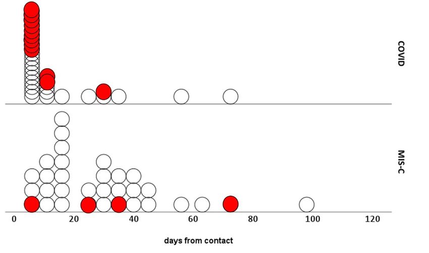
In patients with COVID-19, fecal samples were collected 8 days as median (95% CI
7-13) after the presumed viral exposure and were positive in 12/31 (39%); among children
with MIS-C, stools were collected 27.5 days as median (95% CI 14-34) after presumed
contact and the positivity rate was 12.5% (4/31). (Table 1B). Positive stools of patients
with MIS-C were collected at 27, 36, 43 and 72 days from presumptive primary infection
(Figure 1); in the latter, concomitant serology for SARS-CoV-2 resulted negative, but the
exposure to a confirmed COVID-19 case was ascertained.
3.2. Figures and Tables
Figure 1. Distribution of the samples concerning the esteemed time from the SARS-CoV-2 contact
causing the following COVID-19 or MIS-C and stool positivity (red).
Table 1. Demographic characteristics (A), gastrointestinal symptoms and fecal SARS-CoV-2 col-
lected data (B) of the two patient cohorts. Continuous variables are expressed as median and con-Preprints (www.preprints.org) | NOT PEER-REVIEWED | Posted: 10 May 2021 doi:10.20944/preprints202105.0187.v1
4 of 8
fidence interval (IC 5-95%); the categorical variables are expressed as absolute numbers and per-
centages. Bolded p values are statistically significant
COVID-19 MIS-C p
(n=31) (n=32) value
14 14
Female
(45.2) (43.8)
Gender p>0.05
17 18
Male
(54.8) (56.2)
4 4
Africans
(12.9) (12.5)
26 26
Caucasian
(83.9) (81.25)
Ethnicity p>0.05
1 0
Chinese
(3.2) (0)
0 2
Hispanic
(0) (6.25)
A
5 8
Age years p>0.05
(2-9) (7-11)
27 1
IgG (+)
(96)
Antibodies against Sars-CoV-2 NA
1
IgG (-)
(4)
31 10
Pos (+)
(100) (31.2)
Nasopharyngeal swab p=0.000
0 22
Neg (-)
(0) (68.7)
Esteemed time from the 8 27.5
days p=0.003
Sars-CoV-2 contact (7-13) (14-34)
µg/µL 69.6 22.8
Extracted RNA from stool p>0.05
(33.7- 128.3) (11.6-50.3)
22 6
No
(71) (18.75)
B Gastrointestinal symptoms p=0.000
9 26
Yes
(29) (81.25)
Neg (-) 19 (61.3) 28 (87.5) p=0.017
Sars-CoV-2 RNA in stool
Pos (+) 12 (38.7) 4 (12.5) 2
1 data available only for 28 patients with MIS-C
2 the few data included in any groups can affect the test significantlyPreprints (www.preprints.org) | NOT PEER-REVIEWED | Posted: 10 May 2021 doi:10.20944/preprints202105.0187.v1
5 of 8
4. Discussion
Multisystem inflammatory syndrome related to SARS-CoV-2 infection (MIS-C) is a
severe emerging illness affecting paediatric patients [1,2].
The SARS-CoV-2 attachment site, i.e. the angiotensin-converting enzyme 2 (ACE-2
receptor), is widely expressed also in the gut [15–18]. Previous studies have already
demonstrated the presence of SARS-CoV-2 RNA in faeces of almost one third of patients
with COVID-19 [19–24]. The contagiousness of the virus within the gut and the possible
fecal-oral route of transmission, however, remain unproven [19,25–29].
Multiple studies have reported gastrointestinal symptoms in children with
COVID-19, with diarrhoea, nausea, vomiting, anorexia and abdominal pain being de-
scribed as the main symptoms [6,28]. Children, compared to adults, have a higher ACE-2
expression in the gut [15] and present a higher ACE expression inside the intestine than
in the respiratory tract [24]; these data may explain both the higher proportion of gas-
trointestinal symptoms and the higher percentage of positive faeces in the paediatric than
in the adult form of COVID-19 [15].
MIS-C clinical presentation, characterized by multi-organ involvement, commonly
includes gastrointestinal signs and symptoms. According to literature data [6–8], in our
cohort diarrhoea, nausea, vomiting or abdominal pain were more frequent in patients
with MIS-C compared to children with COVID-19 (respectively more than 75% in the
first group vs less than 30% in the second one); this difference resulted statistically sig-
nificant (p< 0.000).
To the best of our knowledge, data regarding the presence of the virus in the gut of
patients with MIS-C are lacking. In our prospective cohort of pedaitric patients with
MIS-C, more than 10% of patients tested positive (+) for SARS-CoV-2 RNA in faeces. The
rate of patients with acute COVID-19 and fecal SARS-CoV-2 positivity (39%) was con-
sistent with previously reported data [24], confirming the reliability of our methods.
Despite the limits deriving from the small number of patients with MIS-C tested
positive (+) for SARS-CoV-2 fecal RNA, some preliminary conclusions could be drawn.
First, it should be noted that there is no difference in the occurrence rate of gastrointes-
tinal symptoms between patients with MIS-C with a positive (+) or a negative (-) result
for SARS-CoV-2-RNA in the stools. Whether the virus titres in fecal fomites are of suffi-
cient concentration and infectivity for subsequent transmission remains unknown, even
in patients with acute COVID-19 [19,25–29]. Nevertheless, due to the impossibility of
clinically differentiating in our cohort children with positive (+) stools (>10%) from the
others with negative (-) stools, we suggest contact precautions during hospitalization to
be taken in all patients with MIS-C.
In addition, the wide range of days observed between presumed primary viral in-
fection and detection of viral RNA in faeces (72, 43, 36 and 27 days) does not permit to
determine precisely how long these precautions should be applied in children with
MIS-C.
Further sample collection is ongoing to verify our preliminary data on a larger co-
hort.
To further complete the study, gut microbiota analysis on DNA extracted from fecal
samples of our patients with MIS-C will be performed in the next months. In adults, mi-
crobiota modulation linked to SARS-CoV-2-host interaction has been proven to promote
inflammation (through reduction of short-chain fatty acid producers and increase of
pathogenic microorganisms) [30,31]. The quite long pre-symptomatic phase in MIS-C
suggests an additional trigger that likely occurs 2-4 weeks after the initial infection; in
this context, microbiota might represent a crucial key to explain the mechanism of dis-
ease onset. As a consequence, complete microbiota analysis could be an innovative ap-
proach to better understand MIS-C pathogenesis and identify potential early biomarkers
of susceptibility to this emerging paediatric disease.Preprints (www.preprints.org) | NOT PEER-REVIEWED | Posted: 10 May 2021 doi:10.20944/preprints202105.0187.v1
6 of 8
Author Contributions: EP and DT and UR gave substantial contribution to conception and design,
drafted the article, reviewed and critically revised the manuscript; DT and EF made stool sample
processing and extraction and bio-molecular analysis; DT made statistical analysis and bioinfor-
matic; AC, EF, GP, FP and MD contributed to conception and design, collected data, and revised
the manuscript; all authors approved the final version of the manuscript and agree to be account-
able for all aspects of the work.
Funding: This research received no external funding.
Institutional Review Board Statement: The study was conducted according to the guidelines of
the Declaration of Helsinki, and approved by the Ethics Committee of Città della Salute e della
Scienza di Torino, protocol number 00564/2020.
Informed Consent Statement: Written informed consent has been obtained from the parents of the
patients to publish this paper.
Acknowledgments: the Authors thank the University of Torino and all the physicians and nurses
personnel involved in the patients’ care. The Authors make a special acknowledgement to the
children and to their families.
Conflicts of Interest: The authors declare no conflict of interest.
References
[1] Carbajal R, Lorrot M, Levy Y, Grimprel E, Lecarpentier T, Heritier S, et al. Multisystem inflammatory syndrome in children
rose and fell with the first wave of the COVID-19 pandemic in France. Acta Paediatrica, International Journal of Pediatrics
2021;110:922–32. doi:10.1111/apa.15667.
[2] Blumenthal JA, Burns JP. Epidemiology of Multisystem Inflammatory Syndrome in Children: A Step Closer to
Understanding Who, Where, and When. JAMA Pediatrics 2021. doi:10.1001/jamapediatrics.2021.0638.
[3] Gruber CN, Patel RS, Trachtman R, Lepow L, Amanat F, Krammer F, et al. Mapping Systemic Inflammation and Antibody
Responses in Multisystem Inflammatory Syndrome in Children (MIS-C). Cell 2020;183:982-995.e14.
doi:10.1016/j.cell.2020.09.034.
[4] Henderson LA, Canna SW, Friedman KG, Gorelik M, Lapidus SK, Bassiri H, et al. American College of Rheumatology
Clinical Guidance for Multisystem Inflammatory Syndrome in Children Associated With SARS–CoV-2 and
Hyperinflammation in Paediatric COVID-19: Version 1. Arthritis & Rheumatology 2020. doi:10.1002/art.41454.
[5] Rostad CA, Chahroudi A, Mantus G, Lapp SA, Teherani M, Macoy L, et al. Quantitative SARS-CoV-2 Serology in Children
With Multisystem Inflammatory Syndrome (MIS-C). Pediatrics 2020;146.
[6] Lee IC, Huo TI, Huang YH. Gastrointestinal and liver manifestations in patients with COVID-19. Journal of the Chinese
Medical Association 2020;83:521–3. doi:10.1097/JCMA.0000000000000319.
[7] Aronoff SC, Hall A, Del Vecchio MT. The Natural History of Severe Acute Respiratory Syndrome Coronavirus 2-Related
Multisystem Inflammatory Syndrome in Children: A Systematic Review. Journal of the Paediatric Infectious Diseases Society
2020;9:746–51. doi:10.1093/jpids/piaa112.
[8] Radia T, Williams N, Agrawal P, Harman K, Weale J, Cook J, et al. Multi-system inflammatory syndrome in children &
adolescents (MIS-C): A systematic review of clinical features and presentation. Pediatric Respiratory Reviews 2020.
doi:10.1016/j.prrv.2020.08.001.
[9] Lima R, Gootkind EF, De La Flor D, Yockey LJ, Bordt EA, D’Avino P, et al. Establishment of a paediatric COVID-19
biorepository: Unique considerations and opportunities for studying the impact of the COVID-19 pandemic on children.
BMC Medical Research Methodology 2020;20:1–11. doi:10.1186/s12874-020-01110-y.
[10] Mesoraca A, Margiotti K, Viola A, Cima A, Sparacino D, Giorlandino C. Evaluation of SARS-CoV-2 viral RNA in fecalPreprints (www.preprints.org) | NOT PEER-REVIEWED | Posted: 10 May 2021 doi:10.20944/preprints202105.0187.v1
7 of 8
samples. Virology Journal 2020;17:1–3. doi:10.1186/s12985-020-01359-1.
[11] Corman VM, Landt O, Kaiser M, Molenkamp R, Meijer A, Chu DK, et al. Detection of 2019 -nCoV by RT-PCR. Euro Surveill
2020;25:1–8.
[12] Zimmermann P, Curtis N. Coronavirus infections in children including COVID-19: An overview of the epidemiology,
clinical features, diagnosis, treatment and prevention options in children. Pediatric Infectious Disease Journal 2020;39:355–68.
doi:10.1097/INF.0000000000002660.
[13] Centers for Disease Control and Prevention. Information for Healthcare Providers about Multisystem Inflammatory
Syndrome in Children (MIS-C) n.d.
[14] Jiang L, Tang K, Levin M, Irfan O, Morris SK, Wilson K, et al. COVID-19 and multisystem inflammatory syndrome in
children and adolescents. The Lancet Infectious Diseases 2020;3099. doi:10.1016/S1473-3099(20)30651-4.
[15] Canani RB, Comegna M, Paparo L, Cernera G, Bruno C, Strisciuglio C, et al. Comparative Evaluation of Nasal and Small
Intestine Expression of ACE2, TMPRSS2 and ACE1 and in Children and in Adults. SSRN Electronic Journal 2020.
doi:10.2139/ssrn.3714641.
[16] Zhang H, Li HB, Lyu JR, Lei XM, Li W, Wu G, et al. Specific ACE2 expression in small intestinal enterocytes may cause
gastrointestinal symptoms and injury after 2019-nCoV infection. International Journal of Infectious Diseases 2020;96:19–24.
doi:10.1016/j.ijid.2020.04.027.
[17] da Luz BB, de Oliveira NMT, França dos Santos IW, Paza LZ, Braga LLV de M, Platner F da S, et al. An overview of the gut
side of the SARS-CoV-2 infection. Intestinal Research 2020:1–7. doi:10.5217/ir.2020.00087.
[18] Ziegler CGK, Allon SJ, Nyquist SK, Mbano IM, Miao VN, Tzouanas CN, et al. SARS-CoV-2 Receptor ACE2 Is an
Interferon-Stimulated Gene in Human Airway Epithelial Cells and Is Detected in Specific Cell Subsets across Tissues. Cell
2020;181:1016-1035.e19. doi:10.1016/j.cell.2020.04.035.
[19] Ding S, Liang TJ. Is SARS-CoV-2 Also an Enteric Pathogen With Potential Fecal–Oral Transmission? A COVID-19
Virological and Clinical Review. Gastroenterology 2020;159:53–61. doi:10.1053/j.gastro.2020.04.052.
[20] Wong MC, Huang J, Lai C, Ng R, Chan FKL, Chan PKS. Detection of SARS-CoV-2 RNA in fecal specimens of patients with
confirmed COVID-19: A meta-analysis. Journal of Infection 2020;81:e31–8. doi:10.1016/j.jinf.2020.06.012.
[21] Galanopoulos M, Gkeros F, Doukatas A, Karianakis G, Pontas C, Tsoukalas N, et al. COVID-19 pandemic: Pathophysiology
and manifestations from the gastrointestinal tract. World Journal of Gastroenterology 2020;26:4579–88.
doi:10.3748/WJG.V26.I31.4579.
[22] Xiong XL, Wong KKY, Chi SQ, Zhou AF, Tang JQ, Zhou LS, et al. Comparative study of the clinical characteristics and
epidemiological trend of 244 COVID-19 infected children with or without GI symptoms. Gut 2021;70:436–8.
doi:10.1136/gutjnl-2020-321486.
[23] Xiao F, Tang M, Zheng X, Liu Y, Li X, Shan H. Evidence for Gastrointestinal Infection of SARS-CoV-2. Gastroenterology
2020;158:1831-1833.e3. doi:10.1053/j.gastro.2020.02.055.
[24] Zhang JC, Wang S Bin, Xue YD. Fecal specimen diagnosis 2019 novel coronavirus–infected pneumonia. Journal of Medical
Virology 2020;92:680–2. doi:10.1002/jmv.25742.
[25] Mirjalali H, Nazemalhosseini-Mojarad E, Yadegar A, Mohebbi SR, Baghaei K, Shahrokh S, et al. The Necessity of Stool
Examination in Asymptomatic Carriers as a Strategic Measure to Control Further Spread of SARS-CoV-2. Frontiers in Public
Health 2020;8. doi:10.3389/fpubh.2020.553589.
[26] Godoy MG, Kibenge MJT, Kibenge FSB. SARS-CoV-2 transmission via aquatic food animal species or their products: A
review. Aquaculture 2021;536:736460. doi:10.1016/j.aquaculture.2021.736460.
[27] Xu Y, Liu P, Gu J. Gastrointestinal and liver involvement in patients with COVID-19. The Lancet Gastroenterology and
Hepatology 2020;5:798–9. doi:10.1016/S2468-1253(20)30205-3.Preprints (www.preprints.org) | NOT PEER-REVIEWED | Posted: 10 May 2021 doi:10.20944/preprints202105.0187.v1
8 of 8
[28] Scaldaferri F, Ianiro G, Privitera G, Lopetuso LR, Vetrone LM, Petito V, et al. The thrilling journey of sars-cov-2 into the
intestine: From pathogenesis to future clinical implications. Inflammatory Bowel Diseases 2020;26:1306–14.
doi:10.1093/ibd/izaa181.
[29] Heller L, Mota CR, Greco DB. COVID-19 fecal-oral transmission: Are we asking the right questions? Science of the Total
Environment 2020;729. doi:10.1016/j.scitotenv.2020.138919.
[30] Yeoh YK, Zuo T, Lui GCY, Zhang F, Liu Q, Li AYL, et al. Gut microbiota composition reflects disease severity and
dysfunctional immune responses in patients with COVID-19. Gut 2021;70:698–706. doi:10.1136/gutjnl-2020-323020.
[31] Zuo T, Zhang F, Lui GCY, Yeoh YK, Li AYL, Zhan H, et al. Alterations in Gut Microbiota of Patients With COVID-19
During Time of Hospitalization. Gastroenterology 2020;159:944-955.e8. doi:10.1053/j.gastro.2020.05.048.You can also read