Erwinia billingiae causes bacterial Canker of Mango (Mangifera indica) in Costa Rica 1 - Redalyc
←
→
Page content transcription
If your browser does not render page correctly, please read the page content below
Agronomía Mesoamericana
                                                            ISSN: 2215-3608
                                                            pccmca@gmail.com
                                                            Universidad de Costa Rica
                                                            Costa Rica

Erwinia billingiae causes bacterial
Canker of Mango (Mangifera indica) in
Costa Rica
                             1

Vidaurre-Barahona, Daniela; Wang-Wong, Amy; Uribe-Lorío, Lorena
                                                                                      1
Erwinia billingiae causes bacterial Canker of Mango (Mangifera indica) in Costa Rica
Agronomía Mesoamericana, vol. 32, no. 1, 2021
Universidad de Costa Rica, Costa Rica
Available in: https://www.redalyc.org/articulo.oa?id=43765068022
DOI: https://doi.org/10.15517/am.v32i1.40965
© 2020 Agronomía Mesoamericana es desarrollada en la Universidad de Costa Rica bajo una licencia
Creative Commons Atribución-NoComercial-SinDerivar 4.0 Internacional. Para más información escriba a
pccmca@ucr.ac.cr, pccmca@gmail.com

This work is licensed under Creative Commons Attribution-NonCommercial-NoDerivs 4.0 International.

                                       PDF generated from XML JATS4R by Redalyc
                           Project academic non-profit, developed under the open access initiative
Agronomía Mesoamericana, 2021, vol. 32, no. 1, Enero-Abril, ISSN: 2215-3608
Nota técnica
Erwinia billingiae causes bacterial Canker of Mango (Mangifera indica) in Costa
Rica 1
Erwinia billingiae agente causal del cáncer bacteriano del mango (Mangifera indica) en Costa Rica
Daniela Vidaurre-Barahona DOI: https://doi.org/10.15517/am.v32i1.40965
Universidad de Costa Rica, Costa Rica Redalyc: https://www.redalyc.org/articulo.oa?
daniela.vidaurre@ucr.ac.cr id=43765068022
Amy Wang-Wong
Universidad de Costa Rica, Costa Rica
amy.wang@ucr.ac.cr
Lorena Uribe-Lorío
Universidad de Costa Rica, Costa Rica
lorena.uribe@ucr.ac.cr
Received: 04 March 2020
Accepted: 07 July 2020
Abstract:
Introduction. In Costa Rica, bacterial canker of mango has caused economic losses in most of the productive areas since the
mid-1980s. e causal agents have been identified only by phenotypic methods such as Erwinia mangifera and E. herbicola.
Objective. To confirm, using a molecular and phenotypic approach, the species of the Enterobacteriaceae the cause bacterial
canker of mango in Costa Rica. Material and methods. Fruits, branches, and trunks with symptoms were collected in different
orchards in the Alajuela province. Bacterial isolation was performed, and pathogenicity was evaluated by inoculating fruits and
trunks of the Tommy Atkins variety. e positive isolates for the pathogenic test were re-inoculated, isolated, and identified
in order to fulfill Koch’s postulates. e CIBCM-Mg-115 positive isolate that caused symptoms was analyzed by complete
biochemical characterization and molecular identification by phylogenetic analyses of 16S rRNA and the atpD, gyrB, infB, and
rpoB housekeeping genes. Results. According to the data obtained from the biochemical and molecular analysis, the CIBCM-
Mg-115 strain was identified as Erwinia billingiae. Conclusion. E. billingiae corresponds to one of the causal agents of bacterial
canker on mango (M. indica) trees in Costa Rica.
Keywords: Enterobacteriaceae, Tommy Atkins, Erwinia mangifera , Erwinia herbicola .
Resumen:
Introducción. El cáncer del mango ha ocasionado pérdidas económicas en áreas productivas en Costa Rica desde mediados de
los 80. Los agentes causales han sido caracterizados solamente por métodos fenotípicos como Erwinia mangifera y E. herbicola.
Objetivo. Confirmar, utilizando un enfoque molecular y fenotípico las especies de enterobacterias causantes del cáncer bacterial del
mango en Costa Rica. Materiales y métodos. Se tomaron frutas, ramas y troncos con síntomas en diferentes huertos de la provincia
de Alajuela. Se realizó un aislamiento bacteriano y se evaluó la patogenicidad mediante la inoculación de frutos y troncos de la
variedad Tommy Atkins Los aislamientos positivos para la prueba de patogenicidad fueron re-inoculados, aislados e identificados
para cumplir con los postulados de Koch. El aislado positivo CIBCM-Mg-115 que causó los síntomas fue analizado mediante
la caracterización bioquímica completa y la identificación molecular mediante análisis filogenéticos del ARNr 16S y los genes de
mantenimiento atpD, gyrB, infB y rpoB. Resultados. De acuerdo con los datos obtenidos a partir de los análisis bioquímicos y
moleculares se identificó a la cepa CIBCM-Mg-115 como Erwinia billingiae. Conclusión. E. billingiae corresponde a uno de los
agentes causales del cáncer bacteriano del mango (M. indica) en Costa Rica.
Palabras clave: Enterobacteriaceae, Tommy Atkins, Erwinia mangifera , Erwinia herbicola .
Author notes
daniela.vidaurre@ucr.ac.cr
PDF generated from XML JATS4R by Redalyc
Project academic non-profit, developed under the open access initiative 306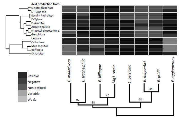
Daniela Vidaurre-Barahona, et al. Erwinia billingiae causes bacterial Canker of Mango (Mangifera ...
Introduction
Mangifera indica crops established in tropical regions are susceptible to different diseases caused by
microorganisms. Mango anthracnose is the most important causing losses in the field and as well as in post-
harvest (Arauz, 2000). Within the common bacterial diseases, Xanthomonas campestris pv. mangiferaeindicae
and Pseudomonas syringae pv. syringae has been reported as causal agents of canker of mango and bacterial
apical necrosis of mango respectively (Pruvost et al., 2012; Yanxiang et al., 2016; Young, 2008).
Bacterial Canker of Mango (M. indica) was first described in South Africa in 1915 by Doidge (Steyn et
al., 1974). In India, the disease emerged in 1948 (Kishun, 1982), while in America, it was found for the first
time in Rio de Janeiro, Brazil in 1955 (Robbs & Rezende, 1978). In Venezuela its presence was reported in
1970 (Rondón & Figueroa, 1970) and named fruit spot. In Costa Rica, it was first observed in 1987 (Sáenz
& Murillo, 1989) and aer that, it has caused premature fruit drop (10-70 %) loss of production (10-85 %)
and rot in storage (5-100 %) (Ureña et al., 2007). It was considered systemic due to its wide distribution
in the plant, causing symptoms in all parts of the trees, and a pre-harvest disease affecting fruits, branches,
inflorescences and peduncles (Viljoen & Kotzë, 1972; Guevara et al., 1980). e main symptom in the trunk
and branches is a resinous light colored (almost beige) exudate that becomes dark brown with time. Fruits
are affected by an internal rot which is difficult to detect in the initial stages and later develops into a black
spot on the fruit surface (Mora et al., 2002).
Bacteriosis caused by Erwinia genus is characterized by reddish brown spots or striations on the phloem
of infested tree branches and trunks where lesions or cankers with rubbery exudate are observed. In the fruits
the lesion is observed externally as an irregular and sunken black spot in the peduncular region, although it
can also appear in other sectors of the fruit and internally necrosis is observed around the seed. In the leaves,
the bacteria are found associated with fungi of the genus Colletotrichum sp. causing small, angular spots of 2
to 3 mm, sometimes large spots on the edges that give it a burnt appearance (Ureña et al., 2007).
e bacteria are perpetuated in epiphytic form in various plant organs, when the environmental conditions
are unfavorable to the development of the disease or the tree is in a vegetative period. Bacteria are spread by
rain, cuttings, and insects (Ureña et al., 2007). e disease is more prevalent in locations with precipitation
greater than 850 mm per year, relative humidity above 80 % and temperatures between 24 and 32 °C. ese
conditions are ideal for pathogen dissemination and infection (Guevara et al., 1985), especially in the Tommy
Atkins variety (Mora et al., 2002).
In Costa Rica, mango production has been decreasing due to different factors (López, 2011), one of which
is bacterial canker caused by an enterobacteria (McMillan & Wang, 1992) and not by Xanthomonas citri pv.
Mangiferae indicae; were Erwinia was isolated from disease trees of Tommy Atkins variety and characterized
by phenotypic traits. e causal agent has been identified by several authors previously using conventional
phenotypic methods as Erwinia mangifera, Erwinia herbicola, and Pectobacterium sp. (Guevara et al., 1985;
Rondón & Figueroa, 1970; Steyn et al., 1974), but there have not been further reports of molecular or
polyphasic identification of this bacteria.
e aim of this study was to confirm, using a molecular and phenotypic approaches the species of the
enterobacteria causing bacterial canker in mango in Costa Rica.
Material and methods
Fruits, branches, and trunks with symptoms previously described as reddish striations and rubbery exudates
on the trunk while the fruits showed a black spot in the peduncular region and fruit internally necrosis near
to the seed (Figure 1), were collected in different orchards from Alajuela province, Costa Rica, during the
second semester of 2008.
PDF generated from XML JATS4R by Redalyc
Project academic non-profit, developed under the open access initiative 307Agronomía Mesoamericana, 2021, vol. 32, no. 1, Enero-Abril, ISSN: 2215-3608
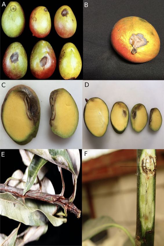
FIGURE 1
Mango plant tissue (M. indica) showing bacterial symptoms. Alajuela, Costa Rica. 2008
A. Young plant with typical symptoms caused by the Erwinia-like bacterium.
Note the black and irregular shape of the lesion. B, C. Striation in the outer cortex.
Figura 1. Tejido de plantas de mango (M. indica) con síntomas bacterianos. Alajuela, Costa Rica. 2008.
A. Planta joven con síntomas causados por una bacteria del género Erwinia. Se observa una
lesión de coloración negruzca y con forma irregular. B, C. Estriación en la corteza exterior.
Bacterial isolation was performed for the three types of tissues. Sterilization procedure was carried out to
7 cm sections from two branches, one trunk and two complete fruits using 1 % sodium hypochlorite for 1
minute followed by sterile distilled water rinse twice.
A piece of each diseased tissue was removed using a sterilized scalpel and placed in nutrient agar (NA
Oxoid, Basingstoke, UK). Plates were incubated for 48 h at 30 ºC or until growth was observed. Colonies
that showed morphological differences by Gram stain were purified using an enterobacteriaceae selective
medium (MacConkey agar, Oxoid, Basingstoke, UK) and the streak plate method to separate bacterial cells
on the agar surface to obtain isolated colonies. Purified strains were further differentiated using preliminary
phenotypic tests (Schaad et al., 2001), and biochemical features as fermentations of dextrose, lactose and
saccharose by TSI (Triple Sugar Iron Agar) and oxidase tests.
Pathogenicity of the bacterial isolates was assessed inoculating by injection two physiological ripeness
fruits and two trunks of the Tommy Atkins variety with 0.5 ml of a calibrated 1 x 10⁷ CFU ml-1 suspension
of each isolate. Sterile distilled water was used as negative control. Inoculation was done using a sterile
needle. When inoculating trunks of young trees, a small injury was performed to facilitate the entrance of the
pathogen. Inoculated material were incubated under greenhouse conditions at 25–30 °C. Aer infection,
the young trees were covered for 24 hours with a plastic bag, large enough to simulate a moist chamber.
Aer this period, the bags were removed, and the trees were kept in the greenhouse for infection to occur
until first symptoms were evident. Only positive isolates that caused symptoms, were analyzed by molecular
identification and complete biochemical characterization and were re-isolated from inoculated into fruit and
identified, thereby, fulfilling Koch’s postulates (Figure 2).
PDF generated from XML JATS4R by Redalyc
Project academic non-profit, developed under the open access initiative 308Daniela Vidaurre-Barahona, et al. Erwinia billingiae causes bacterial Canker of Mango (Mangifera ...
FIGURE 2
Symptoms of infection observed in situ and by inoculation of the CIBCM-
Mg-115 strain, in mango fruits and tissue (M. indica). San José, Costa Rica. 2008.
A, C. Natural infection where a dark lesion with slight sinking is observed at the foot of the fruit. B, D. Symptoms observed by
the artificial inoculation of 0.5 ml of a 1 x 10⁷ cfu ml-1 bacterial suspension. E. Advancement of the lesion when the infection
occurs in the inflorescence or in an outbreak. F. Milky sap flowing through fissures, also described in branches and panicules.
Figura 2. Síntomas de infección observados in situ y mediante inoculación de la cepa
CIBCM-Mg-115, en frutos y tejido de mango (M. indica). San José, Costa Rica. 2008.
A. C. Infección natural donde se observa una lesión oscura y con hundimiento leve en el pie del fruto. B, D. Síntomas observados
por la inoculación artificial de 0,5 ml de una suspensión bacteriana de 1 x 10⁷ cfu ml-1. E. Avance de la lesión cuando la infección
ocurre en la inflorescencia o el brote. F: Savia lechosa fluyendo a través de las fisuras según lo observado en ramas y panículas.
Genomic DNA was extracted using the DNeasy Blood & Tissue Kit (Qiagen®) following the
manufacturer’s instructions. PCR was used to amplify 16S rRNA and recA, gyrB, dnaN, gltX, and rpoB
housekeeping genes (Tailliez et al., 2010). Sequences of all genes were obtained from Macrogen Inc. Contig
assembly and ambiguity resolution were performed with BioEdit (Hall, 1999). Sequences were aligned with
reference sequences from the NCBI database in MEGA (Tamura et al., 2013). Multiple sequence alignments
were performed with ClustalX (Larkin et al., 2007). Sequences corresponding to PCR amplification primers
were removed prior to multiple sequence alignment and phylogenetic analysis. All of the sequenced genes
PDF generated from XML JATS4R by Redalyc
Project academic non-profit, developed under the open access initiative 309Agronomía Mesoamericana, 2021, vol. 32, no. 1, Enero-Abril, ISSN: 2215-3608
were analyzed separately and then in a concatenated tree. Evolutionary distances were calculated by Bayesian
inference and tree topology was evaluated by bootstrap analysis with 10 000 000 resampling.
Phenotypic characterization was based on biochemical tests using the semi-automated identification
systems API 20E, API 50CHE (bioMérieux) and GN microplates (BIOLOG), conducted as recommended
by the manufacturers. Tests known to differentiate between Erwinia species and Pantoea were used to
compare the mango isolate (CIBCM-Mg-115) with type strains from species of the genus Erwinia by
clustering analysis using the statistical program PAST (Hammer et al., 2001).
Results
Five bacterial isolates were obtained and tested. Only CIBCM-Mg-115 strain isolated from fruit caused
symptoms when inoculated on fruits and trunks of young trees. e symptoms were brownish to black
lesion appeared near to the shoulder of the fruit that continued to the internal part of the fruits as well
as milky sap in trunk lesions. Lesions were observed aer 30 and seven days post infections for trunks and
fruits, respectively. Koch’s postulates were accomplished by re-isolating and identifying from fruits the same
pathogen obtained from the same tissue showing symptoms originally.
e Bayesian phylogenetic tree based on 16S rRNA gene sequences (Figure 3) from members of the genera
Erwinia and Pantoea indicated that strain CIBCM-Mg-115 was most closely related to Erwinia billingiae
(DSM 23398T).
FIGURE 3
Bayesian tree based on 16S rRNA gene sequences showing the position of strain CIBCM-Mg-115
in the genus Erwinia. Bootstrap values are based on 10000000 replications. Cronobacter sakazakii
was used as an outgroup. Bar, 0.21 substitutions per nucleotide. San José, Costa Rica. 2018.
Figura 3. Árbol filogenético mediante inferencia bayesiana del gen 16S ARN donde se muestra la posición filogenética
de la cepa CIBCM-Mg-115 dentro del género Erwinia. Los valores bootstrap están basados en 10000000 replicaciones.
Cronobacter sakazakii se utilizó como grupo externo. Bar, 0.02 sustituciones por nucleótido. San José, Costa Rica. 2018.
PDF generated from XML JATS4R by Redalyc
Project academic non-profit, developed under the open access initiative 310Daniela Vidaurre-Barahona, et al. Erwinia billingiae causes bacterial Canker of Mango (Mangifera ...
e phylogenetic tree based on concatenated housekeeping gene sequences (Figure 4) demonstrated that
the Costa Rican strain showed high homology to strains of E. billingiae and formed a cluster supported
by a high bootstrap value. is is consistent with the results of the phylogenetic analysis based on 16S
RNA and suggest that the isolate is related to Erwinia species pathogenic to papaya (Carica papaya): E.
papayae; cantaloupe (Cucumis melo): E. tracheiphyla; guava (Psidium guajava): E. psidii; olive trees (Olea
europaea): E. oleae, and Mallotus japonicus (ornamental tree) E. mallotivora. e genus Pantoea was distantly
related and formed a cluster adjacent to the one composed of Erwinia species. e sequences 16S rRNA,
atpD, gyrB, infB, and rpoB from CIBCM-Mg-115 have been deposited in the Genbank database (accessions
MH643806, MH643807, MH643809, MH643808, and MH643805, respectively).
FIGURE 4
Bayesian analysis of concatenated sequences of the housekeeping genes atpD, gyrB, infB,
and rpoB showing the phylogenetic relationship of isolated CIBCM-Mg115 within the
genus Erwinia. Bootstrap values are based on 10000000 replications. Cronobacter sakazakki
was used as an outgroup. Bar, 0.06 substitutions per nucleotide. San José, Costa Rica. 2018.
Figura 4. Análisis filogenético mediante inferencia bayesiana de las secuencias concatenadas de los genes
de mantenimiento atpD, gyrB, infB, y rpoB donde se muestra la posición filogenética de la cepa CIBCM-
Mg-115 dentro del género Erwinia. Los valores bootstrap están basados en 10000000 replicaciones. Cronobacter
sakazakii se utilizó como grupo externo. Bar, 0,06 sustituciones por nucleótido. San José, Costa Rica. 2018.
e API systems and BIOLOG GN microplates failed to identify the bacterial isolate as E. herbicola
or Pectobacterium carotovora. Results obtained with GN microplates did not show similarity with any of
the Erwinia species in the Biolog database. e CIBCM-Mg-115 isolate from mango is a Gram-negative,
oxidase-negative, catalase-positive motile rod that shows the general characteristics of the Enterobacteriaceae
and of the species Erwinia billingiae as described by Mergaert et al. (1999), except that nitrate is not reduced
to nitrite. As with E. billingiae, colonies were white in Nutrient Agar.
Comparative analysis of seven Erwinia species, P. agglomerans, and CIBCM-Mg-115 (Figure 5) using
the most relevant biochemical characteristics described for E. billingiae (Mergaert et al., 1999) showed the
clustering of CIBCM-Mg1-115 and E. billingiae with a bootstrap value of 97 %, with differences in acid
PDF generated from XML JATS4R by Redalyc
Project academic non-profit, developed under the open access initiative 311Agronomía Mesoamericana, 2021, vol. 32, no. 1, Enero-Abril, ISSN: 2215-3608
production from D-sorbitol, Myo-inositol, Arbutin, and Salicin. e analysis placed P. agglomerans as an
out-group.
FIGURE 5
Cluster analysis based on biochemical characteristics to compare similarity between
species of Erwinia and Pantoea agglomerans genus San José Costa Rica 2018
Figura 5. Análisis de agrupamiento basados en características bioquímicas para comparar
similitud entre especies del género Erwinia y Pantoea agglomerans. San José, Costa Rica. 2018.
Discussion
e symptom observed in the samples collected in this study corresponds to the same disease mentioned by
McMillan & Wang (1992). In this case, the pathogen attacks trunks, stems, leaves, flowers, and fruits, except
the root system that is not susceptible. e infection caused by X. campestris is different, especially on the
symptoms observed in fruits, where the bacterium causes black spots that can become star shaped latter on
(Pruvost et al., 2011), while the lesions caused by Erwinia-like pathogen are irregular, lightly sunken and
in the case of branches and stems, a very distinctive red streak on the wood (cambial region) are observed.
Severe infection will cause canker in the trunk, with a milky colored sap flowing out. Lesions caused by X.
campestris will show a gummy exudate.
Several species from the genus Erwinia have been characterized as pathogens of pome fruit trees. Erwinia
amylovora is the causal agent of the fire blight disease of rosaceous plants (Zwet & Keil, 1979), while E.
pyrifoliae, Japanese Erwinia spp., and E. piriflorinigrans cause bacterial diseases of pear (BSBP and BBSDP).
However, species as Erwinia billingiae and Erwinia tasmaniensis has been described in other regions as non-
pathogenic bacteria (Palacio-Bielsa et al., 2012).
Strains of Erwinia billingiae were initially designated as E. herbicola (Billing & Baker, 1963). Bacteria were
obtained from different lesions on pear, apple, cherry, hawthorn (Crataegus spp.), and elm (Ulmus spp.) trees
and were isolated from symptoms as stem cankers or diseased blossoms and immature fruits.
It was previously classified as white E. herbicola, and was considered to be a secondary invader rather than
a primary pathogen. Later, based on DNA–DNA hybridization assays and 16S rDNA sequence analysis,
the isolate was reclassified as a novel non-pathogenic species, Erwinia billingiae (Mergaert et al., 1999). E.
billingiae tends toinvade necrotic tissue of plants. According to Billing and Baker (1963), English isolates
were considered secondary invaders rather than primary pathogens, possibly helping to extend lesions but
PDF generated from XML JATS4R by Redalyc
Project academic non-profit, developed under the open access initiative 312Daniela Vidaurre-Barahona, et al. Erwinia billingiae causes bacterial Canker of Mango (Mangifera ...
not to initiate them and it has been demonstrated by other authors (Kube et al., 2010). In this study, it
was demonstrate that E. billingiae is a primary pathogen on mango. Artificial inoculation resulted in the
development of symptoms identical to what can be seen in the field. According to mango growers, this disease
can cause serious losses as it affects the fruit.
Strains of this species have been isolated from apple trees in Poland (Geider et al., 2007) and there are
medical reports of dermohypodermitis and bacteremia in a 40-year-old man in Switzerland (Prod’homme
et al., 2017) and knee septic arthritis caused by Erwinia billingiae in France, where an immunocompetent
patient was treated aer a trauma involving a palm tree (Bonnet et al., 2019). e bacterium was identified
through 16S rRNA gene sequencing. Bacteria causing initially human pathologies have been described later
as plant pathogens. Micrococcus luteus SUBG006 was recognized initially as human pathogen, causing
leafspot on M. indica in Rajkot, Gujarat, India (Rakhashiya et al., 2015). e strain was isolated from
leaves with black leaf-spot symptoms and characterized by morphological features and sequencing analysis
(Rakhashiya et al., 2015). ere were no further reports of new isolates of this species as a pathogen or
antagonist prior to this study.
Conclusions
e isolate CIBCM-Mg-115 can be considered as one of the causal agents of bacterial Canker of Mango (M.
indica) trees in Costa Rica. is was demonstrated by pathogenicity test, thereby fulfilling Koch’s postulates.
It was classified as Erwinia billingiae by phylogenetic analysis of the 16S RNA gene and concatenated
housekeeping gene sequences, and by cluster analysis of relevant biochemical characteristics described for
this species.
References
Arauz, L. F. (2000). Mango anthracnose: Economic impact and current options for integrated management. Plant
Disease, 84(6), 600–611. https://doi.org/10.1094/PDIS.2000.84.6.600
Billing, E., & Baker, L. A. (1963). Characteristics of Erwinia-like oganisms found in plant material. Journal of Applied
Bacteriology, 26(1), 58–65. https://doi.org/10.1111/j.1365-2672.1963.tb01155.x
Bonnet, I., Bozzi, B., Fourniols, E., Mitrovic, S., Soulier-Escrihuela, O., Brossier, F., Sougakoff, W., Robert, J.,
Jauréguiberry, S., & Aubry, A. (2019). Erwinia billingiae as unusual cause of septic arthritis, France, 2017.
Emerging Infectious Diseases, 25(8), 1587–1589. https://doi.org/10.3201/eid2508.181073
Geider, K., Jock, S., & Sulikowska. M. (2007). Screening for Erwinia billingiae and E. tasmaniensis in field isolates,
differentiation by sequence analysis and effects as antagonists. Acta Horticulturae, 793, 119–121. https://doi.o
rg/10.17660/ActaHortic.2008.793.13
Guevara, M., Rondón, A., & Solórzano R. (1980). Bacteriosis del mango (Mangifera indica) en Venezuela:
Sintomatología e identificación. Agronomía tropical, 30, 65–76.
Guevara, M., Rondón, A., Arnal, E., & Solórzano, R. (1985). Bacteriosis del mango (Mangifera indica) en Venezuela
II. Distribución, perpetuación, diseminación y evaluación de la resistencia de variedades. Agronomía tropical,
35, 63–75.
Hall, T. A. (1999). BioEdit: a user-friendly biological sequence alignment editor and analysis program for Windows
95/98/NT. Nucleic Acids Symposium Series, 41, 95–98.
Hammer, O., Harper, D. A., & Ryan, P. D. (2001). PAST-Palaeontological statistics, ver. 1,89. Paleontología
Electronica, 4(1), 1–9.
Kishun, R. (1982). Loss in mango fruit due to bacterial canker Xanthomonas mangiferaeindicae. In J. C. Lozano
(Ed.), Proceedings of the fih International Conference on Plant Pathogenic Bacteria (pp. 191–184). Centro
Internacional de Agricultura Tropical.
PDF generated from XML JATS4R by Redalyc
Project academic non-profit, developed under the open access initiative 313Agronomía Mesoamericana, 2021, vol. 32, no. 1, Enero-Abril, ISSN: 2215-3608
Kube, M., Migdoll, A. M., Gehring, I., Heitmann, K., Mayer, Y., Kuhl, H., Knaust, F., Geider, K., & Reinhardt,
R. (2010). Genome comparison of the epiphytic bacteria Erwinia billingiae and E. tasmaniensis with the pear
pathogen E. pyrifoliae. BMC Genomics, 11(1), 393. https://doi.org/10.1186/1471-2164-11-393
Larkin, M. A., Blackshields, G., Brown, N. P., Chenna, R., McGettigan, P. A., McWilliam, H., & ompson, J. D.
(2007). Clustal W and Clustal X version 2.0. Bioinformatics, 23, 2947–2948. https://doi.org/10.1093/bioinf
ormatics/btm404
López, K. (2011). Oportunidades comerciales para las exportaciones de mango. Promotora de Comercio Exterior. ht
tp://servicios.procomer.go.cr/aplicacion/civ/documentos/Mango.pdf
McMillan, Jr. R. T., & Wang, A. (1992). A New disease of mango in Costa Rica causes by an Erwinia-like bacteria.
Proceedings Florida State Horticultural Society, 105, 288–289
Mergaert, J., Hauben, L., Cnockaert, M. C., & Swings, J. (1999). Reclassification of non-pigmented Erwinia herbicola
strains from trees as Erwinia billingiae sp. Nov. International Journal of Systematic and Evolutionary Microbiology,
49, 377–383. https://doi.org/10.1099/00207713-49-2-377
Mora, J., Gamboa-Porras, J., & Elizondo-Porras, R. (2002). Guía para el cultivo del mango (Mangifera indica) en Costa
Rica. Ministerio de Agricultura y Ganadería.
Palacio-Bielsa, A., Roselló, M., Llop, P., & López, M. M. (2012). Erwinia spp. from pome fruit trees: similarities and
differences among pathogenic and non-pathogenic species. Trees, 26(1), 13–29. https://doi.org/10.1007/s004
68-011-0644-9
Prod’homme, M., Micol, L. A., Weitsch, S., Gassend, J.L., Martinet, O., & Bellini, C. (2017). Cutaneous infection and
bactaeremia caused by Erwinia billingiae: a case report. New Microbes and New Infections, 19, 134–136. https:
//doi.org/10.1016/j.nmni.2017.07.006
Pruvost, O., Boyer, C., Vital, K., Verniere, C., Gagnevin, L., & Somda, I. (2011). First report in Burkina Faso of
Xanthomonas citri pv. mangiferaeindicae causing bacterial canker on Mangifera indica. Plant Disease, 95(10),
1312–1312.
Pruvost, O., Boyer, C., Vital, K., Verniere, C., Gagnevin, L., & Traoré, Y. N. (2012). First report in Mali of
Xanthomonas citri pv. mangiferaeindicae causing mango bacterial canker on Mangifera indica. Plant Disease,
96(4), 581–581. https://doi.org/10.1094/PDIS-01-12-0001-PDN
Rakhashiya, P. M., Patel, P. P., & aker, V. S. (2015). First Report of Micrococcus luteus Causing Leafspot on
Mangifera indica in Rajkot, India. Plant Disease, 99(11), 1640. https://doi.org/10.1094/PDIS-12-14-1359-P
DN
Robbs, C. F., & Rezende, H. B. (1978). Necrose cotiledonar da soja no estado de Minas Gerais. Em Centro Nacional de
Pesquisa de Soja (Ed.), Resumos I Seminário Nacional de Pesquisa de Soja (p. 60). Empresa Brasileira de Pesquisa
Agropecuária.
Rondón, G., & Figueroa, R. (1970). Black spot of mango fruits caused by Erwinia mangifera. Agronomia Tropical,
22(4), 271–274.
Sáenz, A., & Murillo, A. (1989). Programa nacional sectorial de Mango. Ministerio de Agricultura y Ganadería. http:
//www.mag.go.cr/bibliotecavirtual/E14-0223.pdf
Schaad, N. W., Jones, J. B., & Chun, W. (2001). Laboratory guide for the identification of plant pathogenic bacteria.
APS Press.
Steyn, P.L., Viljoen, N. M., & Kotze J. M. (1974). e causal organism of bacterial black spot of mangoes.
Phytopathology, 64, 1400–1404.
Tailliez, P., Laroui, C., Ginibre, N., Paule, A., Pagès, S., & Boemare, N. (2010). Phylogeny of Photorhabdus and
Xenorhabdus based on universally conserved protein-coding sequences and implications for the taxonomy of
these two genera. Proposal of new taxa: X. vietnamensis sp. nov., P. luminescens subsp. caribbeanensis subsp.
nov., P. luminescens subsp. hainanensis subsp. nov., P. temperata subsp. khanii subsp. nov., P. temperata subsp.
tasmaniensis subsp. nov., and the reclassification of P. luminescens subsp. thracensis as P. temperata subsp.
thracensis comb. Nov. International Journal of Systematic and Evolutionary Microbiology, 60, 1921–1937. http
s://doi.org/10.1099/ijs.0.014308-0
PDF generated from XML JATS4R by Redalyc
Project academic non-profit, developed under the open access initiative 314Daniela Vidaurre-Barahona, et al. Erwinia billingiae causes bacterial Canker of Mango (Mangifera ...
Tamura, K., Stecher, G., Peterson, D., Filipski, A., & Kumar, S. (2013). MEGA6: molecular evolutionary genetics
analysis version 6.0. Molecular Biology and Evolution, 30(12), 2725–2729. https://doi.org/10.1093/molbev/
mst197
Ureña, A. L., González, J., Meneses, R., & Alvarado E. (2007). Agrocadena de mango. Ministerio de Agricultura y
Ganadería. http://www.mag.go.cr/bibliotecavirtual/E70-4282.pdf
Viljoen, N. M., & Kotzë, J. M. (1972). Bacterial black spot of Mango. e Citrus Grower and Sub-tropical Fruit Journal,
462, 5–8.
Yanxiang, Q., He, Z., Jinji, P., Xin, Z., Quanfang, Y., Ying, L., & Yixian, X. (2016). Characterization of Chinese Isolates
of Mango Bacterial Canker Xanthomonas campestris pv. mangiferaeindicae. Plant Diseases and Pests, 7(3), 33.
Young, A. (2008). Notes on Pseudomonas syringae pv. syringae bacterial necrosis of mango (Mangifera indica) in
Australia. Australasian Plant Disease Notes, 3(1), 138–140. https://doi.org/10.1007/BF03211269.
Zwet, T. V. D., & Keil, H. L. (1979). Fire blight: a bacterial disease of Rosaceous plants. United States Department of
Agriculture.
Notes
1 is work was part of the project number VI-801-A5-520 registered in Vicerrectoría de Investigación of the Universidad
de Costa Rica.
Alternative link
http://www.revistas.ucr.ac.cr/index.php/agromeso (html)
PDF generated from XML JATS4R by Redalyc
Project academic non-profit, developed under the open access initiative 315You can also read