National Institute of Standards and Technology transportable tunable ultraviolet laser irradiance facility for water pathogen inactivation
←
→
Page content transcription
If your browser does not render page correctly, please read the page content below
National Institute of Standards and
Technology transportable tunable
ultraviolet laser irradiance facility for water
pathogen inactivation
Cite as: Rev. Sci. Instrum. 91, 074105 (2020); https://doi.org/10.1063/5.0016500
Submitted: 05 June 2020 . Accepted: 01 July 2020 . Published Online: 21 July 2020

Thomas C. Larason

Rev. Sci. Instrum. 91, 074105 (2020); https://doi.org/10.1063/5.0016500             91, 074105
Review of                                                                                       ARTICLE           scitation.org/journal/rsi
 Scientific Instruments

     National Institute of Standards and Technology
     transportable tunable ultraviolet laser irradiance
     facility for water pathogen inactivation
     Cite as: Rev. Sci. Instrum. 91, 074105 (2020); doi: 10.1063/5.0016500
     Submitted: 5 June 2020 • Accepted: 1 July 2020 •
     Published Online: 21 July 2020

     Thomas C. Larasona)

     AFFILIATIONS
    National Institute of Standards and Technology, Gaithersburg, Maryland 20899, USA

     a)
          Author to whom correspondence should be addressed: thomas.larason@nist.gov

     ABSTRACT
     A method of ultraviolet germicidal irradiation (UVGI) for water pathogen inactivation effectiveness using tunable, narrowband laser light
     is described. A transportable tunable UV (TTUV) laser system for providing a known irradiance (μW/cm2 ) or dose (mJ/cm2 ) suitable for
     irradiating water samples in Petri dishes over the wavelength range of 210 nm–300 nm was developed by the National Institute of Standards
     and Technology. The TTUV facility, consisting of a 1 kHz pulsed UV laser and light-tight enclosure containing the optics necessary to
     uniformly irradiate a water sample, was used in a microbiology laboratory to dose drinking water pathogens and surrogates as part of a Water
     Research Foundation study in the summer and fall of 2012. The approach demonstrated improved accuracy and simplified spectral analysis
     over conventional pathogen inactivation sources consisting of broadband UV sources and bandpass filters. In this work, the TTUV facility
     design and key components are described, including modifications in the field to provide the required irradiance levels. The irradiance and
     dose levels produced by the tunable UV laser during the project are also presented. The transportability of the TTUV system enabled it to be
     brought to a microbiology facility allowing the water samples (microbial suspensions) to be irradiated in a location with experienced staff and
     facilities for preparing, handling, analyzing, storing, and shipping the many samples studied. These results, published elsewhere, established
     that the tunable UV laser system provides unique UVGI capabilities for use with water pathogens and has applications for other pathogen
     experiments, for example, air-purification studies.
     https://doi.org/10.1063/5.0016500., s

     I. INTRODUCTION                                                          micro-organisms often did not exist below 240 nm and particularly
                                                                              not below 220 nm.
           Ultraviolet (UV) radiation effectively inactivates common                In 2011, the National Institute of Standards and Technology
     pathogens found in ground and surface waters, such as Cryp-              (NIST) was asked if they could build on their efforts in UV sen-
     tosporidium, Giardia, and most bacterial pathogens (e.g., E. coli).1–6   sor calibration9,10 and tunable laser systems11–14 to develop a system
     Water treatment facilities are now using UV radiation for disinfec-      to irradiate water samples as part of a Water Research Foundation
     tion of drinking water, supplementing standard chemical treatment.       Project.15 The transportable tunable UV (TTUV) facility was devel-
     These systems typically use either low-pressure (LP) or medium-          oped to provide a known, spatially uniform irradiance (μW/cm2 )
     pressure (MP) mercury vapor lamps. In the 200 nm–300 nm region,          or dose (mJ/cm2 ) to water samples (microbial suspensions) in Petri
     LP lamps emit primarily at 253.7 nm, and MP lamps emit across this       dishes.16 The UV source covered the wavelength range of 210 nm–
     spectral region. Research has been reported showing polychromatic        300 nm and irradiated a 5 cm diameter area with a uniformity of 10%
     light from MP lamps is more effective than that from LP lamps for        or better. By tuning the laser wavelength and controlling the dose
     inactivating certain pathogens, and the wavelengths below 240 nm         exposure, an “action spectrum” or germicidal effectiveness curve is
     are responsible for this enhancement.3,7,8 At the time of this work in   developed to quantify the efficiency of micro-organism inactivation.
     2012, action spectral data for waterborne pathogens and surrogate        The action spectrum is defined as “a plot of a relative biological

Rev. Sci. Instrum. 91, 074105 (2020); doi: 10.1063/5.0016500                                                                               91, 074105-1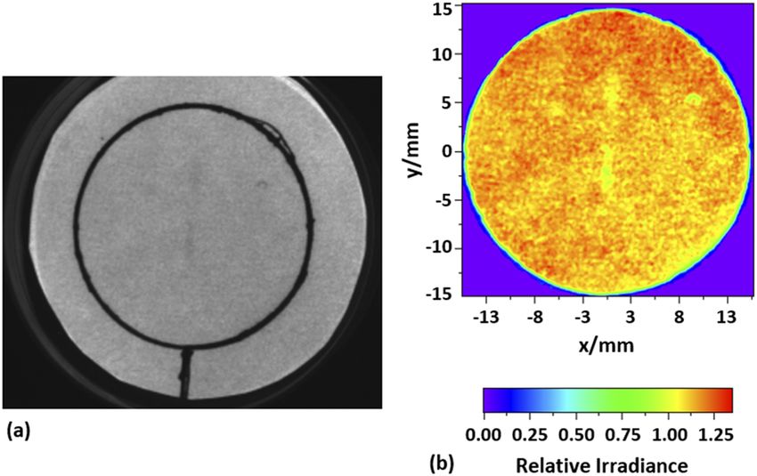
Review of                                                                                          ARTICLE               scitation.org/journal/rsi
 Scientific Instruments

     or chemical photoresponse (= Δy) per number of incident (prior to
     absorption) photons, vs wavelength.”17 Finally, the system was to be
     installed at a microbiology facility in St. Albans, VT. This permit-
     ted the staff and facility experienced with irradiating and analyzing
     water samples with broadband UV sources and bandpass filters to
     work with the TTUV facility.

     II. TTUV LASER IRRADIANCE FACILITY
          The TTUV laser irradiance facility is composed of four major
     parts: the laser, the water sample irradiance system, the sample irra-    FIG. 1. Simplified schematic diagram showing the optical layout of the Ekspla
     diance test position, and a NIST calibrated UV irradiance transfer        NT242 tunable laser (1 kHz pulse rate,
Review of ARTICLE scitation.org/journal/rsi
Scientific Instruments
C. Test (Petri dish) position setup
Each water sample was a microbial suspension in a continu-
ously stirred Petri dish of 36 mm diameter.16,22 Key to these mea-
surements was the ability to repeatably place the Petri dish contain-
ing the water sample in the same position in the optical beam and
to measure the irradiance in the same position. This was done with
an aluminum alignment jig placed on top of the stirring plate. The
height of the stirring plate was also adjusted to measure the irra-
diance at the same plane as the surface of the water in the Petri
dish. Figures 5 (a) and (b), respectively, show the water sample
being placed into the jig and the UV detector in place for irradiance
measurement.
D. Detector
The NIST calibrated UV detector was placed at the water sam-
ple location to measure the irradiance at each wavelength of interest.
The UV detector consisted of an International Radiation Detectors
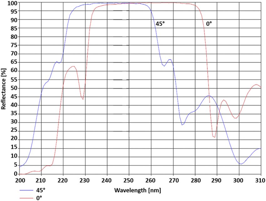
FIG. 3. The reflectance of ARO model MR1520 dielectric mirrors at both 0○ and (now Opto Diode Corp.23 ) SXUV100 silicon photodiode, known to
45○ incidence angles with random polarization. be stable with UV exposure,24 and a precision 8 mm diameter elec-
troformed aperture in a cylindrical aluminum housing. The photo-
diode output was measured with a Keithley 6517 series electrometer.
The irradiance responsivity [A/(μW/cm2 )] of the UV detector was
from the dielectric mirrors. The reflectance of these ARO model calibrated from 200 nm to 400 nm at the NIST in the UV Spectral
MR1520 dielectric mirrors is shown in Fig. 3. Neutral density filters Responsivity Facility.25 The expanded uncertainty (k = 2) for the UV
can be added to the optical path between the shutter and the first detector irradiance was 5%.
mirror, M1, to reduce the irradiance level at the water sample.
Due to low reflectance, the dielectric mirrors were replaced E. Performance
by aluminum front surface reflecting mirrors, a fused silica prism,
and slit to filter out the visible light for 210 nm–230 nm. This The uniformity of the UV irradiance at the water sample (mea-
configuration is shown in Fig. 4. sured separately by the project collaborators in St. Albans, VT) was
The beam then travels through a beam splitter and an engi- calculated as the ratio of the average of the incident irradiance over
neered etched fused silica diffuser.21 The beam splitter sends a small the area of the Petri dish to the irradiance at the center of the dish.
portion of the UV light to a silicon photodiode, which monitored This has been referred to as the Petri Factor16 and was measured
the irradiance level during sample exposure. The diffuser was a at each wavelength before exposing any water samples. To deter-
critical component of the TTUV laser system. Unlike typical opti- mine the Petri factor, a broadband UV-C radiometer with a 1 mm
cal diffusers, this diffuser is specifically engineered to modify the aperture was scanned over the area of the Petri dish along orthogo-
laser beam from a collimated oval shape (1.5 mm by 10 mm) to nal axes in 5 mm steps. The Petri factor was reported to range from
a uniform diverging beam (10○ half-angle) to irradiate the water 0.94 to 1.05,22 which met the design goal of 10%. To reduce the time
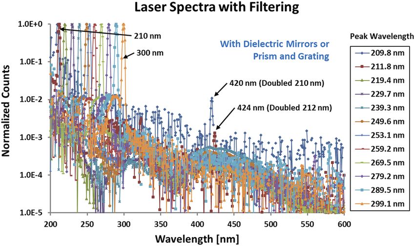
samples. required to determine the Petri factor, a test using digital images and
the fluorescence from typical card stock paper was explored. The
test validated the approach, but time constraints kept it from being
FIG. 4. The NIST tunable UV irradiance laser system optical configuration for 210
nm–230 nm. The irradiance system with the dielectric mirrors replaced by alu- FIG. 5. Photographs of the stirring plate with (a) a water sample being placed
minum mirrors and adding a prism and slit. The UV detector is substituted for the into the Petri dish positioning jig and (b) the UV irradiance detector set for
water sample to measure the irradiance at each wavelength of interest. measurement at the same plane as the surface of the water in the Petri dish.
Rev. Sci. Instrum. 91, 074105 (2020); doi: 10.1063/5.0016500 91, 074105-3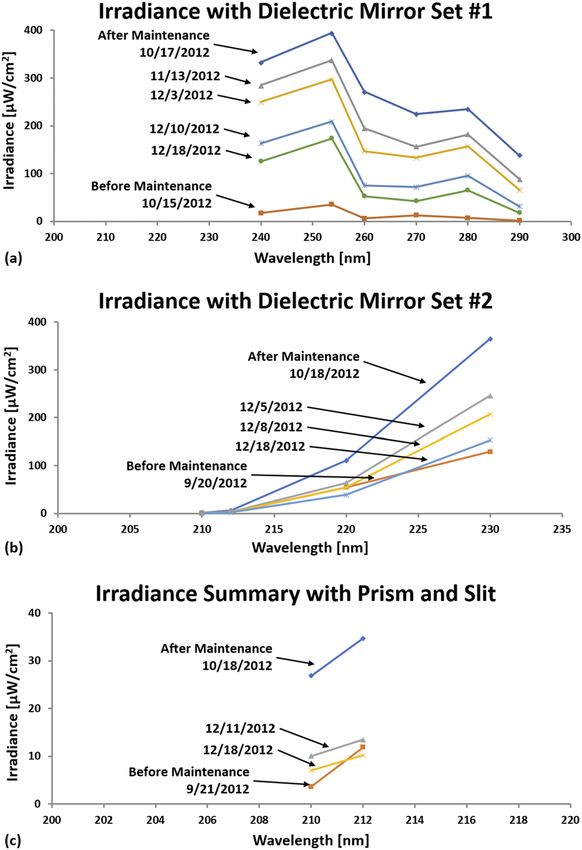
Review of ARTICLE scitation.org/journal/rsi
Scientific Instruments
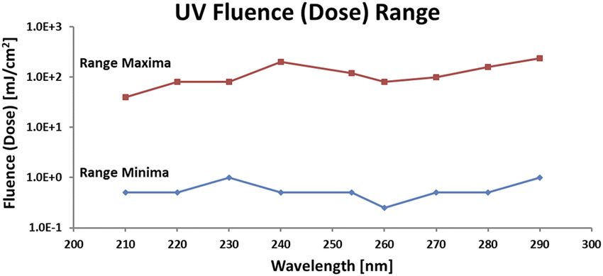
FIG. 8. Graph of the range of fluence or dose (mJ/cm2 ) by the wavelength used
during this project.
FIG. 6. Example of the irradiance uniformity at 253.7 nm. A photograph (a) from the
camera mounted above the water sample imaging the fluorescence from typical studied further during this project. An example of a digital image
card stock paper. The dark circle marks the area of the water sample Petri dish. A from a camera mounted above the water sample and irradiance uni-
plot (b) of the relative irradiance uniformity normalized to the beam center. formity normalized to the beam center at 253.7 nm is shown in
Fig. 6.
The irradiance levels consistently decreased over time due to
unavoidable UV damage to some of the optical components in the
Ekspla laser. Midway through the project, some of the damaged
Ekspla laser optical components were replaced, which increased the
irradiance levels at the UV wavelengths of interest. However, the
UV damage continued to decrease the irradiance levels that could
be provided. This did affect the scheduling of which microbes were
exposed and the sequential order of wavelengths. Figure 7 shows the
decrease in irradiance (μW/cm2 ) over time and the improvements
when some of the damaged Ekspla laser optical components were
replaced. The range of fluence or dose (mJ/cm2 ) by wavelength used
during this project is shown in Fig. 8.
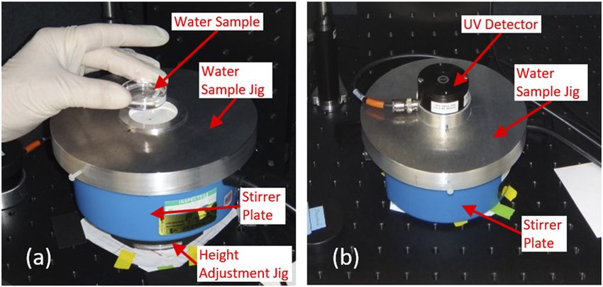
An Instrument Systems26 CAS 140CT array spectrometer was
used to measure the relative spectral irradiance at the water sam-
ple position over the spectral range from 200 nm to 600 nm both
with and without spectral filtering (dielectric mirrors or a prism and
slit). The visible light co-aligned with the laser beam without spec-
tral filtering was lower by one order to one and a half orders of
FIG. 7. Graphs (a)–(c) showing the decrease in irradiance (μW/cm2 ) over time and FIG. 9. Plots of the TTUV facility relative irradiance from 200 nm to 600 nm with
the improvement when some of the damaged Ekspla laser optical components spectral filtering (dielectric mirrors or a prism and slit) at each of the UV wave-
were replaced. Graph (a) is the irradiance with dielectric mirror set 1, (b) mirror set lengths of interest confirming the visible light in the beam was reduced to an
2, and (c) the prism and slit. acceptable level. Each spectral scan is normalized to the peak wavelength.
Rev. Sci. Instrum. 91, 074105 (2020); doi: 10.1063/5.0016500 91, 074105-4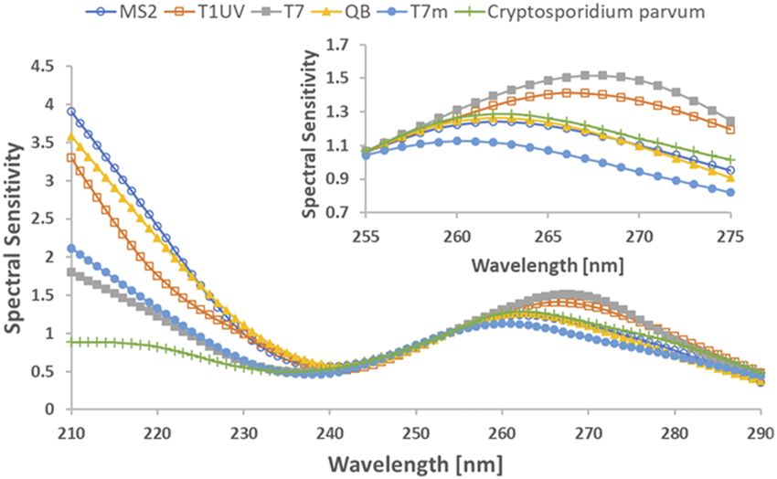
Review of                                                                                                 ARTICLE            scitation.org/journal/rsi
 Scientific Instruments

     magnitude from the selected UV wavelength. Figure 9 shows the                       with the manufacturer to verify the mirror performance. To reduce
     relative spectral irradiance with the spectral filtering at each of the             the uncertainty in the irradiance during a test, a calibrated detector
     UV wavelengths of interest confirming the visible light in the beam                 will be installed to monitor the irradiance during the water sample
     was reduced to an acceptable level. For example, the 420 nm peak in                 exposure. Refinements to the detector signal measurement method
     Fig. 9 is 1% of the UV flux at 210 nm. With the excitation source at                will be explored. Finally, as mentioned above, a camera to analyze
     212 nm and the prism filtering shown in Fig. 4, the flux at 424 nm is               the irradiance uniformity and calculate the Petri factor in real time
     reduced by an order of magnitude, to the 0.1% level.                                will be installed. The camera will reduce the time needed to measure
                                                                                         the Petri factor and provide irradiance uniformity information for
                                                                                         the entire beam and not just along two orthogonal axes through the
     III. DISCUSSION
                                                                                         beam center.
           The types of waterborne pathogens and surrogate micro-                              A static (non-transportable) UV dose irradiance system is
     organisms studied were: Adenovirus—RG 2, also Type 40 and 41,                       under development for NIST’s Remote Sensing Laboratory. The
     Cryptosporidium parvum (Iowa strain)—RG 2, Giardia—RG 2, and                        changes detailed above will be affected for the new static system, and
     Coliphages MS2, T1UV, T7m, Q beta.                                                  the performance of the enhanced system will be evaluated. Changes
           Results and impact of the TTUV laser irradiance facility in the               deemed worthwhile will be introduced into the TTUV system.
     water treatment microbiology are shown in several papers.8,22,27,28
     A fundamental question is the equivalence of UV dose response                       IV. CONCLUSIONS
     using LP UV light and the NIST 1 kHz pulsed laser. The inac-
     tivation of adenovirus using a LP UV light source and the NIST                            Current UV systems rely on lamp sources that are broadband
     laser at 253.7 nm did not show a statistically significant differ-                  and spectrally non-uniform, characteristics that complicate the mea-
     ence in response.27 Figure 10 shows the relative spectral sensitiv-                 surement analysis process and ultimately degrade the accuracy of the
     ity, or action spectra, of several micro-organisms, MS2, T1UV, Q                    pathogen disinfectant action spectra. The NIST TTUV facility has
     Beta, T7m, and T7 Coliphages and C. parvum to UV light, show-                       demonstrated its ability to be deployed to a field site and to provide
     ing differences in the action spectra below 240 nm. Extending the                   irradiance at narrowband, and well defined, UV wavelengths at irra-
     action spectra to 210 nm for selected drinking water pathogens and                  diance levels of 10 μW/cm2 to >100 μW/cm2 and fluences (or doses)
     surrogates used to validate water systems22 is necessary for calcu-                 from 100 mJ/cm2 from 210 nm to 300 nm.
     lating the action spectra correction factors for MP UV water sys-                         Development of action spectra of waterborne micro-organisms
     tem validation. Better understanding of the action spectra improves                 to specific UV wavelengths as part of WaterRF Project 4376 demon-
     the comparison of pathogens and various surrogates used in test-                    strates the unique capabilities of the NIST tunable UV irradiance
     ing UV water treatment systems, the selection of wavelengths                        laser system and its potential application for other biological exper-
     for dose monitoring, and the wavelengths for future technology                      iments, for example, development of action spectra for airborne
     exploration.8                                                                       pathogens. Since the spectral range of the TTUV facility extends
           There are several improvements under way for the TTUV                         from 210 nm to 2500 nm, it can be used as a spectrally tunable
     facility. The simplest improvement to the tunable UV irradiance                     source of radiant flux for a wide variety of applications ranging from
     laser system is to establish computer control of the laser wave-                    UV dose studies to photometric sensor calibration, and more gen-
     length. Another improvement is to develop dielectric mirrors that                   erally, filter radiometer and spectrograph calibrations. The ability to
     can work at 210 nm. This would likely require some collaboration                    transport the system to a facility where subject micro-organisms and
                                                                                         viruses are located allows experienced staff, often with specialized
                                                                                         facilities, to prepare, irradiate, handle, and analyze them.

                                                                                         ACKNOWLEDGMENTS
                                                                                               This work is a part of WaterRF Project 4376 Guidance for
                                                                                         Implementing Action Spectra Correction With Medium Pressure
                                                                                         UV Disinfection. The author thanks Karl Linden, the principal
                                                                                         investigator, of the University of Colorado Boulder (Boulder, CO);
                                                                                         project members Harold Wright of Carollo Engineers (Boise, ID),
                                                                                         Sara Beck of the University of Colorado Boulder (Boulder, CO),
                                                                                         and Tom Hargy of the Environmental Science, Policy, and Research
                                                                                         Institute (Rockland, MA); Keith Lykke, NIST (Gaithersburg, MD),
                                                                                         for help and guidance in setting up and operation of the laser;
                                                                                         and Mike Lin, Jung Research and Development Corp. (Washington,
                                                                                         DC), for help with setup and spectrometer operation.
      FIG. 10. Relative spectral sensitivity, or action spectra, of the studied micro-
      organisms MS2, T1UV, Q Beta, T7m, and T7 Coliphages and Cryptosporidium            DATA AVAILABILITY
      parvum to UV light from the tunable laser. Reproduced with permission from Water
                                                                                             The data that support the findings of this study are available
      Res. 70, 27–37 (2015).8 Copyright 2015 Elsevier.
                                                                                         from the corresponding author upon reasonable request.

Rev. Sci. Instrum. 91, 074105 (2020); doi: 10.1063/5.0016500                                                                                          91, 074105-5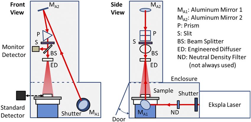
Review of                                                                                                            ARTICLE               scitation.org/journal/rsi
 Scientific Instruments

                                                                                                14
     REFERENCES                                                                                    J. T. Woodward, P.-S. Shaw, H. W. Yoon, Y. Q. Zong, S. W. Brown, and K. R.
     1
                                                                                                Lykke, “Invited article: Advance in tunable laser-based radiometric calibration
       F. L. Gates, “A study of the bactericidal action of ultra violet light: II. The effect   applications at the national institute of standards and technology, USA,” Rev. Sci.
     of various environmental factors and conditions,” J. Gen. Physiol. 13, 249–260             Instrum. 89, 091301 (2018).
     (1929).                                                                                    15
     2                                                                                             K.G. Linden, “Water research foundation project 4376: Guidance for Imple-
       F. L. Gates, “A study of the bactericidal action of ultra violet light: III. The         menting Action Spectra Correction With Medium Pressure UV Disinfection,”
     absorption of ultra violet light by bacteria,” J. Gen. Physiol. 14, 31–42 (1930).          Technical Report No. 4376, Water Research Foundation, 2015.
     3
       K. G. Linden, J. Thurston, R. Schaefer, and J. P. Malley, “Enhanced UV inactiva-         16
                                                                                                   J. R. Bolton and K. G. Linden, “Standardization of methods for fluence
     tion of adenoviruses under polychromatic UV lamps,” Appl. Environ. Microbiol.              (UV dose) determination in bench-scale UV experiments,” J. Environ. Eng. 129,
     73, 7571–7574 (2007).                                                                      209–215 (2003).
     4
       J. L. Clancy, Z. Bukhari, T. M. Hargy, J. R. Bolton, B. W. Dussert, and M. M.            17
                                                                                                   J. R. Bolton, I. Mayor-Smith, and K. G. Linden, “Rethinking the concepts of flu-
     Marshall, “Using UV to inactivate Cryptosporidium,” J. - Am. Water Works Assoc.            ence (uv dose) and fluence rate: The importance of photon-based units - a systemic
     92, 97–104 (2000).                                                                         review,” Photochem. Photobiol. 91, 1252–1262 (2015).
     5
       United States Environmental Protection Agency, “National primary drinking                18
                                                                                                   Ekspla, Savanoriu Ave. 231, LT-02300, Vilnius, Lithuania.
     water regulations: Long term 2 enhanced surface water treatment rule,” Fed.                19
                                                                                                   Figure 4-1, Optical layout of the system, Ekspla NT242-SH Technical Descrip-
     Regist. 71, 653–786 (2006), see https://www.federalregister.gov/d/06-4.
     6                                                                                          tion and User’s Manual, 2010.
       United States Environmental Protection Agency, “Ultraviolet disinfection guid-           20
                                                                                                   Alpine Research Optics, 6810 Winchester Circle, Boulder, CO 80301, USA.
     ance manual for the final long term 2 enhanced surface water treatment rule,”              21
     Technical Report No. EPA 815-R-06-007, United States Environmental Protection                 RPC Photonics, 330 Clay Road, Rochester, NY 14623, USA.
                                                                                                22
     Agency, Office of Water (4601), Washington, DC, 2006.                                         K. Linden, H. Wright, S. Beck, T. Hargy, T. Larason, and R. Mccuin, “UV
     7
       A. C. Eischeid, J. N. Meyer, and K. G. Linden, “UV disinfection of adenoviruses:         action spectra of pathogens and surrogates,” in World Congress and Exposition
     Molecular indications of DNA damage efficiency,” Appl. Environ. Microbiol. 75,             (International Ozone Association, International Ultraviolet Association, 2013).
                                                                                                23
     23–28 (2009).                                                                                 Opto Diode Corporation, 1260 Calle Suerte, Camarillo, CA 93012, USA.
     8                                                                                          24
       S. E. Beck, H. B. Wright, T. M. Hargy, T. C. Larason, and K. G. Linden, “Action             P.-S. Shaw, R. Gupta, and K. R. Lykke, “Stability of photodiodes under
     spectra for validation of pathogen disinfection in medium-pressure ultraviolet             irradiation with a 157-nm pulsed excimer laser,” Appl. Opt. 44, 197–207
     (UV) systems,” Water Res. 70, 27–37 (2015).                                                (2005).
     9                                                                                          25
       T. Larason and Y. Ohno, “Calibration and characterization of UV sensors for                 T. C. Larason and J. M. Houston, “Spectroradiometric detector measurements:
     water disinfection,” Metrologia 43, S151–S156 (2006).                                      Ultraviolet, visible, and near-infrared detectors for spectral power,” Report NIST
     10
        H. Wright, D. Gaithuma, T. Dzurny, C. Schulz, K. McCurdy, T. Bogan, A. Cabaj,           SP 250-41e2008, National Institute of Standards and Technology, 2008.
                                                                                                26
     A. Schmalweiser, Y. Ohno, and T. Larason, “Design and performance guidelines                  Instrument Systems GmbH, Neumarkter Str. 83, 81673 Munich, Germany.
                                                                                                27
     for UV sensor system,” Technical Report No. 91236, Water Research Foundation,                 S. E. Beck, R. A. Rodriguez, K. G. Linden, T. M. Hargy, T. C. Larason, and H. B.
     2009.                                                                                      Wright, “Wavelength dependent UV inactivation and DNA damage of adenovirus
     11                                                                                         as measured by cell culture infectivity and long range quantitative PCR,” Environ.
        S. W. Brown, G. P. Eppeldauer, and K. R. Lykke, “NIST facility for spectral irra-
     diance and radiance responsivity calibrations with uniform sources,” Metrologia            Sci. Technol. 48, 591–598 (2014).
                                                                                                28
     37, 579–582 (2000).                                                                           S. E. Beck, R. A. Rodriguez, M. A. Hawkins, T. M. Hargy, T. C. Larason, and
     12                                                                                         K. G. Linden, “Comparison of UV-induced inactivation and RNA damage in MS2
        S. W. Brown, G. P. Eppeldauer, and K. R. Lykke, “Facility for spectral irradi-
     ance and radiance responsivity calibrations using uniform sources,” Appl. Opt.             phage across the germicidal UV spectrum,” Appl. Environ. Microbiol. 82, 1468–
     45, 8218–8237 (2006).                                                                      1474 (2015).
     13                                                                                         29
        C. W. Stubbs, P. Doherty, C. Cramer, G. Narayan, Y. J. Brown, K. R. Lykke,                 Certain commercial equipment, instruments, or materials are identified in this
     J. T. Woodward, and J. L. Tonry, “Precise throughput determination of the                  paper to foster understanding. Such identification does not imply recommenda-
     PanSTARRS telescope and the gigapixel imager using a calibrated silicon photo-             tion or endorsement by the National Institute of Standards and Technology, nor
     diode and a tunable laser: Initial results,” Astrophys. J., Suppl. Ser. 191, 376–388       does it imply that the materials or equipment identified are necessarily the best
     (2010).                                                                                    available for the purpose.

Rev. Sci. Instrum. 91, 074105 (2020); doi: 10.1063/5.0016500                                                                                                           91, 074105-6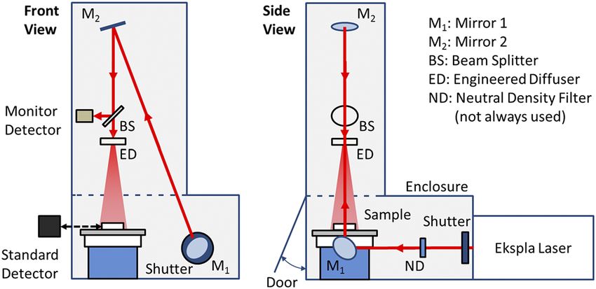
You can also read