Flip Learning: Erase to Segment
←
→
Page content transcription
If your browser does not render page correctly, please read the page content below
Flip Learning: Erase to Segment

                                             Yuhao Huang1,2,3? , Xin Yang1,2,3? , Yuxin Zou1,2,3 , Chaoyu Chen1,2,3 ,
                                        Jian Wang1,2,3 , Haoran Dou4 , Nishant Ravikumar4,5 , Alejandro F Frangi1,4,5,6 ,
                                                             Jianqiao Zhou7 , and Dong Ni1,2,3( )
                                        1
                                          National-Regional Key Technology Engineering Laboratory for Medical Ultrasound,
                                        School of Biomedical Engineering, Health Science Center, Shenzhen University, China
arXiv:2108.00752v1 [cs.CV] 2 Aug 2021

                                                                          nidong@szu.edu.cn
                                          2
                                            Medical Ultrasound Image Computing (MUSIC) Lab, Shenzhen University, China
                                              3
                                                Marshall Laboratory of Biomedical Engineering, Shenzhen University, China
                                            4
                                              Centre for Computational Imaging and Simulation Technologies in Biomedicine
                                                                   (CISTIB), University of Leeds, UK
                                         5
                                           Leeds Institute of Cardiovascular and Metabolic Medicine, University of Leeds, UK
                                                6
                                                  Medical Imaging Research Center (MIRC), KU Leuven, Leuven, Belgium
                                        7
                                           Department of Ultrasound Medicine, Ruijin Hospital, School of Medicine, Shanghai
                                                                       Jiaotong University, China

                                                Abstract. Nodule segmentation from breast ultrasound images is chal-
                                                lenging yet essential for the diagnosis. Weakly-supervised segmentation
                                                (WSS) can help reduce time-consuming and cumbersome manual an-
                                                notation. Unlike existing weakly-supervised approaches, in this study,
                                                we propose a novel and general WSS framework called Flip Learning,
                                                which only needs the box annotation. Specifically, the target in the label
                                                box will be erased gradually to flip the classification tag, and the erased
                                                region will be considered as the segmentation result finally. Our contri-
                                                bution is three-fold. First, our proposed approach erases on superpixel
                                                level using a Multi-agent Reinforcement Learning framework to exploit
                                                the prior boundary knowledge and accelerate the learning process. Sec-
                                                ond, we design two rewards: classification score and intensity distribution
                                                reward, to avoid under- and over-segmentation, respectively. Third, we
                                                adopt a coarse-to-fine learning strategy to reduce the residual errors and
                                                improve the segmentation performance. Extensively validated on a large
                                                dataset, our proposed approach achieves competitive performance and
                                                shows great potential to narrow the gap between fully-supervised and
                                                weakly-supervised learning.

                                                Keywords: Ultrasound · Weakly-supervised segmentation· Reinforce-
                                                ment learning

                                        1     Introduction
                                        Nodule segmentation in breast ultrasound (US) is important for quantitative di-
                                        agnostic procedures and treatment planning. Image segmentation’s performance
                                        ?
                                            Yuhao Huang and Xin Yang contribute equally to this work.
2      Huang et al.

has been significantly advanced by the recent availability of fully-supervised seg-
mentation methods [7, 8]. However, training such methods usually relies on the
availability of pixel-level masks laboriously and manually annotated by sonog-
raphers. Hence, designing an automatic weakly-supervised segmentation (WSS)
based system that only requires coarse labels, e.g. bounding box (BBox), is de-
sirable to ease the pipeline of manual annotation and save time for sonographers.
    Breast cancer occurs in the highest frequency in women among all cancers
and is also one of the leading causes of cancer death worldwide [15]. Thus, ex-
tracting the nodule boundary is essential for detecting and diagnosing breast
cancer at its early stage. As shown in Fig. 1, segmenting the nodule’s boundary
from the US image with weak annotation, i.e., BBox, is still very challenging.
First, nodules of the same histological type may present completely different
US image characteristics because of variances in their disease differentiation and
stage. Second, nodules of different types have extremely high inter-class differ-
ences and often display varied appearance patterns, making designing machine
learning algorithms difficult. The third challenge is that different tissues of US
images have different echo characteristics. Therefore, the intensity distribution
of foreground and background in different US images also has great diversity.

     Fig. 1. Breast nodule images in 2D US with annotated box and boundary.

    In the WSS literature, Class Activation Mapping (CAM) based methods [2,
14,22] were proposed to visualise the most discriminative features and regions of
interest obtained by the classifier. Wei et al. [19] proposed to erase the CAM area
predicted by the classifier constantly to optimise its performance on the WSS
task. However, classifiers are only responsive to small and sparse discriminative
regions from the object of interest, which deviates from the segmentation task
requirement that needs to localise dense, interior and integral regions for pixel-
wise inference. Therefore, the above methods ignored the pairing relationship
between pixels in the image and strongly relied on the CAMs with well posi-
tioning and coverage performance. Except for the image-level WSS approaches
based on inaccurate CAMs, some methods employed box annotations to ob-
tain high-quality prediction masks at a small annotation cost. However, most of
them, such as BoxSup [3], SDI [5] and Box2Seg [6], highly rely on pseudo-mask
generation algorithms (e.g., MCG [10] or GrabCut [12] based on object shape
priors). Thus, they may not suit US image segmentation tasks.
    In this study, we propose a novel and general Flip Learning framework for
BBox based WSS. We believe our proposed framework is totally different from
the current existing WSS methods. Our contribution is three-fold. First, the
erasing process via Multi-agent Reinforcement Learning (MARL) is based on
Flip Learning: Erase to Segment       3

                 Fig. 2. The workflow of our proposed framework.

superpixels, which can capture the prior boundary information and improve
learning efficiency. Second, we carefully design two rewards for guiding the agents
accurately. Specifically, the classification score reward (CSR) is used for pushing
the agents’ erasing for label flipping, while the intensity distribution reward
(IDR) is employed to limit the over-segmentation. Third, we employ a coarse-
to-fine (C2F) strategy to simplify agents’ learning for residuals decreasing and
segmentation performance improvement. Validation experiments demonstrated
that the proposed Flip Learning framework could achieve high accuracy in the
nodule segmentation task.

2   Method
Fig. 2 shows the workflow of our proposed framework for nodule segmentation
in 2D US images. The proposed Flip Learning framework is based on MARL,
in which the agents erase the nodule from its BBox. The classification score of
the nodule will decrease progressively, and the tag will be flipped. The erased
region will be taken as the final segmentation prediction. In our erase-to-segment
system, we first generate a background image to provide the eraser source to fill
the erased region suitably to be indistinguishable from normal tissue. We further
generate superpixels in the BBox for prior boundary extraction and thus improve
learning efficiency. Then, a C2F strategy is employed to obtain an accurate
segmentation result effectively.

Patch-level Copy-Paste for Precise Erasing Source. US images of most
normal tissues display a certain degree of local texture and gray-scale continuity.
To trade-off between algorithm complexity and performance, we present a simple
and feasible method to fill the BBox area. As shown in Fig. 3: (a)-(b) According
4      Huang et al.

                      Fig. 3. Example of patch-level copy-paste.

to the context information, we first obtain the patches (pf and pg with width wf
and wg , respectively) in the original image as region proposals. These patches
are similar to the background around the annotated box. (c) Next, we adopt
a mixup [21] to copy and paste these region proposals to generate the pseudo-
background image (see Equ. 1). (d) Finally, to achieve a higher quality fusion
between the pseudo-background and the real background, we adopt a local-edge
mean filter to optimise the copy-paste edge area ps0 .
                                                                     x
       ps0 (x, y) = α × pf (x, y) + (1 − α) × pg (x, y),   α=                  (1)
                                                                (wf + wg )/2

MARL Framework for Efficient and Accurate Erasing. Recently, RL-
based methods have shown great potential in various medical imaging tasks [20,
23]. To accelerate the learning process, we propose to use a MARL framework
with two agents erasing the object simultaneously. We further define the MARL
erasing framework using these elements:
    -Environment: The Environment is defined as the BBox area of the orig-
inal US image. It contains the object to be segmented and limits the agents’
moving. However, it is noted that erasing based on pixel-level is difficult and
inefficient due to the weak supervised signal. Thus, we use superpixel algorithms
for enhancing the supervised signal, which group pixels with similar proper-
ties into perceptually meaningful atomic regions while considering spatial con-
straints. Additionally, it preserves the edge information of the original image
during enhancing the local consistency. As explained above, in this study, we
generate superpixels for the BBox area to obtain a superpixel-level environment
before erasing. Specifically, we adopt Superpixels Extracted via EnergyDriven
Sampling (SEEDS) to obtain superpixel-blocks (details refer to [1]).
    -Agents: The Agent is to learn a policy for segmentation by interacting
with the superpixel-level environment. In our study, two agents Agentk,k=1,2
share the parameters in the convolution layers for knowledge sharing, and have
independent fully connected layers for decision-making. They traverse the whole
BBox with each of them handling approximately half of the total superpixels,
and the centre superpixel of BBox is set as start point for each agent.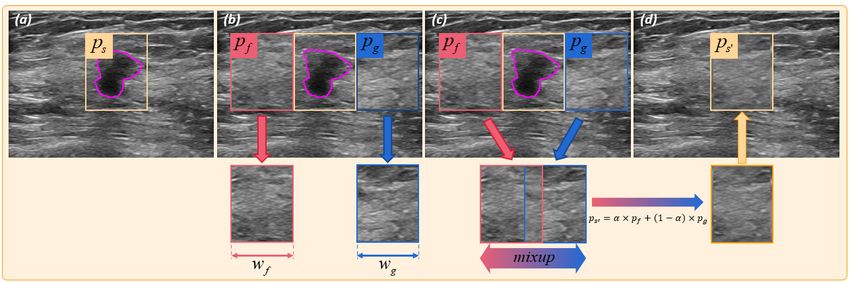
Flip Learning: Erase to Segment          5

Fig. 4. Details of (a) CSR and (b) IDR. Note that we first train a classifier to classify
‘nodule’ and ‘normal tissue’, and obtain CSR by calculating the differences of nodule
scores between stept−1 and stept .

    -States: The State of one agent can be defined as a 64×64 area, with its
centre indicates the agent’s position. We further define States as the last six
areas observed by two agents, with the size of 6×64×64. States concatenating
can provide rich information, which can promote agents’ learning.
    -Actions: The Action space of our framework contains only two operations,
including erasing and passing. Note that the action is taken on the superpixel
level. The action erasing indicates that this superpixel will be erased, and the
region will be filled according to the generated background image. The action
passing represents the agents choose not to erase this superpixel region.
    -Rewards: The Reward guides the agents’ erasing process. As shown in
Fig. 4, we design two rewards, including 1) CSR and 2) IDR. Specifically, CSR
is a basic reward used to guide the agents to erase the object from the BBox for
classification tag flipping. However, using such reward separately may cause over-
segmentation because the agents may tend to fill the whole BBox to make a high
score. Thus, we introduce additional IDR to overcome this problem. Specifically,
if one superpixel is erased in stept and makes the intensity distribution (It ) of
the erased area highly different from that in stept−1 , the agent will be punished.
The difference between the two distributions is calculated using the Wasserstein
distance [18]. The total reward Rk for each Agentk can be defined as:
                   Rk = sgn(Sct−1 − Sct ) + thr(W (It−1 , It ), θ),                  (2)
where sgn and thr are the sign and threshold function, respectively. Sct repre-
sents the nodule scores in stept . W(·) calculates the Wasserstein distance between
the erased region’s intensity distribution in stept−1 and stept , and θ = 25 is the
pre-set threshold. More details can be seen in Fig. 4.
    -Terminal signals: The terminal signal represents when to terminate the
agent-environment interaction. We adopt two types of terminated strategy in our
study: 1) attaining the maximum number of traversals N = 2 and 2) classifica-
tion score of nodule less than a pre-set threshold β = 0.05. Such a termination
strategy can balance efficiency and accuracy during both training and testing.
    In deep Q-network (DQN) [9], both selecting and evaluating an action use
a max operation, which may cause over-estimation for Q-values and lead to
6 Huang et al.
unstable training. Double DQN (DDQN) [17] is then proposed to decouple the
action selection from evaluation to stabilise the learning process. In this study, for
improving learning efficiency, we adopt DDQN with a naive prioritised replay
buffer [13] with the size of M, which contains sequences of state s, action a,
reward r and next state s0 . The qtarget can be calculated by:
qtarget = r + γQ(s0 , argmax Q(s0 , a0 ; ω); ω 0 ), (3)
a0
Q(ω) and Q(ω 0 ) represent current and target Q-networks, respectively. γ is the
discount factor that balances the importance of current and future rewards.
Then, with the uniform sampling U (·), and the loss function can be written by:
L = Es,r,a,s0 ∼U (M) (qtarget − Q(s, a; ω))2 (4)
C2F Learning Strategy. Finer superpixel implies more accurate boundary
prior knowledge, leading to a higher upper bound of segmentation performance.
However, learning on the BBox with fine-superpixel directly may be too difficult
for the agents. The agents may get into local optimality easily due to the weak
supervised signal. Thus, the segmentation result may contain residual errors.
Inspired by [16], we adopt a two-stage strategy for reducing the residual errors
and improving the segmentation performance. Specifically, in the first stage of
learning, the agents learn on a coarse superpixel BBox and output a coarse
prediction (see Stage 1 in Fig. 2). Such coarse predictions are leveraged to make
a channel-wise concatenation with original images as the second stage’s input,
which can provide additional information and help the agent learn on the BBox
with fine-superpixel efficiently (see Stage 2 in Fig. 2).
3 Experimental Result
Materials and Implementation Details. We validated our proposed Flip
Learning method on segmenting the breast nodule in 2D US images. Approved
by the local IRB, a total of 1,723 images (with size 448×320) were collected from
1,129 patients. Each image’s nodule was annotated with the mask and BBox by
sonographers manually (see Fig. 1). The dataset was split into 1278, 100 and
345 images for training, validation and independent testing at the patient level
with no overlap. The classifier and agents used the same training, validation
and testing set. In this study, we implemented our method in P ytorch, using an
NVIDIA TITAN 2080 GPU. We first trained the classifier with architecture of
ResNet18 using AdamW optimiser with learning rate=1e-3 and batch-size=128.
Then, the proposed MARL system (ResNet18 backbone) was trained with Adam
optimiser in 100 epochs, costing about 1.5 days (learning rate=5e-5 and batch-
size=64). The superpixels are generated by OpenCV function and indexed from
1 to S. Both agents start from the centre superpixel with index S/2. One agent
traverses the superpixels from S/2 to S, the other one traverses reversely from
index S/2 to 1. The replay buffer has a size of 8000, and the target Q-network
copied the parameters of the current Q-network every 1200 iterations.Flip Learning: Erase to Segment 7
Table 1. Method comparison (mean±std). The best WSS results are shown in blue.
DICE↑ JAC↑ CON↑ HD↓ ASD↓
U-net 93.44±3.76 87.91±5.02 84.76±9.22 15.22±8.99 2.68±1.79
GrabCut 1.66±8.91 1.07±5.968 Huang et al.
Table 3. Impact of Annotation Box Shift (mean±std).
DICE↑ JAC↑ CON↑ HD↓ ASD↓
0-10 pixels 89.22±4.12 81.22±3.22 72.28±12.18 17.02±7.87 2.88±1.31
10-20 pixels 88.16±6.23 79.56±5.32 69.73±14.11 16.53±7.19 2.92±2.15
20-30 pixels 86.72±8.33 63.44±6.33 53.09±11.28 23.84±9.88 4.12±2.13
Fig. 5. Typical cases of Flip Learning. Mask predictions have been post-processed.
Note that the classification tags of original images are ‘nodule’, and after two-stage
erasing, their tags will flip to ‘normal tissue’. The erase curves show the variation of
classification scores during erasing.
esting to see that interacting with the fine-superpixel-based environment directly
(i.e., one-stage) makes agents learn difficultly. Without enough information for
guiding agents’ action in the huge search space, they may fail to segment accu-
rately. Thus, there will remain many segmentation residuals, which can cause
obvious performance degradation (row 1-6). The contribution of IDR and C2F
can be observed in the last 4 rows: equipping each of them separately can boost
the accuracy, while combining them will obtain a great improvement in all the
evaluation metrics. To test the sensitivity of our approach to box annotation,
we validated it on different box shifting levels, including 1) 0-10 pixels, 2) 10-20
pixels and 3) 20-30 pixels. The results reported in Table 3 indicate that our
methods can perform well though the box’s centre is shifting.
Fig. 5 shows three typical cases of our proposed method. Compared with the
result of stage one, it can be seen that the final result of stage two obtains a more
accurate boundary and overall mask, which is very close to the GT. The erase
curves shown in the last column indicate the relationship among erased area
size (green), DICE (yellow), and classification score (red). It can be observed
that through erasing, the DICE and erased area are gradually increasing, and
their variation is nearly synchronous. Moreover, the classification score curve will
decrease continuously, and the classification tag will be flipped from ‘nodule’ to
‘normal tissue’, which proves the effectiveness of our Flip Learning approach.Flip Learning: Erase to Segment 9
4 Conclusion
We propose a novel Flip Learning framework for nodule segmentation in 2D
US images. We use MARL to erase the nodule from the superpixel-based BBox
to flip its classification tag. We develop two rewards, including CSR and IDR,
for overcoming the under- and over-segmentation, respectively. Moreover, we
propose to adopt a C2F learning strategy in two stages, which can achieve more
accurate results than a one-stage method. Experiments on our large in-house
dataset validate the efficacy of our method. Our patch-level copy-paste filling
strategy is limited in some cases. Thus, in the future, we will explore a more
general background filling approach (e.g. GAN), to generate a more accurate
background for different types of images.
Acknowledgment. This work was supported by the National Key R&D Pro-
gram of China (No. 2019YFC0118300), Shenzhen Peacock Plan (No. KQTD20160
-53112051497, KQJSCX20180328095606003), Royal Academy of Engineering un-
der the RAEng Chair in Emerging Technologies (CiET1919/19) scheme, EPSRC
TUSCA (EP/V04799X/1) and the Royal Society CROSSLINK Exchange Pro-
gramme (IES/NSFC/201380).
References
1. Bergh, M.V.D., Boix, X., Roig, G., Capitani, B.D., Gool, L.V.: Seeds: Superpix-
els extracted via energy-driven sampling. In: European Conference on Computer
Vision. Springer (2012)
2. Chattopadhay, A., Sarkar, A., Howlader, P., Balasubramanian, V.N.: Grad-
cam++: Generalized gradient-based visual explanations for deep convolutional
networks. In: 2018 IEEE Winter Conference on Applications of Computer Vision
(WACV). pp. 839–847. IEEE (2018)
3. Dai, J., He, K., Sun, J.: Boxsup: Exploiting bounding boxes to supervise convolu-
tional networks for semantic segmentation. In: Proceedings of the IEEE interna-
tional conference on computer vision. pp. 1635–1643. IEEE (2015)
4. Hou, X., Zhang, L.: Saliency detection: A spectral residual approach. In: 2007 IEEE
Conference on computer vision and pattern recognition. pp. 1–8. IEEE (2007)
5. Khoreva, A., Benenson, R., Hosang, J., Hein, M., Schiele, B.: Simple does it:
Weakly supervised instance and semantic segmentation. In: Proceedings of the
IEEE conference on computer vision and pattern recognition. pp. 876–885. IEEE
(2017)
6. Kulharia, V., Chandra, S., Agrawal, A., Torr, P., Tyagi, A.: Box2seg: Attention
weighted loss and discriminative feature learning for weakly supervised segmenta-
tion. In: European Conference on Computer Vision. pp. 290–308. Springer (2020)
7. Liu, S., Wang, Y., Yang, X., Lei, B., Liu, L., Li, S.X., Ni, D., Wang, T.: Deep
learning in medical ultrasound analysis: a review. Engineering 5(2), 261–275 (2019)
8. Minaee, S., Boykov, Y., Porikli, F., Plaza, A., Kehtarnavaz, N., Terzopoulos, D.:
Image segmentation using deep learning: A survey. arXiv preprint arXiv:2001.05566
(2020)10 Huang et al.
9. Mnih, V., Kavukcuoglu, K., Silver, D., Rusu, A.A., Veness, J., Bellemare, M.G.,
Graves, A., Riedmiller, M., Fidjeland, A.K., Ostrovski, G., et al.: Human-level
control through deep reinforcement learning. Nature 518(7540), 529–533 (2015)
10. Pont-Tuset, J., Arbelaez, P., Barron, J.T., Marques, F., Malik, J.: Multiscale com-
binatorial grouping for image segmentation and object proposal generation. IEEE
transactions on pattern analysis and machine intelligence 39(1), 128–140 (2016)
11. Ronneberger, O., Fischer, P., Brox, T.: U-net: Convolutional networks for biomedi-
cal image segmentation. In: International Conference on Medical image computing
and computer-assisted intervention. pp. 234–241. Springer (2015)
12. Rother, C., Kolmogorov, V., Blake, A.: “grabcut” interactive foreground extraction
using iterated graph cuts. ACM transactions on graphics (TOG) 23(3), 309–314
(2004)
13. Schaul, T., Quan, J., Antonoglou, I., Silver, D.: Prioritized experience replay. In:
ICLR (Poster) (2016)
14. Selvaraju, R.R., Cogswell, M., Das, A., Vedantam, R., Parikh, D., Batra, D.: Grad-
cam: Visual explanations from deep networks via gradient-based localization. In:
Proceedings of the IEEE international conference on computer vision. pp. 618–626.
IEEE (2017)
15. Siegel, R.L., Miller, K.D., Fuchs, H.E., Jemal, A.: Cancer statistics, 2021. CA: a
Cancer Journal for Clinicians 71(1), 7–33 (2021)
16. Tu, Z.: Auto-context and its application to high-level vision tasks. In: 2008 IEEE
Conference on Computer Vision and Pattern Recognition. pp. 1–8. IEEE (2008)
17. Van Hasselt, H., Guez, A., Silver, D.: Deep reinforcement learning with double
q-learning. In: Thirtieth AAAI conference on artificial intelligence (2016)
18. Villani, C.: Optimal transport: old and new, vol. 338. Springer Science & Business
Media (2008)
19. Wei, Y., Feng, J., Liang, X., Cheng, M.M., Zhao, Y., Yan, S.: Object region min-
ing with adversarial erasing: A simple classification to semantic segmentation ap-
proach. In: Proceedings of the IEEE conference on computer vision and pattern
recognition. pp. 1568–1576. IEEE (2017)
20. Yang, X., Huang, Y., Huang, R., Dou, H., Li, R., Qian, J., Huang, X., Shi, W.,
Chen, C., Zhang, Y., et al.: Searching collaborative agents for multi-plane local-
ization in 3d ultrasound. Medical Image Analysis p. 102119 (2021)
21. Zhang, H., Cissé, M., Dauphin, Y.N., Lopez-Paz, D.: mixup: Beyond empirical risk
minimization. arXiv preprint arXiv:1710.09412 (2017)
22. Zhou, B., Khosla, A., Lapedriza, A., Oliva, A., Torralba, A.: Learning deep features
for discriminative localization. In: Proceedings of the IEEE conference on computer
vision and pattern recognition. pp. 2921–2929. IEEE (2016)
23. Zhou, S.K., Le, H.N., Luu, K., Nguyen, H.V., Ayache, N.: Deep reinforcement
learning in medical imaging: A literature review. arXiv preprint arXiv:2103.05115
(2021)You can also read