Parasitology 2021
MARGIE MORGAN, PHD, D(ABMM)
←
→
Page content transcription
If your browser does not render page correctly, please read the page content below

Parasitology 2021 MARGIE MORGAN, PHD, D(ABMM)
Laboratory Diagnosis
Current diagnosis is predominately   Alternative testing methods:
based on microscopic exam             ◦ Serology for select pathogens
Specimen types:                       ◦ Fluorescent stains of stool for select
 ◦ Stool                                pathogens
 ◦ Non-stool
                                      ◦ Molecular assays of stool for select
   ◦ Perianal specimen
   ◦ Sigmoidoscopic specimen
                                        pathogens
   ◦ Duodenal aspirates
   ◦ Liver abscess
   ◦ Sputum
   ◦ Urine
   ◦ Urogenital
 ◦ Blood
 ◦ Tissue
Diarrheal Disease due to Parasites
Travel history or poor sanitation puts you at the highest risk for
parasitic diarrheal disease
Patients with poor immune status fare worse
Diarrheal disease usually has sporadic symptoms that are chronic in
nature, dysentery not common
Most usual symptoms:
◦ Abdominal pain, cramping, long term nausea, and malaise,
  mucous in stool, and +/- fever
Two-vial Stool Collection Kit
Preservation of stool for parasite exam

10% formalin vial                    PVA with fixative
Concentration of stool performed     Polyvinyl alcohol (PVA)
with ethyl acetate washing to
eliminate fecal debris               Permanent smear prepared
                                     from PVA stool vial and stained
Can use to perform: Wet mount,       with Trichrome stain
Iodine mount, Direct Fluorescent
                                     Detect: Protozoan trophozoites
antibody staining, and NAAT
                                     and cysts
Detect: Helminth eggs, larvae,
microsporidia, and protozoan cysts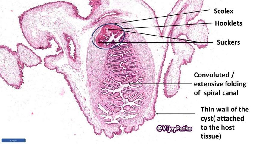
Protozoa: Amebae found in stool
Major pathogen
Entamoeba histolytica/dispar
E. histolytica (pathogen) and E. dispar (nonpathogen) both occur in the large
intestine. Morphologically indistinguishable.
 ◦ Antigen testing or molecular method needed to distinguish the species.
Disease = Amoebiasis, or amoebic dysentery
   ◦Most infections are asymptomatic, invasive intestinal disease may occur
    manifesting with several weeks of cramping, abdominal pain, watery or
    bloody diarrhea, and weight loss
Diagnosis
 ◦ Cysts = infectious form found in stool specimens
   ◦ Found in contaminated water and areas of poor sanitation
 ◦ Trophozoites = invasive form found in the intestine tissue and stool
   ◦ Colon biopsy with “flask-shaped” ulcer and trophozoites in tissue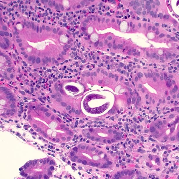
Entamoeba histolytica/dispar cyst and trophozoite

                                     Trophozoite 20-30 um
             Cysts @10-12 um         in diameter
                in diameter          One nucleus with
                                     centrally placed
             Up to 4 nuclei in       karyosome
             the cyst/ central
             karyosome, clean
             chromatin ring

             Chromatoidal body
             present in some cysts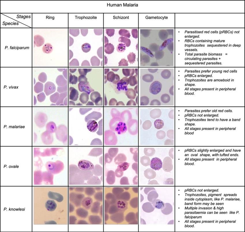
Intestinal Amoebiasis

                        Flask shaped ulcer
                        formed in tissue
                        of the large intestine

                        Trophozoites of
                        E. histolytica
                        observed in intestinal
                        tissue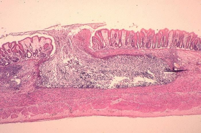
Liver Amebic Abscess
                 Extra-intestinal disease:

                 Trophozoites from large intestine travel through
                 the blood and invades the liver to form abscess
                 Abscess fluid is necrotic and will not contain
                 trophozoites. Must biopsy infected tissue to
                 observe trophozoites.

                 Serology will be positive in >=90% patients with
                 liver abscess compared to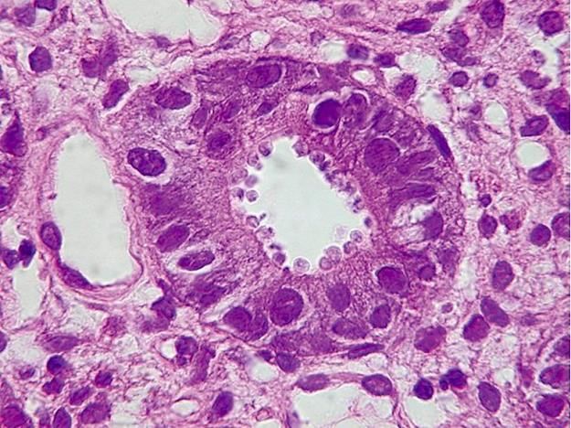
Entamoeba coli – amoeba in large intestine, commensal not
causing disease

                                         Trophozoite is the
                                         invasive form that is
                                         found in the
                                         intestine

                                         Single nucleus with a
                                         large karyosome located
                                         eccentrically with irregular
                                         chromatin ring (differs
Cyst @ 20–25µm                           from E. histolyica).
Up to 8 nuclei                           The cytoplasm appears
Shed from host                           dirty and vacuolated
Lives in environment
Iodamoeba butschlii Cysts, 10 – 12 µm Unique large starch inclusion (glycogen mass) Usually not considered a pathogen Iodine-stained stool preparation – glycogen inclusions stain dark color with iodine
Protozoa Found in Stool: Flagellates

               Pathogen
Giardia lamblia (G. intestinalis,
and G. duodenalis)
CYSTS
Found in contaminated water & undercooked foods
Mild diarrhea to severe malabsorption, waxing and waning
symptoms
Foul smelling , watery diarrhea TROPHOZOITE
“falling leaf”
Traveler’s diarrhea (Russia and Mexico) motility
Day-care center outbreaks reported
Cysts/trophozoites may be irregularly shed in stool, and be
difficult to detect; Fluorescent stains and NAAT for provide
more sensitive detection
Confined to intestine / Therapy Flagyl (Metronidazole)Giardia lamblia
Trophozoite Cysts
Clearing between the
cell wall and the cell
membraneGiardia lamblia invades intestinal tissue – shown are duodenal biopsies with trophozoites near the surface epithelium
Chilomastix mesnili cyst
                        Nonpathogen
                        Morphology mimics Giardia
                        lamblia cyst – except for the clear
                        space at end of cyst
                        Internal structure looks like
                        “shepherd’s crook” or safety pin

                   C. mesnili trophozoite – seldom
                   seen in clinical specimens
Trichomonas vaginalis Urogenital protozoan STD in both men and women Infection leads to Scant, watery vaginal discharge in women Infection in urethral orifice in men Four flagella, short undulating membrane
Protozoa Found in Stool:
Ciliates, Coccidia, Blastocystis   Primary Human Pathogens
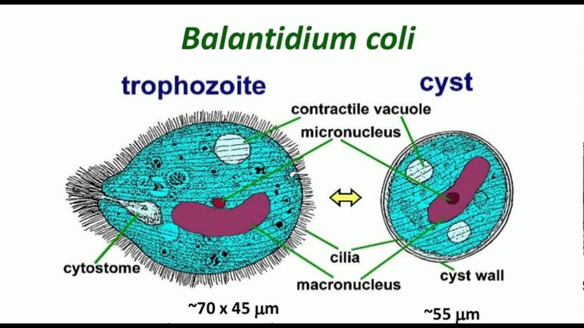
Balantidium coli
◦ Humans infected by contact with swine
◦ Poor hygiene
◦ Only ciliate that’s pathogenic to humans
◦ Amebiasis like disease
◦ Largest (50-200 um) trophozoite in parasitology
  ◦ Surface covered with cilia; macronucleus
◦ Cyst 40-60 um
◦ Readily identified in fresh stools and wet mounts
◦ Can cause flask shaped ulcer in intestine like that of
  Entamoeba histolytica
Coccidia: Isospora (Cystoisospora) belli
 Contaminated food/water, oral-anal route of infection
 Found most commonly in HIV/AID patients
 Infects small intestine epithelium
 Malabsorption syndrome mimicking giardiasis
 Egg is oval and 10 - 33 um in length
 Stains positive with modified acid-fast staining

                                      (+) Modified acid fast stain
Cryptosporidium spp
Contaminated water is source of infection
◦ Resistant to usual water-purification procedures
(chlorination, ozone)
Watery diarrhea -
◦ More severe in HIV/AIDS – chronic/debilitating
infection
◦ Daycare center outbreaks (fecal-oral transmission)
Not detected in routine O & P exams (left)
Modified acid-fast stains (PAF) aid in detection,
Oocysts measure 4-6 µm
Stool antigen, Direct Fluorescence antibody staining
and Molecular assays aid detection.Cryptosporidia located in intestinal tissue just below the plasma membrane
Cyclospora cayetanensis
Contaminated fruits and vegetables – particularly ones with plant hairs
Watery diarrhea; fatigue, anorexia, weight loss, flu like symptoms. More severe in
immune suppressed, can last for months
Infects upper small bowel
Treatment: Oral Trimethoprim/sulfamethoxazole
Found in vacuoles in cytoplasm of jejunal epithelium, villous atrophy, crypt
hyperplasia
                 Modified acid
                                                 UV autofluorescence
                 fast positive
                 Cysts 8-10
                                                 Also positive on Calcofluor
                 microns
                                                 white stain
Blastocystis hominis
Trichrome stain
Blastocystis hominis is a genetically diverse
unicellular organism (algae) of variable size found
in the stools of people who have ingested
contaminated food or water Nuclear blobs around Iodine wet mount
the cell periphery
It can be found in healthy people who aren't
having digestive symptoms, and sometimes
found in the stools of people who have diarrhea,
abdominal pain or other gastrointestinal
problems.
The role in disease is not well understood /
perhaps some forms are more pathogenic.Microsporidia Enterocytozoon and Encephalitozoon species most common genera Infection by ingestion of spores (1-4 um) Chronic diarrhea in HIV/AIDS patients Can disseminate and be found in cases of Myositis, hepatitis, peritonitis, keratitis, gastrointestinal and biliary tract infection Stain on modified trichrome stain Tissue histopathology (H&E) Minimal to no changes in intestinal tissue with 3 - 5 micron, rounded, basophilic structures in enterocytes, often surrounded by a halo
Nematodes/ Roundworms
Enterobius vermicularis (pinworm)
Humans are the only host
Most common helminth in US
Worms: Females 8-13mm, males 2-5 mm
 ◦Appear like strings in the stool
Worms dwell in the cecum
Migrate to perineum at night
 ◦Deposit eggs
 ◦Diagnosis- Scotch tape test or anal swab in AM
Eggs 0val with flattened side: 50-60um by 20-30um
Charcot Leydon
Ascaris lumbricoides                                               Crystals

1-1.2 billion people infected in Africa, the Americas, China
and East Asia, common in children
Most common in developing countries with poor
sanitation/ feces contaminated soil, humans ingest eggs
Largest helminth to affect humans
 ◦ Worms: Females 20-35cm long and straight, males 15-30cm
   with a curved tale
 ◦   Bolus of worms can cause intestinal obstruction
 ◦   Egg – thick-walled shell with internal larvae
 ◦   Charcot Leyden crystals can be seen in stool mounts
 ◦   Loeffler’s syndrome – pulmonary infiltration and
     eosinophilia from worm migration through lung to reach
     the intestine

                                                  Bolus of worms
Trichuris trichuria (whipworm) Soil transmitted/due to fecal contamination Amebiasis-like disease Adult worm attaches to large intestine, rarely recovered – best diagnosis by detecting egg in stool specimens Head is the thinnest part of worm Egg is barrel shaped, golden brown with knobs on both ends
Necator americanus and Ancylostoma duodenale
(Hookworms)
Soil transmission – filiform larvae
penetrates the skin causing a slight skin
rash then migrates to the intestine where it
can cause abdominal pain and diarrhea
2nd most common helminth infection after
Ascaris
Necator and Ancylostoma are known as
Hookworms.
Hookworm egg, brownish colored with
visible larvae, 60 X 40 um
Strongyloides stercoralis
Life cycle occurs in humans
Strongyloides larvae
Larval form only
Larvae penetrates intact skin from contact
with contaminated soil, then migrates to
intestine
Majority do not have symptoms, some
non-specific complaints or minor
abdominal pain and diarrhea from the
larvae’s presence in intestine
In immune suppressed can evolve into a
massive intestinal infection or with larvae
migration to the respiratory tract causing
an eosinophilic pneumoniae termed
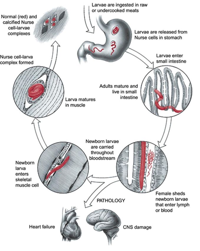
“autoinfection”Anisakis simplex Infective larvae are ingested from fish or squid that humans eat raw or undercooked. Nematode worm present in the fish flesh invades the stomach wall or intestine of humans. Infection can occur without symptoms. This infection is treated by removal of the larvae via endoscopy or surgery. Rare cases cause granuloma formation and bowel obstruction.
Dracunculus medinensis
Dracunculiasis (Guinea worm disease)
Due to eradication campaign, now isolated to narrow belt in Africa
Infected by drinking unfiltered water containing copepods (small crustaceans)
which are infected with larvae of D. medinensis
Following ingestion, the copepods die, release the larvae, which penetrate the
stomach and intestinal wall and the abdominal cavity and retroperitoneal space
The male worm dies, the female migrates in the subcutaneous tissue toward the
skin surface
The clinical manifestations are localized but incapacitating. The worm emerges as
a whitish filament (duration of emergence: 1 to 3 weeks) in the center of a
painful ulcer, accompanied by inflammation and frequently by secondary
bacterial infection.
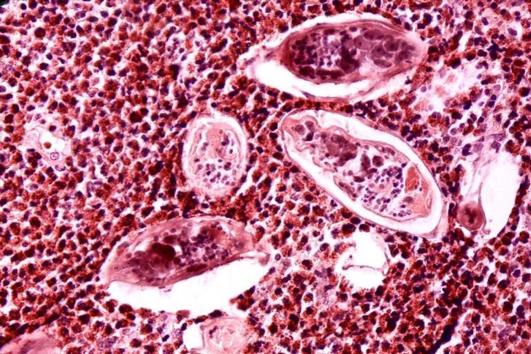
Trichinella spiralis

-Tissue nematode
-Infection from the ingestion of
undercooked meat
-Usually only an incidental finding
when viewing histopathology of
muscle
Naegleria fowleri
 Primary amoebic meningoencephalitis
(PAM)
Acquired by swimming or diving into fresh-
water pools and water goes up nose thru
cribriform plate into the brain
Usually, a fatal infection

                                             N. fowleri
                                             trophozoite
Acanthamoeba species
◦ Contact lens keratitis
◦ Primary diagnosis: Wright stain of corneal scrapings to visualize
amoeba
◦ Can culture for Acanthamoeba. Corneal scrapings placed in a lawn of
E.coli. Visible tracts made in E. coli growth. Do wet mount to look for
amoeba
Wright’s Amoeba in
stain wet mountTrypanosoma brucei → Sleeping sickness
(African trypanosomiasis)
Vector: Tse Tse fly
The two T. brucei species that cause African trypanosomiasis are
indistinguishable morphologically
◦ T. brucei gambiense
◦ T. brucei rhodesiense
A typical trypomastigote has:
◦ A small kinetoplast located at the posterior end
◦ A centrally located nucleus
◦ An undulating membrane and flagellum
◦ 14 to 33 µm in length
Trypomastigote is the only stage found in clinical specimens nucleus
kinetoplastTrypanosoma cruzi → Chagas disease
(American trypanosomiasis)
Vector: Reduvid/Triatoma (kissing) bug
Trypomastigote is the form in the blood of an infected person and
may be seen in CSF in CNS infections
Motile circulating trypomastigotes are readily seen on slides of fresh
anticoagulated blood in acute infection Trypomastigote
A typical trypomastigote has:
◦ Large, subterminal or terminal kinetoplast,
◦ Centrally located nucleus,
◦ Undulating membrane, and flagellum
◦ 12 to 30 µm in length.
◦ Can appear C shaped
Amastigote stage parasite may be seen in histopathology specimens
from affected organs.
AmastigoteLeishmania – Clinical Disease Cutaneous – L. tropica and L. brasiliensis ◦ Single or few chronic, ulcerating lesions; many species ◦ Latin America, southern Europe, Middle east, southern Asia, Africa ◦ Mucocutaneous in Latin America Visceral – L. donovani complex most common ◦ primarily L. donovani complex (Asia), L. infantum/chagasi (Africa and Latin America) ◦ Hepatosplenomegaly, anemia, cytopenias, systemic symptoms ◦ India, Bangladesh, Nepal, Sudan, and Brazil ◦ Important opportunistic infection in HIV infection Vector – Female sand fly
Leishmania
Diagnosis
◦ Biopsy of infected tissue (skin, bone marrow)
  ◦ Multiple, tiny 2-5 um amastigotes within histiocytes
  ◦ Distinct kinetoplast (bar-like structure adjacent to nucleus)
◦ PCR methods
◦ Urinary antigens (visceral)
DDx of multiple tiny intracellular organisms
◦ Leishmania – kinetoplast
◦ Histoplasma – budding
◦ Toxoplasma – somewhat curved, mostly extracellular
Babesia
Two species: B. microti, B. divergens                                    Ixodes tick
Zoonosis (deer, cattle, rodents; humans accidental host)
Transmission vector: Ixodes tick bite
Infects red blood cells
Found world-wide
B. microti along the Northeast US
 ◦ Nantucket Island, Martha’s vineyard, Shelter Island
Malaria-like syndrome
◦ Fever but without periodicity, night sweats, weight loss,
  hemolytic anemia, hemoglobinuria, renal failure
Diagnosis; Blood smear examination                       Maltese cross
  ◦ Ring form only (mimics P. falciparum)                  (tetrads)
  ◦ Tetrads (unlike P. falciparum)
MALARIA
Protozoan parasite
Transmitted by the anopheles mosquito
Endemic in tropical areas
Malaria Physical Diagnosis
Physical exam findings for most uncomplicated malarial cases:
 ◦ Fever and splenomegaly

P. falciparum - most pathogenic species** lethal malignant tertian fever
  ◦ Jaundice
  ◦ Hepatomegaly
  ◦ Increase in respiratory rate
  ◦ Possible CNS involvement
  ◦ Blackwater fever – hemolysis, renal failure

 Tertian = fever every 48 hours / Quartan = fever every 48 hours
Malarial Slide Preparations – standard tool for
diagnosis
Thick smear                      Thin smear
Drop of blood on slide (non-     Feather edge smear
anticoagulate blood is best)
                                 For optimal morphology, stain
Water rinse to eliminate rbc’s   with Giemsa (not Wright-
                                 Giemsa) stain with proper pH
Stain with Giemsa stain (not
Wright-Giemsa) proper pH         Speciation of malaria
Need the proper pH to stain      Parasitemia (%)
the Schuffner’s granules
Concentrated to spot malaria
parasites
Notes of importance:
P. falciparum
1. Calculation of % or parasitemia is
Used to assess seriousness of infection
2. Only ring and gametocyte seen in blood smears
P. vivax
1. Duffy negative red blood cell protect from P. vivax
infection
2. Untreated infections can last several years and
remain dormant in the liver. Recurrent and chronic
infection can lead to brain, kidney and liver
damage
P. ovale
1. Fimbriated edge of red blood cellToxoplasma gondii Toxoplasmic
lymphadenitis
Toxoplasmosis is considered to be a leading cause of death
attributed to foodborne illness in the United States.
Caused by eating undercooked, contaminated meat (especially
pork, lamb, and venison) and accidental ingestion of oocysts from
cat’s litter box, esp a problem in the immunodeficient patient.
Immunocompetent, mostly solitary posterior cervical
lymphadenopathy.
Histology of toxoplasmic lymphadenitis: Usually lack of necrosis
or well-formed granulomas. Follicular hyperplasia, interfollicular Tachyzoites
epitheloid histiocytes, singly or in small clusters. Usually involves
monocytoid B cells in a sinusoidal and parasinusoidal pattern.Microfilariae
• Identification is based on the
microfilariae being either
sheathed or non-sheathed
• Sheathed / thin covering over
the larvae
• Wucheria bancrofti and
• Loa loa
• Not sheathed
Onchocerca volvulus
Identification of microfilariae is based on the presence of a sheath covering the larvae, as well
as the distribution of nuclei in the tail region
A, W. bancrofti. B, B. malayi. C, L. loa. D, O. volvulus. E, Mansonella perstans. F, Mansonella
streptocerca. G, Mansonella ozzardi.Wuchereria bancrofti • Filarial nematode (roundworm) that is the major cause of lymphatic filariasis. • Filarial worms are spread by a variety of mosquito vector species. • W. bancrofti is the most common filarial worm and affects over 120 million people, primarily in Central Africa and the Nile delta, South and Central America, the tropical regions of Asia including southern China, and the Pacific islands. • If left untreated, the infection can develop into a chronic disease called lymphatic filariasis
Loa loa

• Loiasis (African eye worm)
• It is passed on to humans through the repeated bites of
  deerflies of the genus Chrysops. The flies that pass on the
  parasite breed in certain rain forests of West and Central
  Africa.
• Infection with the parasite can also cause repeated
  episodes of itchy swellings of the body known as Calabar
  swellings.
• There may be more than 29 million people who are at risk
  of getting loaisis in affected areas of Central and West
  Africa.
Black fly

Onchocerciasis
                                                         Microfiliarea of Onchocerca
• Onchocerciasis, or river blindness, is caused by the   from skin nodule
  parasitic worm Onchocerca volvulus.
• It is transmitted through repeated bites by
  blackflies of the genus Simulium.
• The disease is called river blindness because the
  blackfly that transmits the infection lives and
  breeds near fast-flowing streams and rivers, mostly
  near remote rural villages. The infection can result
  in visual impairment and sometimes blindness.
• Onchocerciasis can also cause skin disease,
  including intense itching, rashes, or nodules under
  the skin.
Diphyllobothrium latum Infected by ingesting poorly-cooked fresh-water fish (salmon particularly problematic) Scandinavian, Russia, Canada, N. USA, Alaska Known as the Broad fish tapeworm Scolex has a Longitudinal sucker Eggs have non-shouldered operculum and knob ◦ They are not embryonated Infection causes VitaminB12 deficiency
Diphyllobothrium latum

       Broad proglottid

                                          Longitudinal
Operculum                                 sucking plate
door

                      knob
Taenia saginata Taenia solium
▪ Beef tapeworm ▪Pig tapeworm
▪ 4 suckers on scolex ▪Ring of thorns/crown on
▪ >12 uterine branches in scolex
proglottids ▪ ▪Intestinal tapeworm
Non-human pathogen ▪Ingestion of eggs ->
No disease CysticercosisTaenia eggs Identical eggs for the two species
Taenia saginata                    Taenia solium

Proglottid > 12 uterine branches     Proglottid
Cysticercosis

Parasitic tissue infection caused by larval
cysts of the tapeworm Taenia solium from
ingestion of T. solium eggs.
The larval cysts infect brain, muscle, or other
tissue, and are a major cause of adult onset
seizures.
Hymenolepis nana
Dwarf Tapeworm
Definitive host: Human and rodent
Infected by rodent contaminating food and water
Worm is 2-4 cm, smallest tapeworm
Egg 30-45 um in diameter, thick shell covering
with 6 hooks
Echinococcus granulosus
Hydatid cyst disease
Most commonly found in Africa, Europe, Asia,
the Middle East, and Central and South
America.

Highest prevalence in populations that raise
sheep
Dogs(definitive host) ingest cysts in organs of
infected sheep (intermediate host) and dog
produces eggs that are ingested by human

Ingestion of embryonated egg leads to
hydatic cyst formation
Echinococcus – hydatid cyst

Sand like material
contained in the
hydatid cyst results from
the inverted folded
tapeworms

                                     Hydatid cyst in liver
Relative size and appearance of Helminth eggs
http://www2.bc.cc.ca.us/bio16/pal/Parasitology.htmBot fly bites human,
larvae develops in
subcutaneous area, Bot fly
Maggots/ matures and then
House fly larvae extrudes from the
skin
Native to central and
south America
Bot fly larvaeHard Ticks
Ticks
Soft tick -
Expands with blood mealSpiders
Hour glass
on underside
Black Widow
spiderBody louse
Flea Hair nit – Body
louse egg on
hair shaft
Crab louseScabies Mite Eggs
Mite
Mite
Bumps created from Mites
Burrowing under the skinAdditional slides for study/ not included in recorded lecture
Trematodes (Flatworms)
Intestinal and Liver flukes
◦ Fasciolopsis buski
◦ Fasciola hepatica

Liver flukes
◦ Clonorchis sinensis (Chinese liver fluke)

Paragonimus westermani – oriental lung fluke
Schistosomes
◦ S mansoni – intestinal bilharziasis
◦ S haematobium - urinary
◦ S japonicum – blood fluke, found in intestines
Fasciola hepatica, "common liver fluke" or “
sheep liver fluke.

• Associated with raising sheep and cattle, they
                                                         Distinct nose
  contaminate water with feces
• Infected by eating raw watercress or other water
  plants contaminated with immature larvae.
• The immature larval flukes migrate through the
  intestinal wall, the abdominal cavity, and the liver
  tissue, into the bile ducts. The pathology is most
  pronounced in the bile ducts and liver.
• Fasciola infection is both treatable and preventable
• Eggs – ellipsoidal, operculated and large 140 X
  80µm
Fasciolopsis buski, is the largest intestinal fluke of humans. • Prevented by cooking aquatic plants before ingestion • Found in south and southeastern Asia. • Many people do not have symptoms from Fasciolopsis infection. However, abdominal pain and diarrhea can occur 1 or 2 months after infection. • With heavy infections Fasciolopsis flukes can cause intestinal obstruction, abdominal pain, nausea, vomiting, and fever. • Treatable infection
Clonorchis sinensis
                             knobbin
• Liver fluke that infects
  the liver, gallbladder
  and bile duct.
• Found across parts of
  Asia, it is also known
  as the Chinese or
  oriental liver fluke.
                                  Shoulders
                                  operculates
Paragonimus westermani is
a parasitic lung fluke.
Infections occur after a person eats raw
or undercooked infected crab or
crayfish.
Infection also can be quite serious if
the fluke travels to the central nervous
system, where it can cause symptoms
of meningitis.
Egg is operculate, not
embryonated, thick shell,
asymmetrical and largeSchistosoma species, Schistosomiasis • Also known as bilharzia, more than 200 million people are infected worldwide. Second only to malaria as the most devastating parasitic disease. • Live in certain types of freshwater snails. • The infectious form of the parasite, known as 1 2 3 cercariae, emerge from the snail, and contaminate water. • You become infected when your skin comes in contact with contaminated freshwater. • Most human infections are caused by (1)Schistosoma mansoni, (2) S. haematobium, or (3) S. japonicum.
Schistosoma japonicum
                                Intestine tissue
Schistosoma mansoni
Liver tissue

             Schistosoma
             haematobium –
             bladder tissue
             Intense
             inflammation
             with eosinophils
You can also read