Diaporthe species causing stem gray blight of red fleshed dragon fruit (Hylocereus polyrhizus) in Malaysia - Nature
←
→
Page content transcription
If your browser does not render page correctly, please read the page content below
www.nature.com/scientificreports
OPEN Diaporthe species causing stem
gray blight of red‑fleshed dragon
fruit (Hylocereus polyrhizus)
in Malaysia
Abd Rahim Huda‑Shakirah, Yee Jia Kee, Kak Leong Wong, Latiffah Zakaria &
Masratul Hawa Mohd*
This study aimed to characterize the new fungal disease on the stem of red-fleshed dragon fruit
(Hylocereus polyrhizus) in Malaysia, which is known as gray blight through morphological, molecular
and pathogenicity analyses. Nine fungal isolates were isolated from nine blighted stems of H.
polyrhizus. Based on morphological characteristics, DNA sequences and phylogeny (ITS, TEF1-α, and
β-tubulin), the fungal isolates were identified as Diaporthe arecae, D. eugeniae, D. hongkongensis, D.
phaseolorum, and D. tectonendophytica. Six isolates recovered from the Cameron Highlands, Pahang
belonged to D. eugeniae (DF1 and DF3), D. hongkongensis (DF9), D. phaseolorum (DF2 and DF12), and
D. tectonendophytica (DF7), whereas three isolates from Bukit Kor, Terengganu were recognized as
D. arecae (DFP3), D. eugeniae (DFP4), and D. tectonendophytica (DFP2). Diaporthe eugeniae and D.
tectonendophytica were found in both Pahang and Terengganu, D. phaseolorum and D. hongkongensis
in Pahang, whereas D. arecae only in Terengganu. The role of the Diaporthe isolates in causing
stem gray blight of H. polyrhizus was confirmed. To date, only D. phaseolorum has been previously
reported on Hylocereus undatus. This is the first report on D. arecae, D. eugeniae, D. hongkongensis, D.
phaseolorum, and D. tectonendophytica causing stem gray blight of H. polyrhizus worldwide.
Red-fleshed dragon fruit (Hylocereus polyrhizus) is one of the most highly demand varieties, grown in Malaysia
owing to its nutritional value and attractive color. It belongs to the Cactaceae family. This exotic fruit is locally
known as “buah naga” or “buah mata naga”1. It is also known as pitaya, strawberry pear, and night-blooming
cereus2. In 1999, dragon fruit was first introduced in Setiawan, Perak, and Kuala Pilah, Negeri Sembilan, Malaysia.
The fruit was named “dragon fruit” owing to the dragon-like scales or bracts on its s urface3. Aside from having
an attractive color and a pleasant taste, it is considered as a healthy fruit containing excessive amounts of vitamin
C and water-soluble fiber4.
Like other fruit crops in Malaysia, dragon fruit has been infected with a number of fungal diseases, thus
jeopardizing its future. Several cases of fungal attacks on dragon fruit have been documented worldwide, namely,
Alternaria sp.5, Bipolaris cactivora6, Botryosphaeria dothidea7, Colletotrichum gloeosporioides8, Colletotrichum
siamense9,10, and Colletotrichum truncatum11, Diaporthe phaseolorum12, Fusarium oxysporum13, and Fusarium
solani14, Gilbertella persicaria15, Lasiodiplodia theobromae16, Monilinia fructicola17, Neoscytalidium dimidiatum18,19,
Nigrospora sphaerica20, and Sclerotium rolfsii21. In Malaysia, previous studies have identified a range of fungal
diseases on dragon fruit, including anthracnose22–24, stem necrosis25,26, stem rot27,28, stem blight29, and reddish-
brown spot30.
Dragon fruits with stem gray blight were found in two locations, namely, Bukit Kor, Terengganu, Malaysia,
and the Cameron Highlands, Pahang, Malaysia, in November 2017 and July 2018, respectively. These fruits
exhibited irregular gray chlorotic lesion on the stem surface and black pycnidia on the infected part. In both
locations, of the 50 dragon fruit plants, 20 (40% disease incidence) had been infected with the stem gray blight
disease, which may result in its reduced production. This study could provide insights into the management
of plant diseases. This study aimed to identify the causal pathogen of the stem gray blight of H. polyrhizus via
morphological, molecular, and pathogenicity analyses.
School of Biological Sciences, Universiti Sains Malaysia, 11800 Penang, Malaysia. *email: masratulhawa@usm.my
Scientific Reports | (2021) 11:3907 | https://doi.org/10.1038/s41598-021-83551-z 1
Vol.:(0123456789)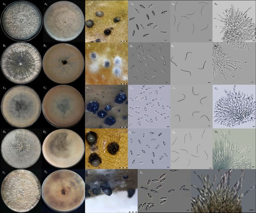
www.nature.com/scientificreports/

                                            Figure 1.  Morphological characteristics of Diaporthe species isolated from stem gray blight of H. polyrhizus.
                                            Group 1 (A1–A6): (A1) colony appearance, (A2) pigmentation, (A3) pycnidial conidiomata, (A4) α-conidia, (A5)
                                            β-conidia, (A6) conidiogenous cell for α-conidia; Group 2 (B1–B6): (B1) colony appearance, (B2) pigmentation,
                                            (B3) pycnidial conidiomata, (B4) α-conidia, (B5) β-conidia, (B6) conidiogenous cell for α-conidia; Group 3
                                            (C1–C6): (C1) colony appearance, (C2) pigmentation, (C3) pycnidial conidiomata, (C4) α-conidia, (C5) β-conidia,
                                            (C6) conidiogenous cell for α-conidia; Group 4 (D1–D6): (D1) colony appearance, (D2) pigmentation, (D3)
                                            pycnidial conidiomata, (D4) α-conidia, (D5) β-conidia, (D6) conidiogenous cell for α-conidia; Group 5 (E1–E5):
                                            (E1) colony appearance, (E2) pigmentation, (E3) pycnidial conidiomata, (E4) α-conidia, (E5) conidiogenous cell
                                            for α-conidia. Scale bar: A3–E3 = 1000 µm; A4–A6, B4–B6, C4–C6, D4–D6, E4–E5: 0.5 µm.

                                            Results
                                            Fungal isolation and morphological identification. A total of nine fungal isolates were recovered
                                            from nine gray blighted stems obtained from the different plants of H. polyrhizus. Of these, three isolates (DFP2,
                                            DFP3, and DFP4) were recovered from Bukit Kor, Terengganu and six isolates (DF1, DF2, DF3, DF7, DF9, and
                                            DF12) from the Cameron Highlands, Pahang, Malaysia. A species or isolate was recovered from a single lesion.
                                            In general, the fungal isolates produced whitish, grayish, or brownish colonies on potato dextrose agar (PDA)
                                            plates. Two types of conidia, namely, α- and β-conidia, were produced from the formation of pycnidial conidi-
                                            omata on carnation leaf agar (CLA). α-conidia were characterized as aseptate, hyaline, and fusiform with bi- or
                                            multi-guttulate, meanwhile, β-conidia were characterized as aseptate, hyaline, filiform, straight, or more often
                                            hamate, and lack guttule. The conidiogenous cells of α-conidia were phialidic, cylindrical, terminal, hyaline, and
                                            slightly tapered toward the end. However, in this study, the structure of the conidiogenous cells for β-conidia
                                            was not observed. Conidiophore was characterized as hyaline, branched, multiseptate, and filiform. Based on
                                            the described characteristics, the fungal isolates were tentatively identified as Diaporthe species. By sorting their
                                            morphological similarities and differences, the fungal isolates were classified into five groups of Diaporthe spe-
                                            cies (Fig. 1, Table 1).

          Scientific Reports |   (2021) 11:3907 |                https://doi.org/10.1038/s41598-021-83551-z                                                    2

Vol:.(1234567890)
www.nature.com/scientificreports/
Morphological characteristics
Pycnidial conidiomata Conidiophore of Conidiogenous cell of
Group/isolate Colony on PDA on CLA A
α-conidia A
β-conidia α-conidia α-conidia
Fusiform, slightly
tapered end, aseptate,
Filiform to hamate, asep-
Group 1 Abundant and whitish- Black and globose and hyaline Cylindrical phialides,
tate, and hyaline
DF1 brown aerial mycelia Presence of whitish Conidia with size of Hyaline, branched, and terminal, hyaline, and
Conidia with size of
DF3 Whitish-brown on the conidial masses exuda- 6.33 ± 0.68a × 1.98 ± 0.25a straight to slightly curve slightly tapered towards
24.57 ± 2.77b × 1.33 ± 0.29a
DFP4 lower surface tion µm end
µm
Bi/multi-guttulate with
size of 0.41 ± 0.07a µm
Ovoid with bluntly
rounded base end, asep-
Filiform to hamate, asep-
Cottony and whitish tate, and hyaline Cylindrical phialides,
Group 2 tate, and hyaline
aerial mycelium Conidia with size of Hyaline, branched, and terminal, hyaline, and
DF2 Black and globose Conidia with size of
Brownish-white on the 6.43 ± 0.55a × 2.38 ± 0.21b straight to slightly curve slightly tapered towards
DF12 17.34 ± 2.17a × 1.49 ± 0.34a
lower surface µm end
µm
Bi-guttulate with size of
1.53 ± 0.17c µm
Fusoid with bluntly
rounded on both ends,
Filiform to hamate, asep-
Cottony and brownish- aseptate, and hyaline Cylindrical phialides,
Group 3 tate, and hyaline
white aerial mycelia Conidia with size of Hyaline, branched, and terminal, hyaline, and
DFP2 Black and globose Conidia with size of
Brownish colour on the 6.00 ± 0.81a × 2.39 ± 0.35b straight to slightly curve slightly tapered towards
DF7 16.29 ± 4.22a × 1.20 ± 0.44a
lower surface µm end
µm
Bi-guttulate with size of
1.55 ± 0.13c µm
Fusiform with tapering
towards both ends,
Cottony and grayish- Filiform to hamate, asep-
Black and globose aseptate, and hyaline Cylindrical phialides,
white aerial mycelium tate, and hyaline
Group 4 Presence of whitish Conidia with size of Hyaline, branched, and terminal, hyaline, and
Whitish with gray- Conidia with size of
DF9 conidial masses exuda- 6.28 ± 0.64a × 2.57 ± 0.22b straight to slightly curve slightly tapered towards
patches on the lower 18.29 ± 2.26a × 1.21 ± 0.26a
tion µm end
surface µm
Bi-guttulate with size of
0.58 ± 0.07b µm
Fusiform with slightly
pointed ends, aseptate,
Cottony and brownish- Black and globose and hyaline Cylindrical phialides,
Group 5 white aerial mycelia Presence of whitish Conidia with size of Hyaline, branched, and terminal, hyaline, and
Not observed
DFP3 Yellowish-brown on the conidial masses exuda- 7.06 ± 0.55b × 2.47 ± 0.34b straight to slightly curve slightly tapered towards
lower surface tion µm end
Bi-guttulate with size of
0.40 ± 0.07a µm
Table 1. Morphological characteristics of five different groups of Diaporthe isolates recovered from stem gray
blight of H. polyrhizus. A Means ± standard deviation followed by different letters within the column are
significantly different (p < 0.05) according to Tukey’s test.
Molecular identification and phylogenetic analysis. The comparison of DNA sequences based on
ITS, TEF1-α, and β-tubulin demonstrated that the isolates were similar to the reference sequences of D. eugeniae,
D. phaseolorum, D. tectonendophytica, D. hongkongensis, and D. arecae from the Genbank database. The phylo-
genetic trees generated from each single gene had the same topology as the tree generated from the combined
genes of ITS, TEF1-α, and β-tubulin (Fig. 2) (Supplementary Information). The groupings of each single tree
demonstrated that all the isolates were clustered in the same clades as their respective species of Diaporthe
(D. eugeniae, D. phaseolorum, D. tectonendophytica, D. hongkongensis, and D. arecae). Isolates DF1, DF3, and
DFP4 were grouped with D. eugeniae CBS 444.82; isolates DF2 and DF12 with D. phaseolorum CBS113425 and
BDKHADRA-2; isolates DFP2 and DF7 with D. tectonendophytica MFLUCC 13-0471; and isolates DF9 and
DFP3 with D. hongkongensis CBS 115448 and D. arecae CBS 161.64, respectively. The result of the phylogenetic
analysis was in accordance with the molecular identification based on DNA sequences [Basic Local Alignment
Search (BLAST)], thus resolving the morphological identification. The isolates from group 1 were confirmed to
be D. eugeniae, group 2 was D. phaseolorum, group 3 was D. tectonendophytica, group 4 was D. hongkongensis,
and group 5 was D. arecae. The combined sequence matrix and phylogenetic tree were deposited in TreeBASE
(http://purl.org/phylo/treebase/phylows/study/TB2:S27649).
Pathogenicity test and comparative aggressiveness among Diaporthe isolates. The result of
pathogenicity test indicated that all isolates of the Diaporthe species recovered from the stem gray blight of H.
polyrhizus were pathogenic, exhibiting similar symptoms to those in the field (Fig. 3A1–A5). The tested isolates
showed typical symptoms of gray blight on the inoculated stems of H. polyrhizus. Initially, irregular yellowish
lesion surrounded by reddish border appeared on the wounded point (Fig. 3B1), which gradually turned into a
dark-brown sunken lesion and demonstrated dampening (Fig. 3B2). As the disease progressed, the lesion became
apparently dry and turned gray (Fig. 3B3). Then, it expanded periodically, and tiny black pycnidia appeared on
the area of the lesion (Fig. 3B4–B5). No symptoms developed on the control points.
Isolate DF1 (D. eugeniae) recorded the highest lesion length (10.25 ± 0.35 cm), whereas isolate DFP3 (D.
arecae) had the lowest (3.25 ± 0.35 cm) (Table 2). The means of the length lesion of the tested isolates were
Scientific Reports | (2021) 11:3907 | https://doi.org/10.1038/s41598-021-83551-z 3
Vol.:(0123456789)www.nature.com/scientificreports/
Diaporthe unshiuensis ZJUD52
87
Diaporthe unshiuensis ZJUD50
99 Diaporthe unshiuensis ZJUD51
Diaporthe sojae FAU644
62 100
Diaporthe sojae FAU599
DF7
Diaporthe tectonendophytica MFLUCC 13-0471
100 Group 3
93 DFP2
Diaporthe ueckerae FAU659
85
Diaporthe ueckerae FAU658
Diaporthe ueckerae FAU656
84
Diaporthe miriciae BRIP 56918a
100 Diaporthe miriciae BRIP 55662c
76 Diaporthe miriciae BRIP 54736j
78 Diaporthe novem CBS 127269
100 Diaporthe novem CBS 127270
52 Diaporthe novem CBS 127271
Diaporthe schini LGMF910
97
100 Diaporthe schini CBS 133181
Diaporthe helianthi CBS 344.94
100 Diaporthe helianthi CBS 592.81
100 Diaporthe masirevicii BRIP 57330
Diaporthe masirevicii BRIP 57892a
DF12
99
DF2
Group 2
99 Diaporthe phaseolorum CBS 113425
100
Diaporthe phaseolorum CBS 139281
Diaporthe phaseolorum BDKHADRA-2
100 Diaporthe sennae CFCC 51636
84 Diaporthe sennae CFCC 51637
68
Diaporthe pascoei BRIP 54847
Diaporthe litchicola BRIP 54900
Diaporthe fraxini-angustifoliae BRIP 54781
67
Diaporthe musigena CBS 129519
Diaporthe perseae CBS 151.73
DF1
DF3 Group 1
100
DFP4
Diaporthe eugeniae CBS 444.82
93
Diaporthe pseudomangiferae CBS 101339
97 Diaporthe pseudomangiferae CBS 388.89
Diaporthe arengae CBS 114979
67
100 Diaporthe pseudophoenicicola CBS 462.69
Diaporthe pseudophoenicicola CBS 176.77
DFP3
61 100
Diaporthe arecae CBS 535.75 Group 5
99 Diaporthe arecae CBS 161.64
59 Diaporthe pescicola MFLUCC 16-0106
99
Diaporthe pescicola MFLUCC 16-0105
100
Diaporthe pescicola MFLUCC 16-0107
55 DF9
Diaporthe hongkongensis CBS 115448
100 Group 4
Diaporthe hongkongensis ZJUD74
65
Diaporthe hongkongensis ZJUD78
80 Diaporthe oncostoma CBS 100454
Diaporthe oncostoma CBS 589.78
100
Diaporthe oncostoma CBS 109741
Diaporthe vaccinii CBS 160.32
68 100 Diaporthe vaccinii CBS 118571
92 Diaporthe vaccinii CBS 122112
98 Diaporthe amygdali CBS 126679
100 Diaporthe amygdali CBS 115620
Diaporthe amygdali CBS 111811
57
100 Diaporthe brasiliensis CBS 133183
Diaporthe brasiliensis LGMF926
65 Diaporthe caulivora CBS 127268
100 Diaporthe caulivora CBS 178.55
Diaporthe oxe CBS 133186
100 Diaporthe oxe CBS 133187
Diaporthella corylina CBS 121124
Paraphoma chlamydocopiosa BRIP 65168
Lasiodiplodia pseudotheobromae CBS116459
Nigrospora musae CBS 319.34
100 Arthrinium obovatum LC4940
0.1
Figure 2. Maximum-likelihood tree of Diaporthe species isolated from stem gray blight of H. polyrhizus
based on combined dataset of ITS, TEF1-α, and β-tubulin using Tamura and Nei model with 1000 bootstrap
replications. Isolates of the present study are presented in bold and other fungal genera are used as an outgroup.
Bootstrap values are shown at the nodes and the scale bar indicates the number of substitutions per position.
Scientific Reports | (2021) 11:3907 | https://doi.org/10.1038/s41598-021-83551-z 4
Vol:.(1234567890)www.nature.com/scientificreports/
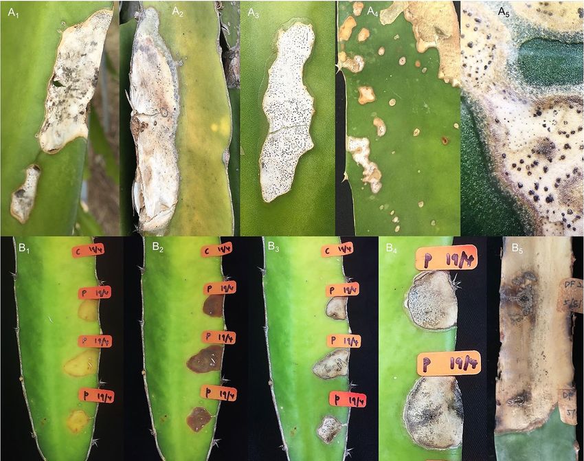
Figure 3. Stem gray blight of H. polyrhizus. (A1–A5) Disease symptoms observed in the fields. (B1) After 2 days
of inoculation, irregular yellowish lesions surrounded by reddish borders appeared. (B2) The lesions became
sunken and turned darker. (B3) The lesions apparently dry and turned to gray. (B4–B5) At later stage, the lesions
expanded resulting in the appearance of blighted stem with formation of tiny black pycnidia. C denotes control
and P represents treatment.
Species Isolate A
Lesion length (cm)
DF1 10.25 ± 0.35e
D. eugeniae DF3 5.50 ± 0.70c
DFP4 5.10 ± 0.84bc
DF2 7.50 ± 0.00d
D. phaseolorum
DF12 7.75 ± 0.35d
DF7 8.25 ± 0.35d
D. tectonendophytica
DFP2 3.45 ± 0.70ab
D. hongkongensis DF9 3.50 ± 0.00ab
D. arecae DFP3 3.25 ± 0.35a
Control 0.00 ± 0.00f
Table 2. Lesion length recorded by Diaporthe isolates after 3 weeks of inoculation on stems of H. polyrhizus.
A
Mean ± standard deviation followed by different letters within the column is significantly different (p < 0.05)
according to Tukey’s test.
Scientific Reports | (2021) 11:3907 | https://doi.org/10.1038/s41598-021-83551-z 5
Vol.:(0123456789)www.nature.com/scientificreports/
significantly different compared with the control at p < 0.05. The tested isolates of Diaporthe exhibited variability
in length lesion after 3 weeks of inoculation on the stems of H. polyrhizus. The same Diaporthe species were reiso-
lated from the symptomatic inoculated stems of H. polyrhizus, and their identities were reconfirmed by compar-
ing the macroscopic and microscopic characteristics with the original cultures, thus fulfilling Koch’s postulates.
Discussion
The present study reported on stem gray blight, which is a new emerging disease infecting H. polyrhizus plan-
tations in Malaysia. The five species of Diaporthe, namely, D. eugeniae (group 1), D. phaseolorum (group 2),
D. tectonendophytica (group 3), D. hongkongensis (group 4), and D. arecae (group 5), were identified to be the
causal agents of the disease. The Diaporthe species may act as a plant pathogen or a saprophyte or an endophytic
symbiont31–34, however, several studies have reported that it is the genus responsible for multiple destructive
diseases, such as root and fruit rots, dieback, stem cankers, leaf spots, leaf and pod blights, and seed decay31,33,35–39.
A total of nine Diaporthe isolates were recovered from the blighted stem of H. polyrhizus. Based on their
morphological characteristics, all the isolates produced both α-conidia and β-conidia, except for the D. arecae
isolate, of which β-conidia was not observed. α- and β-conidia are the key characteristics for the identification
of Diaporthe33,40. The formation of β-conidia can sometimes be rare or absent in certain species of Diaporthe41.
According to Tuset and P ortilla42 and Diogo et al.43, for some Diaporthe species (e.g. Phomopsis amygdali), the
formation of β-conidia can only be observed in pycnidia on the host but not in pycnidia in the culture plate.
Based on the similarities and differences of their macroscopic and microscopic characteristics, the isolates
were assigned to five different groups. Among the groups, significant differences were observed in the number of
α-conidia guttules and their size (Table 1). Gomes et al.34 revealed that both characteristics can be varied among
the Diaporthe species. The isolates from group 1 (D. eugeniae) tended to produce bi- and multi-guttules, whereas
the other isolates only produced bi-guttules of α-conidia. The size of the guttules of α-conidia varied among the
groups. The isolates from groups 1 and 5 (D. eugeniae and D. arecae) produced significantly smaller guttules
compared with those produced by isolates from groups 2, 3, and 5 (D. phaseolorum, D. tectonendophytica, and
D. hongkongensis) (Table 1). The guttule is defined as a small drop or particle in a spore resembling a n ucleus44.
Moreover, the morphology of α-conidia of the D. eugeniae, D. hongkongensis, and D. arecae isolates was tapered
toward the ends compared with the D. phaseolorum and D. tectonendophytica isolates, the ends of which were
bluntly rounded (Fig. 1). This finding was in agreement with those of Santos et al.38, Dissanayake et al.45, Doilom
et al.46, and Lim et al.47. A significant difference was also observed in the length of β-conidia, of which the D.
eugeniae isolates produced longer β-conidia than other isolates from different groups. Conidial mass exudation
can be observed in the isolates of D. eugeniae, D. hongkongensis, and D. arecae. Contrarily, it was not observed in
the isolates of D. phaseolorum and D. tectonendophytica. According to Machowicz-Stefaniak et al.48, the Diaporthe
species require temperatures ranging from 22 to 28 °C for the optimal growth, sporulation, and rate of conidia
release of conidiomata. As applied in the present study, the addition of carnation leaves to the growing medium
as substrates has been recommended to improve the sporulation of the Diaporthe species49,50.
Aside from the microscopic characteristic, the cultural characteristics of all isolates in this study also varied
among the groups. The color of the colonies ranged from whitish, grayish, brownish, to olive green. Due to this
inconsistency, cultural characteristic is commonly considered as a less important criterion in distinguishing spe-
cies within Diaporthe as it can be influenced by several environmental factors, such as light and temperature34.
Based on the results obtained, morphological characteristics alone were insufficient to identify all the isolates
up to the species level due to the complexity of the genus. This finding was in agreement with that of Lim et al.47
who revealed that the morphological method alone is not informative for the species identification of Diaporthe
due to pleomorphism and overlapping c haracteristics43,51,52.
With the advances in molecular techniques, DNA sequences and multigene phylogenetic analysis of ITS,
TEF1-α, and β-tubulin were employed to support the morphological identification of the Diaporthe isolates in
this study. The result of the BLAST search and phylogenetic inference indicated that the use of all the three genes
resolved identification of the Diaporthe isolates. Aside from the present study, ITS, TEF1-α, and β-tubulin were
extensively applied to delineate species within Diaporthe46,53,54. The ITS region served as an identification guide
for the Diaporthe species33. It was also considered as a fungal barcode in distinguishing genera and species owing
to its easy amplification and ability to provide preliminary screening of fungal classification55,56. However, the tree
constructed based on ITS sequences alone may be doubtful and not demonstrate clear phylogenetic relationships
due to the lack of interspecific variation or even deceptive in some f ungi57. Thus, TEF1-α and β-tubulin were
added to support the phylogenetic analysis of ITS in delimiting the species of the Diaporthe isolates. TEF1-α
comprises an essential part of the protein translation machinery, and highly informative at the species level;
moreover, non-orthologous copies have not been detected in Diaporthe58. β-tubulin was utilized as an alterna-
tive phylogenetic marker to specify Diaporthe as it contains fewer ambiguously aligned regions and exhibits less
homoplasy among the genus59. Collectively, phylogenetic analysis of a combined dataset of ITS, TEF1-α, and
β-tubulin was conducted in this study to overcome the ambiguity that could have emerged in the single gene
analysis. Santos et al.60 stated that the combined phylogenetic tree commonly provides a better resolution for
the identification of the Diaporthe species compared with the single gene analysis.
All the tested isolates of Diaporthe exhibited varying lengths of lesion on the inoculated stems of H. polyrhi-
zus, of which isolate DF1 (D. eugeniae) was found to be the most virulent. The fungus can act as a pathogen or a
saprophyte and was reported to cause stem-end rot on mango (Mangifera indica)47. It also occurs as a saprophyte
on cloves (Eugenia aromatica)34. This study discovered a new host and disease caused by D. eugeniae. The asso-
ciation of D. phaseolorum with dragon fruit was not new, because recently, this pathogen was reported to cause
stem rot on Hylocereus undatus in Bangladesh12. However, the symptoms described were slightly different from
those observed in the present study. It appeared as a yellow spot with a chlorotic halo in the previous report, but
Scientific Reports | (2021) 11:3907 | https://doi.org/10.1038/s41598-021-83551-z 6
Vol:.(1234567890)www.nature.com/scientificreports/
in the present study, chlorotic halo was not observed; rather, a reddish border surrounded the lesion. Similarly,
gray to black pycnidia were scattered on the surface of the lesion. Aside from the dragon fruit, D. phaseolorum
was reported as a causal agent of pod and stem blight, stem canker, and seed rot on soybean and trunk disease
on grapevine38,45,61,62. It was also found to be an endophyte on Kandelia candel by Cheng et al.63.
Similar to D. eugeniae, the present study highlighted H. polyrhizus as a new host associated with D. tectonen-
dophytica as it causes stem gray blight. Contrarily, a study by Doilom et al.46 demonstrated the role of D. tectonen-
dophytica as an endophyte occurring on teak (Tectona grandis) in Thailand. The capability of D. hongkongensis
to act as a pathogen is undeniable as the fungus has been reported to cause severe diseases on a number of host
plants, such as stem-end rot on kiwifruit64, dieback on grapevine45, and shoot canker on pear65. Meanwhile, D.
arecae has been reported to be pathogenic on M. indica47, Areca catechu34, and Citrus66. D. hongkongensis and D.
arecae were first reported on H. polyrhizus worldwide especially in Malaysia.
The occurrence of the disease in two different locations in Malaysia indicates its possibility to spread world-
wide. Aside from Diaporthe, dragon fruits in Malaysia also suffer from multiple diseases caused by other fungi.
Among these diseases are anthracnose caused by C. gloeosporioides22,23 and C. truncatum24; stem necrosis by
Curvularia lunata25; stem canker by N. dimidiatum26; stem rot by Fusarium proliferatum27 and Fusarium fuji-
kuroi28; reddish brown spot by Nigrospora lacticolonia and N. sphaerica30; and stem blight by F. oxysporum29.
This study provides overview of the five different species of Diaporthe causing stem gray blight on H. polyrhi-
zus in Malaysia. It improves our knowledge on the symptomatology of the disease and identity of the pathogens
through morphological and molecular analyses. The findings may be essential to strategize effective disease
management for stem gray blight on H. polyrhizus and for quarantine restrictions.
Materials and methods
Fungal isolation. In November 2017 and July 2018, nine gray blighted stems from the different plants of H.
polyrhizus were collected from Bukit Kor, Terengganu, Malaysia, and the Cameron Highlands, Pahang, Malaysia.
The symptomatic samples were brought back to the laboratory for isolation. One lesion per stem exhibiting the
same symptom was selected for fungal isolation. The lesion consisting of diseased and healthy parts was excised
(1.5 cm2) and surface-sterilized with 70% ethanol for 3 min. Then, the samples were soaked in 10% sodium
hypochlorite (1% NaOCl) for 3 min and rinsed with sterile distilled water three times consecutively for 1 min
each. The sterilized samples were air-dried on the sterile filter papers before being transferred to PDA plates. The
inoculated plates were incubated at 25 °C ± 2 °C for 2 to 3 days. Pure cultures of fungal isolates were obtained via
hyphal tip isolation and were used for morphological and molecular analyses.
Morphological identification. Each fungal isolate obtained was cultured on PDA and incubated at
25 °C ± 2 °C for 7 days. Macroscopic characteristics, such as colony appearance and pigmentation, were recorded.
CLA was utilized to induce the formation of pycnidial conidiomata, and the inoculated plates were incubated at
25 °C ± 2 °C for 7 days. The morphology of α- and β-conidia was observed from the pycnidial conidiomata. The
other microscopic characteristics observed were conidiophores and conidiogenous cells. The length and width
of 30 randomly selected conidia and the size of the guttules of 30 randomly selected α-conidia were measured
and recorded. The differences in the length and width of conidia and the size of the guttules of α-conidia were
evaluated via one-way ANOVA. In addition, the means of both parameters were compared via Tukey’s test
(p < 0.05) using the IBM SPSS Statistics software version 24.
Molecular identification and phylogenetic analysis. The identity of all the fungal isolates was further
confirmed by molecular characterization. The isolates were grown in potato dextrose broth (PDB) and incubated
at 25 °C ± 2 °C for 7 days. Fungal mycelia from PDB were homogenized under liquid nitrogen to obtain fine
powder. A total of 60 mg fine powder was transferred into a 1.5 mL microcentrifuge tube, and the genomic DNA
of the fungal isolates was extracted using the Invisorb Spin Plant Mini Kit (Stratec Biomedical AG, Birkenfeld,
Germany), following the manufacturer’s protocols. The primers of ITS5/ITS467, EF1-728/EF1-98668, and BT2a/
BT2b69 were used for the amplification of ITS, TEF1-α, and β-tubulin, respectively. A total of 50 µL reaction
mixture was prepared, which contained 8 µL of green buffer (Promega, USA), 8 µL of MgCl2 (Promega, USA), 1
µL of deoxynucleotide triphosphate polymerase (dNTP) (Promega, USA), 8 µL of each primer (Promega, USA),
0.3 µL of Taq polymerase (Promega, USA), 1 µL of genomic DNA, and sterile distilled water. Polymerase chain
reaction (PCR) was performed using MyCycler Thermal Cycler (BioRad, Hercules, USA) under the following
conditions: initial denaturation at 95 °C for 4 min, followed by 35 cycles of denaturation at 95 °C for 35 s, anneal-
ing at 54 °C (ITS)/57 °C (TEF1-α)/58 °C (β-tubulin) for 1 min, extension at 72 °C for 90 s, and final extension
at 72 °C for 10 min. The PCR product was separated by running it in 1.0% agarose gel (Promega, USA) stained
with HealthView Nucleic Acid Stain (Genomics, Taiwan) at 90 V and 400 mA for 90 min. The 100 bp DNA lad-
der (Thermo Scientific, USA) was used as a marker to estimate the size of the amplified PCR products. The PCR
products were sent to a service provider (First BASE Laboratories Sdn Bhd, Seri Kembangan, Malaysia) for DNA
sequencing.
The obtained sequences were aligned using the Molecular Evolutionary Genetic Analysis software (MEGA7)70.
After pairwise alignment, the BLAST algorithm (https://blast.ncbi.nlm.nih.gov/Blast.cgi) was used to compare
the generated consensus sequences with other sequences in the GenBank database. The sequences obtained were
deposited in the GenBank database.
The isolates in the present study and reference sequences used in the phylogenetic analysis are presented in
Table 3. Multiple sequence alignments of fungal isolates and reference isolates were generated using the MEGA7
software. Phylogenetic analysis was conducted using the maximum likelihood (ML) method in MEGA7. The
Scientific Reports | (2021) 11:3907 | https://doi.org/10.1038/s41598-021-83551-z 7
Vol.:(0123456789)www.nature.com/scientificreports/
GenBank accession no.
Species Isolate Host Locality ITS TEF1-α β-tubulin References
D. amygdali CBS 126679EP Prunus dulcis Portugal KC343022 KC343748 KC343990 Gomes et al.34
D. amygdali CBS 111811 Vitis vinifera South Africa KC343019 KC343745 KC343987 Gomes et al.34
D. amygdali CBS 115620 Prunus persica USA KC343020 KC343746 KC343988 Gomes et al.34
D. arecae CBS 161.64 EI
Arecae catechu India KC343032 KC343758 KC344000 Gomes et al.34
D. arecae CBS 535.75 Citrus sp. Suriname KC343033 KC343759 KC344001 Gomes et al.34
Bukit Kor, Terengganu,
Diaporthe sp. (Group 5) DFP3 Hylocereus polyrhizus MN862382 MN889938 MN889947 This study
Malaysia
D. arengae CBS 114979ET Arenga engleri Hong Kong KC343034 KC343760 KC344002 Gomes et al.34
D. brasiliensis CBS 133183ET Aspidosperma tomentosum Brazil KC343042 KC343768 KC344010 Gomes et al.34
D. brasiliensis LGMF 926 Aspidosperma tomentosum Brazil KC343043 KC343769 KC344011 Gomes et al.34
D. caulivora CBS 127268EN Glycine max Croatia KC343045 KC343771 KC344013 Gomes et al.34
D. caulivora CBS 178.55 Glycine soja Canada KC343046 KC343772 KC344014 Gomes et al.34
D. eugeniae CBS 444.82 Eugenia aromatica Indonesia KC343098 KC343824 KC344066 Gomes et al.34
Cameron Highlands,
Diaporthe sp. (Group 1) DF1 Hylocereus polyrhizus MN862375 MN889932 MN889940 This study
Pahang, Malaysia
Cameron Highlands,
Diaporthe sp. (Group 1) DF3 Hylocereus polyrhizus MN862377 MN889935 MN889944 This study
Pahang, Malaysia
Bukit Kor, Terengganu,
Diaporthe sp. (Group 1) DFP4 Hylocereus polyrhizus MN862383 MN889939 MN889948 This study
Malaysia
D. fraxini-angustifoliae BRIP 54781EI Fraxinus angustifolia Australia JX862528 JX862534 KF170920 Tan et al.73
D. helianthi CBS 592.81ET Helianthus annuus Serbia KC343115 KC343841 KC344083 Gomes et al.34
D. helianthi CBS 344.94 Helianthus annuus – KC343114 KC343840 KC344082 Gomes et al.34
D. hongkongensis CBS 115448ET Dichroa febrifuga Hong Kong KC343119 KC343845 KC344087 Gomes et al.34
D. hongkongensis ZJUD74 Citrus unshiu China KJ490609 KJ490488 KJ490430 Huang et al.66
D. hongkongensis ZJUD78 Citrus unshiu China KJ490613 KJ490492 KJ490434 Huang et al.66
Cameron Highlands,
Diaporthe sp. (Group 4) DF9 Hylocereus polyrhizus MN862379 MN889933 MN889941 This study
Pahang, Malaysia
D. litchicola BRIP 54900 EH
Litchi chinensis Australia JX862533 JX862539 KF170925 Tan et al.73
D. masirevicii BRIP 57892aEH Helianthus annuus Australia KJ197276 KJ197239 KJ197257 Thompson et al.74
D. masirevicii BRIP 57330 Chrysanthemoides monilifera Australia KJ197275 KJ197237 KJ197255 Thompson et al.74
D. miriciae BRIP 54736jEH Helianthus annuus Australia KJ197282 KJ197244 KJ197262 Thompson et al.74
D. miriciae BRIP 55662c Glycine max Australia KJ197283 KJ197245 KJ197263 Thompson et al.74
D. miriciae BRIP 56918a Vigna radiata Australia KJ197284 KJ197246 KJ197264 Thompson et al.74
D. musigena CBS 129519ET Musa sp. Australia KC343143 KC343869 KC344111 Gomes et al.34
D. novem CBS 127270 ET
Glycine max Croatia KC343156 KC343882 KC344124 Gomes et al.34
D. novem CBS 127269 Glycine max Croatia KC343155 KC343881 KC344123 Gomes et al.34
D. novem CBS 127271 Glycine max Croatia KC343157 KC343883 KC344125 Gomes et al.34
D. oncostoma CBS 589.78 Robinia pseudoacacia France KC343162 KC343888 KC344130 Gomes et al.34
D. oncostoma CBS 100454 Robinia pseudoacacia Germany KC343160 KC343886 KC344128 Gomes et al.34
D. oncostoma CBS 109741 Robinia pseudoacacia Russia KC343161 KC343887 KC344129 Gomes et al.34
D. oxe CBS 133186 ET
Maytenus ilicifolia Brazil KC343164 KC343890 KC344132 Gomes et al.34
D. oxe CBS 133187 Maytenus ilicifolia Brazil KC343165 KC343891 KC344133 Gomes et al.34
D. pascoei BRIP 54847EI Perseae americana Australia JX862532 JX862538 KF170924 Tan et al.73
D. perseae CBS 151.73 Perseae americana Netherlands KC343173 KC343899 KC344141 Gomes et al.34
D. pescicola MFLUCC 16-0105EH Prunus persica China KU557555 KU557623 KU557579 Dissanayake et al.75
D. pescicola MFLUCC 16-0106 Prunus persica China KU557556 KU557624 KU557580 Dissanayake et al.75
D. pescicola MFLUCC 16-0107 Prunus persica China KU557557 KU557625 KU557581 Dissanayake et al.75
D. phaseolorum CBS 139281 EP
Phaseolus vulgaris USA KJ590738 KJ590739 KJ610893 Udayanga et al.76
D. phaseolorum CBS 113425 Olearia cf. rani New Zealand KC343174 KC343900 KC344142 Gomes et al.34
D. phaseolorum BDKHADRA-2 Hylocereus undatus Bangladesh MH714560 KC343902 KC344144 Karim et al.12
Cameron Highlands,
Diaporthe sp. (Group 2) DF2 Hylocereus polyrhizus MN862376 MN889931 MN889942 This study
Pahang, Malaysia
Cameron Highlands,
Diaporthe sp. (Group 2) DF12 Hylocereus polyrhizus MN862380 MN889936 MN889945 This study
Pahang, Malaysia
D. pseudomangiferae CBS 101339ET Mangifera indica Dominican Republic KC343181 KC343907 KC344149 Gomes et al.34
D. pseudomangiferae CBS 388.89 Mangifera indica Mexico KC343182 KC343908 KC344150 Gomes et al.34
D. pseudophoenicicola CBS 462.69ET Phoenix dactylifera Spain KC343184 KC343910 KC344152 Gomes et al.34
D. pseudophoenicicola CBS 176.77 Mangifera indica Iraq KC343183 KC343909 KC344151 Gomes et al.34
Continued
Scientific Reports | (2021) 11:3907 | https://doi.org/10.1038/s41598-021-83551-z 8
Vol:.(1234567890)www.nature.com/scientificreports/
GenBank accession no.
Species Isolate Host Locality ITS TEF1-α β-tubulin References
D. schini CBS 133181ET Schinus terebinthifolius Brazil KC343191 KC343917 KC344159 Gomes et al.34
D. schini LGMF 910 Schinus terebinthifolius Brazil KC343192 KC343918 KC344160 Gomes et al.34
D. sennae CFCC 51636 EH
Senna bicapsularis China KY203724 KY228885 KY228891 Yang et al.77
D. sennae CFCC 51637 Senna bicapsularis China KY203725 KY228886 KY228892 Yang et al.77
D. sojae FAU 599 EH
Glycine max USA KJ590728 KJ590767 KJ610883 Udayanga et al.76
D. sojae FAU 644 Glycine max USA KJ590730 KJ590769 KJ610885 Udayanga et al.76
D. tectonendophytica MFLUCC 13-0471EH Tectona grandis Thailand KU712439 KU749367 KU743986 Doilom et al.46
Cameron Highlands,
Diaporthe sp. (Group 3) DF7 Hylocereus polyrhizus MN862378 MN889934 MN889943 This study
Pahang, Malaysia
Bukit Kor, Terengganu,
Diaporthe sp. (Group 3) DFP2 Hylocereus polyrhizus MN862381 MN889937 MN889946 This study
Malaysia
D. ueckerae FAU 656 EH
Cucumis melo USA KJ590726 KJ590747 KJ610881 Udayanga et al.76
D. ueckerae FAU 659 Cucumis melo USA KJ590724 KJ590745 KJ610879 Udayanga et al.76
D. ueckerae FAU 658 Cucumis melo USA KJ590725 KJ590746 KJ610880 Udayanga et al.76
D. unshiuensis ZJUD 52 Citrus unshiu China KJ490587 KJ490466 KJ490408 Huang et al.66
D. unshiuensis ZJUD 50 Citrus japonica China KJ490585 KJ490464 KJ490406 Huang et al.66
D. unshiuensis ZJUD 51 Citrus japonica China KJ490586 KJ490465 KJ490407 Huang et al.66
D. vaccinii CBS 160.32ET Oxycoccus macrocarpos USA KC343228 KC343954 KC344196 Gomes et al.34
D. vaccinii CBS 118571 Vaccinium corymbosum USA KC343223 KC343949 KC344191 Gomes et al.34
D. vaccinii CBS 122112 Vaccinium macrocarpon USA KC343224 KC343950 KC344192 Gomes et al.34
Diaporthella corylina CBS 121124 Corylus sp. China KC343004 KC343730 KC343972 Gomes et al.34
Lasiodiplodia pseudothe-
CBS 116459 ET
Gmelina arborea Costa Rica EF622077 EF622057 EU673111 Alves et al.78
obromae
Nigrospora musae CBS 319.34EH Musa paradisiaca Australia KX986076 KY019419 KY019455 Wang et al.79
Arthrinium obovatum CGMCC 3.18331 EH
Lithocarpus sp. China KY494696 KY705095 KY705166 Wang et al.80
Paraphoma chlamydocopiosa BRIP 65168 EH
Tanacetum cinerariifolium Australia KU999072 KU999080 KU999084 Moslemi et al.81
Table 3. Isolates in the present study and reference isolates used in the phylogenetic analysis. EP ex-epitype
culture, EI ex-isotype culture, ET ex-type culture, EN ex-neotype culture, EH ex-holotype culture.
Tamura-Nei model71 was used to generate the ML trees based on a single and combined genes of ITS, TEF1-α,
and β-tubulin with 1000 bootstrap r eplications72.
Pathogenicity test. The pathogenicity test was conducted on 18 healthy stems of H. polyrhizus for all the
obtained fungal isolates. Conidial suspension was prepared by flooding the 7-day-old PDA culture with sterile
distilled water, and the concentration was adjusted to 1 × 10⁶ conidia/mL using a hemocytometer (Weber, Ted-
dington, UK). The stems were surface-sterilized with 70% ethanol, and 0.1 mL of conidial suspension was uti-
lized for inoculation using a disposable needle and syringe. Likewise, the control points were treated with sterile
distilled water. On each stem, three points were used to inoculate fungal isolate and one point for control. Each
fungal isolate was tested in three replicates, and the pathogenicity tests were conducted twice. All the inocu-
lated plants were placed in a plant house in the School of Biological Sciences, USM, and incubated at 26–32 °C
for 21 days. The progression of the disease symptom was observed daily. The lesion length was measured and
recorded after 3 weeks of inoculation. The differences in the lesion length were evaluated via one-way ANOVA,
and the means were compared via Tukey’s test (p < 0.05) using the IBM SPSS Statistics software version 24. For
the fulfillment of Koch’s postulates, the fungal isolates were reisolated from symptomatic inoculated stems and
reidentified by morphological characteristics.
Received: 26 October 2020; Accepted: 3 February 2021
References
1. Ismail, N. S. M., Ramli, N., Hani, N. M. & Meon, Z. Extraction and characterization of pectin from dragon fruit (Hylocereus
polyrhizus) using various extraction conditions. Sains Malays. 41, 41–45 (2012).
2. Abdul Razak, U. N. A., Taha, R. M., Che Musa, S. A. N. I. & Mohamed, N. Detection of betacyanins pigment stability from Hyloce-
reus polyrhizus (Weber) Britton & Rose fruit pulp and peel for possible use as natural coating. Pigm. Resin Technol. 46, 303–308
(2017).
3. Hoa, T. T., Clark, C. J., Waddell, B. C. & Woolf, A. B. Postharvest quality of dragon fruit (Hylocereus undatus) following disinfesting
hot air treatments. Postharvest Biol. Technol. 41, 62–69 (2006).
4. Ruzainah, A. J., Ahmad, R., Nor, Z. & Vasudevan, R. Proximate analysis of dragon fruit (Hylocereus polyhizus). Am. J. Appl. Sci. 6,
1341–1346 (2009).
Scientific Reports | (2021) 11:3907 | https://doi.org/10.1038/s41598-021-83551-z 9
Vol.:(0123456789)www.nature.com/scientificreports/
5. Patel, J. S. & Zhang, S. First report of Alternaria blight of pitahaya (Hylocereus undatus) caused by Alternaria sp. in South Florida
of the United States. Plant Dis. 101, 1046 (2017).
6. He, P. F., Ho, H., Wu, X. X., Hou, M. S. & He, Y. Q. Bipolaris cactivora causing fruit rot of dragon fruit imported from Vietnam.
Plant Pathol. Quar. 2, 31–35 (2012).
7. Valencia-Botín, A. J., Sandoval-Islas, J. S., Cárdenas-Soriano, E., Michailides, T. J. & Rendón-Sánchez, G. Botryosphaeria dothidea
causing stem spots on Hylocereus undatus in Mexico. Plant Pathol. 52, 803 (2003).
8. Ma, W. J. et al. First report of anthracnose disease on young stems of Bawanghua (Hylocereus undatus) caused by Colletotrichum
gloeosporioides in China. Plant Dis. 98, 991 (2014).
9. Zhao, H. J. et al. First report of red dragon fruit (Hylocereus polyrhizus) anthracnose caused by Colletotrichum siamense in China.
Plant Dis. 102, 1175 (2018).
10. Abirammi, K. et al. Occurrence of anthracnose disease caused by Colletotrichum siamense on dragon fruit (Hylocereus undatus)
in Andaman Islands, India. Plant Dis. 103, 768 (2019).
11. Guo, L. W. et al. First report of dragon fruit (Hylocereus undatus) anthracnose caused by Colletotrichum truncatum in China. J.
Phytopathol. 162, 272–275 (2014).
12. Karim, M. M. et al. Occurrence of stem rot disease of Hylocereus undatus in Bangladesh. Indian Phytopathol. 72, 545–549 (2019).
13. Wright, E. R., Rivera, M. C., Ghirlanda, A. & Lori, G. A. Basal rot of Hylocereus undatus caused by Fusarium oxysporum in Buenos
Aires, Argentina. Plant Dis. 91, 323 (2007).
14. Rita, W. S., Suprapta, D. N., Sudana, I. M. & Swantara, I. M. D. First report on Fusarium solani, a pathogenic fungus causing stem
rot disease on dragon fruits (Hylocereus sp.) in Bali. J. Biol. Agric. Healthc. 3, 93–99 (2013).
15. Guo, L. W., Wu, Y. X., Mao, Z. C., Ho, H. H. & He, Y. Q. Storage rot of dragon fruit caused by Gilbertella persicaria. Plant Dis. 96,
1826 (2012).
16. Briste, P. S. et al. First report of dragon fruit stem canker caused by Lasiodiplodia theobromae in Bangladesh. Plant Dis. 103, 2686
(2019).
17. Awang, Y. B., Abdul Ghani, M. A., Sijam, K., Mohamad, R. B. & Hafiza, Y. Effect of postharvest application of calcium chloride on
brown rot and quality of red-flesh dragon fruit (Hylocereus polyrhizus). In International Symposium on Underutilized Plant Species:
Crops for the Future-Beyond Food Security, vol. 1. (ed. Massawe, F., Mayes, S. & Alderson, P.) 763–771 (International Society for
Horticultural Science, Leuven, 2013).
18. Yi, R. H., Mo, J. J., Wu, F. F. & Chen, J. Fruit internal brown rot caused by Neoscytalidium dimidiatum on pitahaya in Guangdong
province, China. Aust. Plant Dis. Notes 10, 13 (2015).
19. Sanahuja, G., Lopez, P. & Palmateer, A. J. First report of Neoscytalidium dimidiatum causing stem and fruit canker of Hylocereus
undatus in Florida. Plant Dis. 100, 1499 (2016).
20. Liu, F., Wu, J. B., Zhan, R. L. & Ou, X. C. First report of reddish-brown spot disease on pitaya caused by Nigrospora sphaerica in
China. Plant Dis. 100, 1792 (2016).
21. Zheng, F. et al. First report of southern blight in pitaya (Hylocereus undatus) caused by Sclerotium rolfsii in China. Plant Dis. 102,
441 (2018).
22. Mohd, M. H., Hew, P. Y., Maziah, Z., Nagao, H. & Salleh, B. Aethiology and symptomatology of anthracnose caused by Colle-
totrichum gloeosporioides on dragon fruit (Hylocereus polyrhizus) in Malaysia. In The Sixth Regional IMT-GT Uninet Conference
(Penang, Malaysia, 2008).
23. Masyahit, M., Sijam, K., Awang, Y. & Satar, M. G. M. The first report of the occurrence of anthracnose disease caused by Colletotri-
chum gloeosporioides (Penz.) Penz. & Sacc. on dragon fruit (Hylocereus spp.) in Peninsular Malaysia. Am. J. Appl. Sci. 6, 902–912
(2009).
24. Iskandar Vijaya, S., Mohd Anuar, I. S. & Zakaria, L. Characterization and pathogenicity of Colletotrichum truncatum causing stem
anthracnose of red-fleshed dragon fruit (Hylocereus polyrhizus) in Malaysia. J. Phytopathol. 163, 67–71 (2015).
25. Mohd, M. H., Salleh, B. & Latiffah, Z. First report of Curvularia lunata on red-fleshed dragon fruit (Hylocereus polyrhizus) in
Malaysia. Plant Dis. 93, 971 (2009).
26. Mohd, M. H., Salleh, B. & Zakaria, L. Identification and molecular characterizations of Neoscytalidium dimidiatum causing stem
canker of red-fleshed dragon fruit (Hylocereus polyrhizus) in Malaysia. J. Phytopathol. 161, 841–849 (2013).
27. Mohd, M. H., Salleh, B. & Latiffah, Z. Characterization and pathogenicity of Fusarium proliferatum causing stem rot of Hylocereus
polyrhizus in Malaysia. Ann. Appl. Biol. 163, 269–280 (2013).
28. Mohd, M. H., Nurul Faziha, I., Nik Mohamad Izham, M. N. & Latiffah, Z. Fusarium fujikuroi associated with stem rot of red-fleshed
dragon fruit (Hylocereus polyrhizus) in Malaysia. Ann. Appl. Biol. 170, 434–446 (2017).
29. Mohd Hafifi, A. B., Kee, Y. J. & Mohd, M. H. First report of Fusarium oxysporum as a causal agent of stem blight of red-fleshed
dragon fruit (Hylocereus polyrhizus) in Malaysia. Plant Dis. 103, 1040 (2019).
30. Kee, Y. J. et al. First report of reddish-brown spot disease of red-fleshed dragon fruit (Hylocereus polyrhizus) caused by Nigrospora
lacticolonia and Nigrospora sphaerica in Malaysia. Crop Prot. 122, 165–170 (2019).
31. Uecker, F. A. A World list of Phomopsis names with notes on nomenclature, morphology and biology. Mycol. Mem. 13, 1–231
(1988).
32. Rossman, A. Y., Farr, D. F. & Castlebury, L. A. A review of the phylogeny and biology of the Diaporthales. Mycoscience 48, 135–144
(2007).
33. Udayanga, D. et al. The genus Phomopsis: Biology, applications, species concepts and names of common phytopathogens. Fungal
Divers. 50, 189 (2011).
34. Gomes, R. R. et al. Diaporthe: A genus of endophytic, saprobic and plant pathogenic fungi. Persoonia 31, 1–41 (2013).
35. Mostert, L., Crous, P. W., Kang, J. C. & Phillips, A. J. Species of Phomopsis and a Libertella sp. occurring on grapevines with specific
reference to South Africa: Morphological, cultural, molecular and pathological characterization. Mycologia 93, 146–167 (2001).
36. van Rensburg, J. C. J., Lamprecht, S. C., Groenewald, J. Z., Castlebury, L. A. & Crous, P. W. Characterisation of Phomopsis spp.
associated with die-back of rooibos (Aspalathus linearis) in South Africa. Stud. Mycol. 55, 65–74 (2006).
37. Rehner, S. A. & Uecker, F. A. Nuclear ribosomal internal transcribed spacer phylogeny and host diversity in the coelomycete
Phomopsis. Can. J. Bot. 72, 1666–1674 (1994).
38. Santos, J. M., Vrandečić, K., Ćosić, J., Duvnjak, T. & Phillips, A. J. L. Resolving the Diaporthe species occurring on soybean in
Croatia. Persoonia 27, 9–19 (2011).
39. Díaz, G. A. et al. Identification and characterization of Diaporthe ambigua, D. australafricana, D. novem, and D. rudis causing a
postharvest fruit rot in kiwifruit. Plant Dis. 101, 1402–1410 (2017).
40. Sutton, B. C. The Coelomycetes. Fungi imperfecti with pycnidia, acervuli and stromata. (Commonwealth Mycological Institute, Kew,
England, 1980).
41. Hilário, S. et al. Diaporthe species associated with twig blight and dieback of Vaccinium corymbosum in Portugal, with description
of four new species. Mycologia 112, 293–308 (2020).
42. Tuset, J. J. & Portilla, M. A. T. Taxonomic status of Fusicoccum amygdali and Phomopsis amygdalina. Can. J. Bot. 67, 1275–1280
(1989).
43. Diogo, E. L., Santos, J. M. & Phillips, A. J. Phylogeny, morphology and pathogenicity of Diaporthe and Phomopsis species on almond
in Portugal. Fungal Divers. 44, 107–115 (2010).
Scientific Reports | (2021) 11:3907 | https://doi.org/10.1038/s41598-021-83551-z 10
Vol:.(1234567890)www.nature.com/scientificreports/
44. Morris, R. A. C., Coley-Smith, J. R. & Whipps, J. M. Isolation of mycoparasite Verticillium biguttatum from sclerotia of Rhizoctonia
solani in the United Kingdom. Plant Pathol. 41, 513–516 (1992).
45. Dissanayake, A. J. et al. Morphological and molecular characterization of Diaporthe species associated with grapevine trunk disease
in China. Fungal Biol. 119, 283–294 (2015).
46. Doilom, M. et al. Microfungi on Tectona grandis (teak) in Northern Thailand. Fungal Divers. 82, 107–182 (2017).
47. Lim, L., Mohd, M. H. & Zakaria, L. Identification and pathogenicity of Diaporthe species associated with stem-end rot of mango
(Mangifera indica L.). Eur. J. Plant Pathol. 155, 687–696 (2019).
48. Machowicz-Stefaniak, Z., Zalewska, E. & Król, E. Pathogenicity of Phomopsis diachenii Sacc. isolates to caraway Carum carvi L.
(Apiaceae). Acta Sci. Pol-Hortoru. 11, 185–202 (2012).
49. Castillo-Pando, M. S., Nair, N. G., Emmett, R. W. & Wicks, T. J. Inhibition in pycnidial viability of Phomopsis viticola on canes in
situ as an aid to reducing inoculum potential of cane and leaf blight disease of grapevines. Aust. Plant Pathol. 26, 21–25 (1997).
50. Król, E. Influence of some chemicals on the viability of Phomopsis viticola Sacc. spores. J. Plant Prot. Res. 45, 195–203 (2005).
51. Alexopoulos, C. J., Mims, C. W. & Blackwell, M. Introductory Mycology (Wiley, New York, 1996).
52. Gao, Y., Liu, F., Duan, W., Crous, P. W. & Cai, L. Diaporthe is paraphyletic. IMA Fungus 8, 153–187 (2017).
53. Hosseini, B., El-Hasan, A., Link, T. & Voegele, R. T. Analysis of the species spectrum of the Diaporthe/Phomopsis complex in
European soybean seeds. Mycol. Prog. 19, 455–469 (2020).
54. Yang, Q., Jiang, N. & Tian, C. M. Three new Diaporthe species from Shaanxi Province, China. MycoKeys 67, 1–18 (2020).
55. Gardes, M. & Bruns, T. D. ITS primers with enhanced specificity for basidiomycetes-application to the identification of mycor-
rhizae and rusts. Mol. Ecol. 2, 113–118 (1993).
56. Schoch, C. L. et al. Nuclear ribosomal internal transcribed spacer (ITS) region as a universal DNA barcode marker for fungi. Proc.
Nat. Acad. Sci. 109, 6241–6246 (2012).
57. Udayanga, D., Castlebury, L. A., Rossman, A. Y., Chukeatirote, E. & Hyde, K. D. Insights into the genus Diaporthe: Phylogenetic
species delimitation in the D. eres species complex. Fungal Divers. 67, 203–229 (2014).
58. Geiser, D. M. et al. FUSARIUM-ID v. 1.0: A DNA sequence database for identifying Fusarium. Eur. J. Plant Pathol. 110, 473–479
(2004).
59. Udayanga, D. et al. A multi-locus phylogenetic evaluation of Diaporthe (Phomopsis). Fungal Divers. 56, 157–171 (2012).
60. Santos, T. T. et al. High genetic variability in endophytic fungi from the genus Diaporthe isolated from common bean (Phaseolus
vulgaris L.) in Brazil. J. Appl. Microbiol. 120, 388–401 (2015).
61. Hobbs, T. W. & Phillips, D. V. Identification of Diaporthe and Phomopsis isolates from soybean. Phytopathology 75, 500 (1985).
62. Pioli, R. N. et al. Morphologic, molecular, and pathogenic characterization of Diaporthe phaseolorum variability in the core
soybean-producing area of Argentina. Phytopathology 93, 136–146 (2003).
63. Cheng, Z. S. et al. First report of an endophyte (Diaporthe phaseolorum var. sojae) from Kandelia candel. J. For. Res. 19, 277–282
(2008).
64. Erper, I., Turkkan, M., Ozcan, M., Luongo, L. & Belisario, A. Characterization of Diaporthe hongkongensis species causing stem-
end rot on kiwifruit in Turkey. J. Plant Pathol. 99, 779–782 (2017).
65. Guo, Y. S. et al. High diversity of Diaporthe species associated with pear shoot canker in China. Persoonia 45, 132–162 (2020).
66. Huang, F. et al. Endophytic Diaporthe associated with Citrus: A phylogenetic reassessment with seven new species from China.
Fungal Biol. 119, 331–347 (2015).
67. White, T. J., Bruns, T., Lee, S. & Taylor, J. W. Amplification and direct sequencing of fungal ribosomal RNA genes for phylogenet-
ics. In PCR protocols: A Guide to Methods and Applications. (eds. Innis, M. A., Gelfand, D. H., Sninsky, J. J. & White, T. J.) 315–322
(Academic Press, New York, 1990).
68. Carbone, I. & Kohn, L. M. A method for designing primer sets for speciation studies in filamentous ascomycetes. Mycologia 91,
553–556 (1999).
69. Glass, N. L. & Donaldson, G. C. Development of primer sets designed for use with the PCR to amplify conserved genes from
filamentous ascomycetes. Appl. Environ. Microbiol. 61, 1323–1330 (1995).
70. Kumar, S., Stecher, G. & Tamura, K. MEGA7: Molecular evolutionary genetics analysis version 7.0 for bigger datasets. Mol. Biol.
Evol. 33, 1870–1874 (2016).
71. Tamura, K. & Nei, M. Estimation of the number of nucleotide substitutions in the control region of mitochondrial DNA in humans
and chimpanzees. Mol. Biol. Evol. 10, 512–526 (1993).
72. Felsenstein, J. Confidence limits on phylogenies: An approach using the bootstrap. Evolution 39, 783–791 (1985).
73. Tan, Y. P., Edwards, J., Grice, K. R. E. & Shivas, R. G. Molecular phylogenetic analysis reveals six new species of Diaporthe from
Australia. Fungal Divers. 61, 251–260 (2013).
74. Thompson, S. M. et al. Green and brown bridges between weeds and crops reveal novel Diaporthe species in Australia. Persoonia
35, 39–49 (2015).
75. Dissanayake, A. J., Phillips, A. J. L., Hyde, K. D., Yan, J. Y. & Li, X. H. The current status of species in Diaporthe. Mycosphere 8,
1106–1156 (2017).
76. Udayanga, D., Castlebury, L. A., Rossman, A. Y., Chukeatirote, E. & Hyde, K. D. The Diaporthe sojae species complex: Phylogenetic
re-assessment of pathogens associated with soybean, cucurbits and other field crops. Fungal Biol. 119, 383–407 (2015).
77. Yang, Q., Fan, X. L., Du, Z. & Tian, C. M. Diaporthe species occurring on Senna bicapsularis in southern China, with descriptions
of two new species. Phytotaxa 302, 145–155 (2017).
78. Alves, A., Crous, P. W., Correia, A. & Phillips, A. J. L. Morphological and molecular data reveal cryptic speciation in Lasiodiplodia
theobromae. Fungal Divers. 28, 1–13 (2008).
79. Wang, M., Liu, F., Crous, P. W. & Cai, L. Phylogenetic reassessment of Nigrospora: Ubiquitous endophytes, plant and human
pathogens. Persoonia 39, 118 (2017).
80. Wang, M., Tan, X. M., Liu, F. & Cai, L. Eight new Arthrinium species from China. MycoKeys 34, 1–24 (2018).
81. Moslemi, A. et al. Paraphoma chlamydocopiosa sp. nov. and Paraphoma pye sp. nov., two new species associated with leaf and crown
infection of pyrethrum. Plant Pathol. 67, 124–135 (2018).
Author contributions
A.R.H.-S.: Conceptualization, methodology, formal analysis, investigation, writing-original draft preparation.
Y.J.K.: Methodology and investigation. K.L.W.: Methodology and investigation. L.Z.: Writing-review & editing.
M.H.M.: Writing-review & editing, supervision.
Funding
This study was funded by Research University Grant (RUI) from Universiti Sains Malaysia (1001/
PBIOLOGI/8011061).
Scientific Reports | (2021) 11:3907 | https://doi.org/10.1038/s41598-021-83551-z 11
Vol.:(0123456789)www.nature.com/scientificreports/
Competing interests
The authors declare no competing interests.
Additional information
Supplementary information The online version contains supplementary material available at https://doi.
org/10.1038/s41598-021-83551-z.
Correspondence and requests for materials should be addressed to M.H.M.
Reprints and permissions information is available at www.nature.com/reprints.
Publisher’s note Springer Nature remains neutral with regard to jurisdictional claims in published maps and
institutional affiliations.
Open Access This article is licensed under a Creative Commons Attribution 4.0 International
License, which permits use, sharing, adaptation, distribution and reproduction in any medium or
format, as long as you give appropriate credit to the original author(s) and the source, provide a link to the
Creative Commons licence, and indicate if changes were made. The images or other third party material in this
article are included in the article’s Creative Commons licence, unless indicated otherwise in a credit line to the
material. If material is not included in the article’s Creative Commons licence and your intended use is not
permitted by statutory regulation or exceeds the permitted use, you will need to obtain permission directly from
the copyright holder. To view a copy of this licence, visit http://creativecommons.org/licenses/by/4.0/.
© The Author(s) 2021
Scientific Reports | (2021) 11:3907 | https://doi.org/10.1038/s41598-021-83551-z 12
Vol:.(1234567890)You can also read