Investigating the efficacy and safety of mineral smectite granules on wound healing
←
→
Page content transcription
If your browser does not render page correctly, please read the page content below
EXPERIMENTAL AND THERAPEUTIC MEDICINE 21: 160, 2021

                          Investigating the efficacy and safety of mineral
                               smectite granules on wound healing
                  JIANKUN WANG*, MIN WANG*, LILI ZHAO, LI LIU, XIANG WANG and ZHINING FAN

                Digestive Endoscopy Department and General Surgery Department, The First Affiliated Hospital with
                  Nanjing Medical University and Jiangsu Province Hospital, Nanjing, Jiangsu 210029, P.R. China

                                      Received August 3, 2019; Accepted September 8, 2020

                                                   DOI: 10.3892/etm.2020.9591

Abstract. Mineral smectite granules have traditionally been          key research focus in the surgical field. Wound healing research
used for diarrhea and gastrointestinal bleeding; however,            involves investigation at the cellular, molecular and gene level.
the wound healing ability of mineral smectite granules has           The healing process is conducted by a combination of cytokines,
not been investigated in detail. Therefore, the present study        inflammatory cells and repair cells (2), and consists of three
explored the efficacy and safety of mineral smectite granules        phases: Inflammatory response, granulation tissue formation,
on wound healing. An excision wound rat model was estab‑             re‑epithelialization and tissue remodeling, which form an ordered
lished. The experimental group was treated with smectite             metabolic process (3). Although it was reported that several drugs,
granules, the positive control group was treated with starch         including fibroblast growth factor, could effectively promote
and the blank control group was left untreated. Wound closure        wound healing, some limitations restrict their application, such
rates, wound healing times and histopathological analysis            as high cost, complex production or poor efficacy (4‑6); hence,
were compared among the three groups. The mechanism                  further research is required to identify a more appropriate drug.
underlying the effects of mineral smectite granules on wound             Smectite is a common clinical drug that has a strong
healing was assessed by performing picrosirius red staining          covering ability in the gastrointestinal mucosa and can activate
and immunohistochemical staining and measuring hydroxy‑              coagulation factors (7,8). Due to its pharmacological proper‑
proline content. Based on wound closure rates, wound healing         ties, smectite has been used for diarrhea (9) was also reported
times and histopathological analysis, the results indicated          to display promising results for the treatment of hemor‑
that rats treated with smectite granules displayed increased         rhages (10,11). The aforementioned effects of smectite have
wound healing efficacy compared with the other two groups.           been indicated in previous research, which also indicated that
Investigation of the underlying mechanism suggested that             mineral smectite granules may promote wound healing (12).
mineral smectite granules could promote wound healing by             As hemostasis is a part of wound healing, accelerating hemo‑
inducing collagen fiber synthesis and increasing the number of       stasis may be helpful for wound healing (13); therefore, it was
neovessels. The present study indicated the efficacy and safety      hypothesized that smectite could accelerate stages of wound
of mineral smectite granules on wound healing and provided a         healing process.
theoretical basis for its application in a clinical setting.             To the best of our knowledge, few studies have evaluated
                                                                     the application of mineral smectite granules for wound healing.
Introduction                                                         Therefore, a cutaneous wound rat model was established to
                                                                     assess wound healing responses. The present study aimed to
Wound healing is a complex natural body process involving            explore the efficacy and safety of mineral smectite granules in
structural reconstruction and functional recovery (1), which is a    a cutaneous wound rat model.

                                                                     Materials and methods

                                                                     Animal preparation. A total of 48 Sprague‑Dawley rats
Correspondence to: Professor Zhining Fan, Digestive Endoscopy        (male; weight, 320‑350 g; age, 8 weeks) were obtained from
Department and General Surgery Department, The First Affiliated      the Animal Center of Nanjing Medical University (Nanjing,
Hospital with Nanjing Medical University and Jiangsu Province
                                                                     China). The rats were maintained in the animal experiment
Hospital, 300 Guangzhou Road, Nanjing, Jiangsu 210029, P.R. China
                                                                     center of Nanjing Medical University. Each rat was housed
E‑mail: fan_Zhining@163.com
                                                                     in a separate cage at 22±2˚C with 50% humidity and 12‑h
*
    Contributed equally                                              light/dark cycles. The animals had free access to food and
                                                                     water. The present study was approved by the Animal Ethical
Key   words: mineral smectite granules,          collagen   fiber,   and Welfare Committee of Nanjing Medical University, China
hydroxyproline, angiogenesis, wound healing                          (IACUC approval no. 1601136). The animal experiments
                                                                     were conducted according to the relevant guidelines and
                                                                     regulations (14).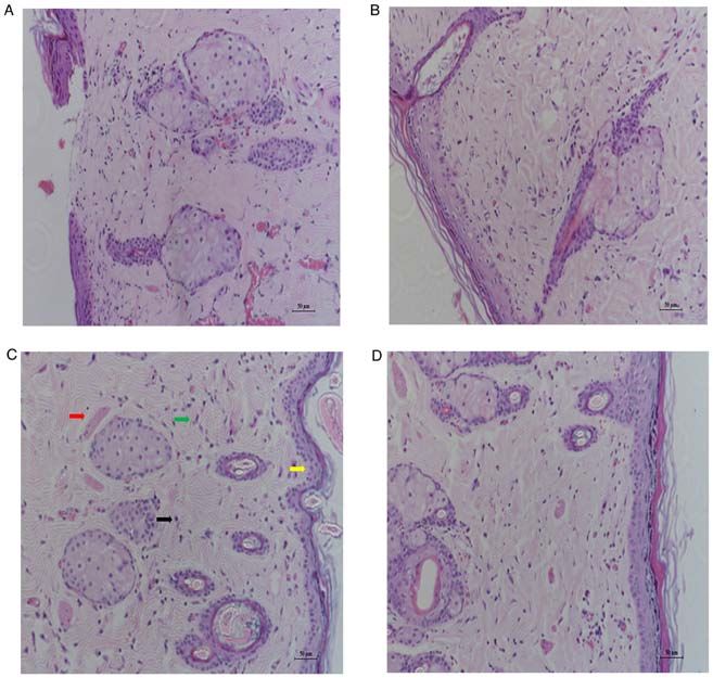
2 WANG et al: SMECTITE GRANULES ON WOUND HEALING
Wound model and topical management. A total of 12 rats thick) were stained with celestine blue solution for 5‑10 min
were grouped as the normal skin group to provide normal skin and with picrosirius red solution for 15‑30 min, both at 4˚C.
tissues. The other 36 rats were anesthetized with intraperito‑ Stained sections were observed using a light microscope (x200)
neal injection of 40 mg/kg pentobarbital sodium. The dorsal to evaluate collagen fiber distribution. For further evaluation,
fur was shaved using an electrical clipper and the skin was the content of hydroxyproline, the basic constituent of collagen,
disinfected with iodine. A full thickness wound area (2x2 cm) was measured. The healing tissue was dried at 60‑70˚C for 24 h
was created in the dorsal region using a scalpel. Subsequently, and weighed. Dried tissue was hydrolyzed in 6 N HCl at 120˚C
the rats were randomly divided into three groups (n=12 per for 18 h in sealed tubes. The hydrolyzed samples were adjusted
group): i) The smectite group was treated topically with to pH 7.0 and subjected to chloramine‑T oxidation for 20 min
0.5 mg/mm 2 mineral smectite granules (cat. no. 151207; at 37˚C. The reaction was terminated by addition of 3.15 M
Shandong Xianhe Pharmaceutical Co., Ltd.). Prior to experi‑ perchloric acid and para‑dimethylaminobenzaldehyde at 60˚C
ments, a simple pretest to study different concentrations (0.1, 0.5 to develop a pink color. The absorbance of each sample was
and 1.0 mg/mm 2) of mineral smectite granules for wound measured at a wavelength of 557 nm using an SMA4000 spec‑
healing was performed, and the results showed that the time trophotometer (Merinton Instrument, Ltd.) (16). The normal
for complete wound closure was shortest when the concentra‑ skin tissues, which were obtained from the normal skin group,
tion was 0.5 mg/mm2 (Table I); ii) the positive control group were also subjected to picrosirius red staining and hydroxypro‑
was treated topically with 0.5 mg/mm 2 starch; and iii) the line content evaluation.
blank control group was left untreated. The mineral smectite
granules and starches were in powder form, which was applied Neovascularization evaluation. Paraffin‑embedded tissue
directly to the wound without being ground or dissolved. All sections were maintained in xylene at room temperature.
wounds were covered with sterilized dressing. The wounding Endogenous peroxidase activity was blocked using hydrogen
day was considered as day 0. peroxide. Antigen retrieval was performed using ethylenedi‑
aminetetraacetic acid at 95˚C. Then the sections were washed
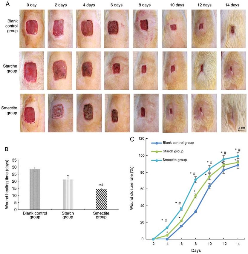
Wound healing evaluation. During the wound healing period, with phosphate buffer saline for 3 times. Subsequently, the
the wound boundary was photographed every two days until sections were incubated with a mouse monoclonal anti‑CD31
the wound was completely recovered. Different colors of the (1:100; cat. no. WH0005175M1; Sigma‑Aldrich; Merck KGaA)
wound represented different conditions: Bright red=blood primary antibody for 1 h at 37˚C. Subsequently, the sections
covering the wound; dark red=coagulation of blood in the were incubated with a biotinylated secondary antibody (1:300;
epidermis and red=granulation tissue and pink, which repre‑ cat. no. B9904; IgG; Sigma‑Aldrich; Merck KGaA) for 1 h at
sented the epithelialization phase. Wound areas were measured 37˚C. In addition, Streptavidin/HRP reagent (cat. no. DY998;
using ImageJ software (v1.8.0; National Institutes of Health). Sigma‑Aldrich; Merck KGaA) was added to each section.
The wound was considered to be completely closed when Immunoreactivity was visualized by placing sections in 0.1%
the wound was covered by new epithelial tissue. The wound 3.3'‑diaminobenzidine and 0.02% hydrogen peroxide solution
closure rate was defined as a percentage of reduction of the (DAB chromogenic system; cat. no. CTS002; Sigma‑Aldrich;
initial wound size according to the following formula: Wound Merck KGaA). The sections were counterstained with hema‑
closure (%)=(original wound size‑wound size at a specific toxylin for 30 sec at room temperature (17). Stained sections
day)/original wound size x100. Wound healing time was were observed using a light microscope (x200) and neovas‑
recorded when the wound displayed complete epithelialization. cularization was quantified using ImageJ software (v1.8.0;
National Institutes of Health). Neovascularization evaluation
Histological examination. When complete wound healing of the normal skin sections was also performed.
was observed, rats in the blank control, starch, smectite and
normal skin groups were sacrificed with intravenous injec‑ Statistical analysis. Data are presented as the mean ± SD.
tion of excess pentobarbital sodium (200 mg/kg). Death of The wound closure rate was calculated as a percentage of
rats were verified when breath and heartbeat of the animals the original wound area. Differences among multiple groups
were not detected for more than 3 min. The healing tissue in were compared by one‑way ANOVA or the χ2 test. The post
the middle of the wound area from the blank control, starch hoc analysis was performed using Tukey's test. Statistical
and smectite groups and the normal skin tissue in the dorsal analyses were performed using SPSS software (version 19.0;
region from the normal skin group were excised for histo‑ IBM Corp.). P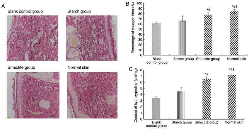
EXPERIMENTAL AND THERAPEUTIC MEDICINE 21: 160, 2021                                                                                                                            3

Table I. Wound healing times of differenct smectite concentrations.

                                                                                           Wound closure rate (%), mean ± SD
                     ‑‑‑‑‑‑‑‑‑‑‑‑‑‑‑‑‑‑‑‑‑‑‑‑‑‑‑‑‑‑‑‑‑‑‑‑‑‑‑‑‑‑‑‑‑‑‑‑‑‑‑‑‑‑‑‑‑‑‑‑‑‑‑‑‑‑‑‑‑‑‑‑‑‑‑‑‑‑‑‑‑‑‑‑‑‑‑‑‑‑‑‑‑‑‑‑‑‑‑‑‑‑‑‑‑‑‑‑‑‑‑‑‑‑‑‑‑‑‑‑‑‑‑‑‑‑‑‑‑‑‑‑‑‑‑‑‑‑‑‑‑‑‑‑‑‑‑‑‑‑‑‑‑‑‑‑‑‑‑‑‑‑‑‑‑‑‑‑‑‑‑‑‑‑‑‑‑‑‑‑‑‑‑‑‑‑‑‑‑‑‑‑‑‑‑‑‑‑‑‑‑‑‑‑‑‑‑‑‑‑
Group                2 days                    4 days                           6 days                           8 days                          10 days                          12 days                          14 days

Blank control          0.00                 0.00                         15.51±2.48                       33.23±3.12                       63.31±3.11                       82.50±2.45                       88.72±1.61
Starch                 0.00              4.74±0.96a                      21.87±1.46a                      51.73±2.61a                      74.76±2.24a                      88.40±1.02a                      91.92±1.50a
Smectite               0.00             13.92±1.83a,b                    36.01±2.07a,b                    71.05±1.64a,b                    83.86±1.04a,b                    95.33±1.59a,b                    98.88±1.20a,b

P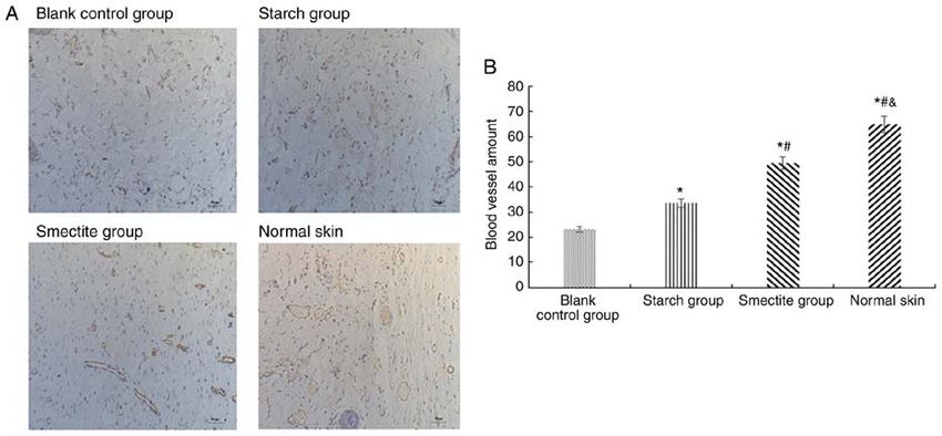
4                                           WANG et al: SMECTITE GRANULES ON WOUND HEALING

Figure 2. Hematoxylin and eosin staining of wound tissues in blank control, starch, smectite and normal skin groups. The red arrow indicates neovasculariza‑
tion, the green arrow indicates fibroblasts, the yellow arrow indicates the epidermal layer and the black arrow indicates inflammatory cells, Magnification,
x200. Scale bar, 50 µm. (A) Blank control group. (B) Starch group. (C) Smectite group. (D) Normal skin group.

Table Ⅱ Wound closure rate in the blank control, starch and                     and starch groups (60.84±2.42 and 67.35±3.05%, respectively),
smectite groups.                                                                and most closely resembled the collagen content of normal
                                                                                skin tissues (83.60±3.06%; Fig. 3B).
Smectite                                          Wound healing                     Similar results were obtained for hydroxyproline
concentration (mg/mm2)                         time (day), mean ± SD            levels. The hydroxyproline content of the smectite group
                                                                                (6.51±0.10 µm/mg) was significantly higher compared with the
0.1                                                   16.17±1.47                starch (4.54±0.14 µm/mg) and blank control (3.46±0.16 µm/mg)
0.5                                                   14.00±0.89a               groups (Fig. 3C). The hydroxyproline content of the smectite
1.0                                                   14.17±0.75a               group most closely resembled the hydroxyproline content of
                                                                                normal skin tissues (7.14±0.08 µm/mg; Fig. 3C). The results indi‑
P
EXPERIMENTAL AND THERAPEUTIC MEDICINE 21: 160, 2021 5 Figure 3. Smectite granules increase the percentage of collagen fibers and hydroxyproline content. (A) Picrosirius red staining of wound tissues in blank control, starch, smectite and normal skin groups. Magnification, x200. Scale bar, 50 µm. (B) The percentage of (B) collagen fibers and (C) hydroxyproline content in blank control, starch, smectite and normal skin groups. *P
6                                     WANG et al: SMECTITE GRANULES ON WOUND HEALING

inflammatory cells decreases, and a large number of collagen        neovascularization and maturation of the extracellular matrix.
fibers are produced by fibroblasts (27). Collagen has a key         The results provided a potential explanation for how smectite
function in wound healing and is the main component of              granules may enhance the wound healing process. The present
extracellular matrix, which serves a vital role in cell differen‑   study suggested that mineral smectite granules displayed
tiation, tissue repair and organ nourishing (28). Additionally,     wound healing potential; however, further studies are required
collagen can also activate macrophage phagocytosis, enhance         to improve the experimental scheme and identify the under‑
the immune function and decrease the infectious rate of             lying molecular mechanisms.
wounds (29). Sasaki et al (30) reported that collagen deposi‑
tion was accelerated and the density of collagen fibers was         Acknowledgements
increased upon application of Mg‑smectite in a rat cutaneous
wound model. In the present study, the collagen was mainly          Not applicable.
located in the site of application of mineral smectite granules.
Therefore, it was hypothesized that the collagen localization       Funding
may be changed according to the distribution of mineral
smectite granules. Hydroxyproline is a degradation product          No funding was received.
of collagen, which is an essential amino acid for cell repair,
providing abundant nourishment and promoting wound                  Availability of data and materials
healing (31). In the present study, the wound healing effect of
smectite granules was evaluated using a rat wound model. The        The datasets used and/or analyzed during the present study are
number of blood vessels, collagen fiber content and hydroxy‑        available from the corresponding author on reasonable request.
proline content of the smectite group were significantly higher
compared with the blank control and starch groups, which indi‑      Authors' contributions
cated that smectite may promote rapid wound healing. During
the experimental period, assessment rat weight suggested that       JW and MW designed the study and drafted the manuscript.
there were no significant differences among the groups and no       LZ and LL acquired and analyzed the data. XW and ZF
side effects were observed, which indicated that smectite was       constructed the animal model and revised the manuscript. All
safe and biocompatible in rats. Furthermore, the H&E staining       authors read and approved the final manuscript.
results indicated that the smectite group presented with fewer
inflammatory cells compared with the blank control and starch       Ethics approval and consent to participate
groups, which suggested that smectite granules could form
a barrier against microbial contamination. The results from         This animal experimental was performed according to the
previous antibacterial activity assays indicated that smectite      Guidelines for Animal Experimentation of Nanjing Medical
could prevent the Gram‑positive bacterial infection, which are      University and approved by the Animal Ethical and Welfare
pathogens that are usually involved in skin infections (32,33).     Committee of Nanjing Medical University, China (IACUC
Starches were used as the positive control group due to its low     approval no. 1601136).
cost, wide availability, biocompatibility and wound healing
properties (34). In addition, starches display similar physical     Patient consent for publication
characteristics to smectite granules (35). Compared with the
blank control group, the wound in the starch group recovered        Not applicable.
more quickly, which suggested that starch may also promote
wound healing, as reported in previous studies (36).                Competing interests
    The process of wound healing is not only associated with
cell regrowth, but also with dissolving and absorbing necrotic      The authors declare that they have no competing interests.
tissues (27). The present study did not investigate whether smec‑
tite granules influenced the dissolving or absorbing processes      References
of necrotic tissue; therefore, the molecular mechanism
underlying smectite‑induced wound healing requires further           1. Han G and Ceilley R: Chronic wound healing: A review of
investigation. The present study had a number of limitations.           current management and treatments. Adv Ther 34: 599‑610, 2017.
                                                                     2. Yamakawa S and Hayashida K: Advances in surgical applica‑
Firstly, the present study used an animal wound model, but              tions of growth factors for wound healing. Burns Trauma 7: 10,
as rat skin differs from human skin, the results of the present         2019.
study need to be verified in human skin. Secondly, only one          3. Schaffer CJ and Nanney LB: Cell biology of wound healing. Int
                                                                        Rev Cytol 169: 151‑181, 1996.
prominent angiogenic marker (CD31) was used in the present           4. Fu X, Shen Z, Chen Y, Xie J, Guo Z, Zhang M and Sheng Z:
study. More valid markers are needed to confirm the present             Randomised placebo‑controlled trial of use of topical recom‑
findings, such as EGF and TGF‑β1. Thirdly, some studies have            binant bovine basic fibroblast growth factor for second‑degree
                                                                        burns. Lancet 352: 1661‑1664, 1998.
indicated that smectite granules can lead to distal thrombosis       5. Ammar I, Bardaa S, Mzid M, Sahnoun Z, Rebaii T, Attia H
in a vascular injury wound model (11,37), but the present study         and Ennouri M: Antioxidant, antibacterial and in vivo dermal
did not investigate the long‑term safety of smectite granules.          wound healing effects of Opuntia flower extracts. Int J Biol
                                                                        Macromol 81: 483‑490, 2015.
    In conclusion, the topical application of mineral smectite       6. Fikru A, Makonnen E, Eguale T, Debella A and Abie Mekonnen G:
granules increased the percentage of wound contraction, inhib‑          Evaluation of in vivo wound healing activity of methanol extract
ited infection, accelerated re‑epithelialization and stimulated         of Achyranthes aspera L. J Ethnopharmacol 143: 469‑474, 2012.
EXPERIMENTAL AND THERAPEUTIC MEDICINE 21: 160, 2021 7
7. Pérez‑Gaxiola G, Cuello‑García CA, Florez ID and Pérez‑Pico VM: 23. Donauerová A, Bujdák J, Smolinská M and Bujdáková H:
Smectite for acute infectious diarrhoea in children. Cochrane Photophysical and antibacterial properties of complex systems
Database Syst Rev 4: CD011526, 2018. based on smectite, a cationic surfactant and methylene blue.
8. Gerlach T, Grayson JK, Pichakron KO, Sena MJ, DeMartini SD, J Photochem Photobiol B 151: 135‑141, 2015.
Clark BZ, Estep JS and Zierold D: Preliminary study of the effects 24. Gurtner GC, Werner S, Barrandon Y and Longaker MT: Wound
of smectite granules (WoundStat) on vascular repair and wound repair and regeneration. Nature 453: 314‑321, 2008.
healing in a swine survival model. J Trauma 69: 1203‑1209, 2010. 25. Long KB, Burgwin CM, Huneke R, Ar tlett CM and
9. Hou FQ, Wang Y, LI J, Wang GQ and Liu Y: Management of Blankenhorn EP: Tight skin 2 mice exhibit delayed wound
acute diarrhea in adults in China: A cross‑sectional survey. BMC healing caused by increased elastic fibers in fibrotic skin. Adv
Public Health 13: 41, 2013. Wound Care (New Rochelle) 3: 573‑581, 2014.
10. Pourshahrestani S, Zeimaran E, Djordjevic I, Kadri NA and 26. Reinke JM and Sorg H: Wound repair and regeneration. Eur Surg
Towler MR: Inorganic hemostats: The state‑of‑the‑art and recent Res 49: 35‑43, 2012.
advances. Mater Sci Eng C Mater Biol Appl 58: 1255‑1268, 2016. 27. Martin P and Nunan R: Cellular and molecular mechanisms of
11. Kheirabadi BS, Mace JE, Terrazas IB, Fedyk CG, Estep JS, repair in acute and chronic wound healing. Br J Dermatol 173:
Dubick MA and Blackbourne LH: Safety evaluation of new 370‑378, 2015.
hemostatic agents, smectite granules, and kaolin‑coated gauze in 28. Kallis PJ and Friedman AJ: Collagen powder in wound healing.
a vascular injury wound model in swine. J Trauma 68: 269‑278, J Drugs Dermatol 17: 403‑408, 2018.
2010. 29. Chattopadhyay S and Raines RT: Review collagen‑based bioma‑
12. Wang J, Zhao L, Liu W, He K, Wang M and Fan Z: Treatment terials for wound healing. Biopolymers 101: 821‑833, 2014.
outcomes of mineral smectite granules in the hemorrhage rat 30. Sasaki Y, Sathi GA and Yamamoto O: Wound healing effect of
model. Jinagsu Med J 44: 484‑487, 2018 (In Chinese). bioactive ion released from Mg‑smectite. Mater Sci Eng C Mater
13. Guo S and Dipietro LA: Factors affecting wound healing. J Dent Biol Appl 77: 52‑57, 2017.
Res 89: 219‑229, 2010. 31. El‑Mesallamy HO, Diab MR, Hamdy NM and Dardir SM:
14. Jaykamn, Yadav P and Kantharia ND: Ethics in animal experi‑ Cell‑based regenerative strategies for treatment of diabetic skin
ments. Indian J Med Ethics 9: 70‑71, 2012. wounds, a comparative study between human umbilical cord
15. Rittié L: Method for picrosirius red‑polarization detection blood‑mononuclear cells and calves' blood haemodialysate.
of collagen fibers in tissue sections. Methods Mol Biol 1627: PLoS One 9: e89853, 2014.
395‑407, 2017. 32. Li S, Guo Y, Zhao C, Chen H, Hu B, Chu Y, Zhang Z, Hu Y,
16. Colgrave ML, Allingham PG and Jones A: Hydroxyproline quan‑ Liu Z, Du Y, et al: In vitro activities of tedizolid compared with
tification for the estimation of collagen in tissue using multiple other antibiotics against Gram‑positive pathogens associated
reaction monitoring mass spectrometry. J Chromatogr A 1212: with hospital‑acquired pneumonia, skin and soft tissue infection
150‑153, 2008. and bloodstream infection collected from 26 hospitals in China.
17. Márquez WH, Gómez‑Hoyos J, Woodcock S, Arias LF, J Med Microbiol 65: 1215‑1224, 2016.
Sampson TG and Gallo JA: The regional microvascular density 33. Huang DB, Magnet S, De Angelis S, Holland TL, File TM Jr,
of the gluteus medius tendon determined by immunohistochem‑ Dryden M, Corey GR, Torres A and Wilcox MH: Surveillance of
istry with CD31 staining: A cadaveric study. Hip Int 25: 168‑171, iclaprim activity: In vitro susceptibility of Gram‑positive skin infec‑
2015. tion pathogens collected from 2015 to 2016 from North America
18. de Almeida CM, de Jesus SF, Poswar Fde O, Gomes ES, and Europe. Diagn Microbiol Infect Dis 93: 154‑158, 2019.
Fraga CA, Farias LC, Santos SH, Feltenberger JD, de Paula AM 34. Waghmare VS, Wadke PR, Dyawanapelly S, Deshpande A,
and Guimarães AL: Increasing demonstration of angiogenic Jain R and Dandekar P: Starch based nanofibrous scaffolds for
markers in skin neoplastic lesions. Pathol Res Pract 212: 101‑105, wound healing applications. Bioact Mater 3: 255‑266, 2017.
2016. 35. Baghaie S, Khorasani MT, Zarrabi A and Moshtaghian J: Wound
19. Rizzi SC, Upton Z, Bott K and Dargaville TR: Recent advances healing properties of PVA/starch/chitosan hydrogel membranes
in dermal wound healing: Biomedical device approaches. Expert with nano zinc oxide as antibacterial wound dressing material.
Rev Med Devices 7: 143‑154, 2010. J Biomater Sci Polym Ed 28: 2220‑2241, 2017.
20. Huang W, Shao M, Liu H, Chen J, Hu J, Zhu L, Liu F, Wang D, 36. Amal B, Veena B, Jayachandran VP and Shilpa J: Preparation
Zou Y, Xiong Y and Wang X: Fibroblast growth factor 21 and characterisation of Punica granatum pericarp aqueous
enhances angiogenesis and wound healing of human brain micro‑ extract loaded chitosan‑collagen‑starch membrane: Role in
vascular endothelial cells by activating PPARγ. J Pharmacol wound healing process. J Mater Sci Mater Med 26: 181, 2015.
Sci 140: 120‑127, 2019. 37. Kheirabadi BS, Edens JW, Terrazas IB, Estep JS, Klemcke HG,
21. Maddaluno L, Urwyler C and Werner S: Fibroblast growth factors: Dubick MA and Holcomb JB: Comparison of new hemostatic
Key players in regeneration and tissue repair. Development 144: granules/powders with currently deployed hemostatic products
4047‑4060, 2017. in a lethal model of extremity arterial hemorrhage in swine.
22. Tsala DE, Habtemariam S, Simplice FH, Martin Thierry BN, J Trauma 66: 316‑326, 2009.
Abraham JA and Theophile D: Topically applied Tetrapleura
tetraptera stem‑bark extract promotes healing of excision and This work is licensed under a Creative Commons
incision wounds in rats. J Intercult Ethnopharmacol 3: 63‑67, Attribution-NonCommercial-NoDerivatives 4.0
2014. International (CC BY-NC-ND 4.0) License.You can also read