PLACENTAL PATHOLOGY FINDINGS DURING AND AFTER SARS-COV-2 INFECTION: FEATURES OF VILLITIS AND MALPERFUSION - KARGER PUBLISHERS
←
→
Page content transcription
If your browser does not render page correctly, please read the page content below
Research Article

                                                     Pathobiology 2021;88:69–77                                         Received: July 1, 2020
                                                                                                                        Accepted: August 31, 2020
                                                     DOI: 10.1159/000511324                                             Published online: September 18, 2020

Placental Pathology Findings during
and after SARS-CoV-2 Infection: Features
of Villitis and Malperfusion
Thomas Menter a Kirsten Diana Mertz b Sizun Jiang c Han Chen c
Cécile Monod d Alexandar Tzankov a Salome Waldvogel e Sven M. Schulzke e
Irene Hösli d Elisabeth Bruder a
a Pathology, Institute of Medical Genetics and Pathology, University Hospital Basel, University of Basel,
Basel, Switzerland; b Institute of Pathology, Cantonal Hospital Baselland, Liestal, Switzerland; c Department of
Pathology, Stanford University, Stanford, CA, USA; d Department of Obstetrics and Antenatal Care, University
Hospital Basel, University of Basel, Basel, Switzerland; e Department of Neonatology, University Children’s Hospital
Basel UKBB, Basel, Switzerland

Keywords                                                                  delivery of 3/5 women, and their placentas did not show in-
Placenta · Malperfusion · COVID-19 · Chronic villitis                     creased inflammatory infiltrates. Signs of maternal and/or
                                                                          foetal malperfusion were present in 100% and 40% of cases,
                                                                          respectively. There was no transplacental transmission to the
Abstract                                                                  infants. In our cohort, we can document different time points
Since the outbreak of coronavirus disease 2019 (COVID-19),                regarding SARS-CoV-2 infection. In acute COVID-19, promi-
there has been a debate whether pregnant women are at a                   nent lymphohistiocytic villitis may occur and might poten-
specific risk for COVID-19 and whether it might be vertically             tially be attributable to SARS-CoV-2 infection of the placenta.
transmittable through the placenta. We present a series of                Furthermore, there are histopathological signs of maternal
five placentas of SARS coronavirus 2 (SARS-CoV-2)-positive                and foetal malperfusion, which might have a relationship to
women who had been diagnosed with mild symptoms of                        an altered coagulative or microangiopathic state induced by
COVID-19 or had been asymptomatic before birth. We pro-                   SARS-CoV-2, yet this cannot be proven considering a pletho-
vide a detailed histopathologic description of morphological              ra of confounding factors.                  © 2020 S. Karger AG, Basel
changes accompanied by an analysis of presence of SARS-
CoV-2 in the placental tissue. All placentas were term deliver-
ies (40th and 41st gestational weeks). One SARS-CoV-2-posi-
tive patient presented with cough and dyspnoea. This pla-                     Introduction
centa showed prominent lymphohistiocytic villitis and
intervillositis and signs of maternal and foetal malperfusion.              Coronavirus disease 2019 (COVID-19) caused by
Viral RNA was present in both placenta tissue and the um-                 SARS coronavirus 2 (SARS-CoV-2) has evolved into a
bilical cord and could be visualized by in situ hybridization in          worldwide pandemic within few months since its first
the decidua. SARS-CoV-2 tests were negative at the time of                documented appearance. Although severe courses and

karger@karger.com       © 2020 S. Karger AG, Basel                        Thomas Menter or Elisabeth Bruder
www.karger.com/pat                                                        Institute of Medical Genetics and Pathology
                                                                          Schönbeinstrasse 40
                                                                          CH–4031 Basel (Switzerland)
                                                                          Thomas.Menter @ usb.ch or Elisabeth.Bruder @ usb.ch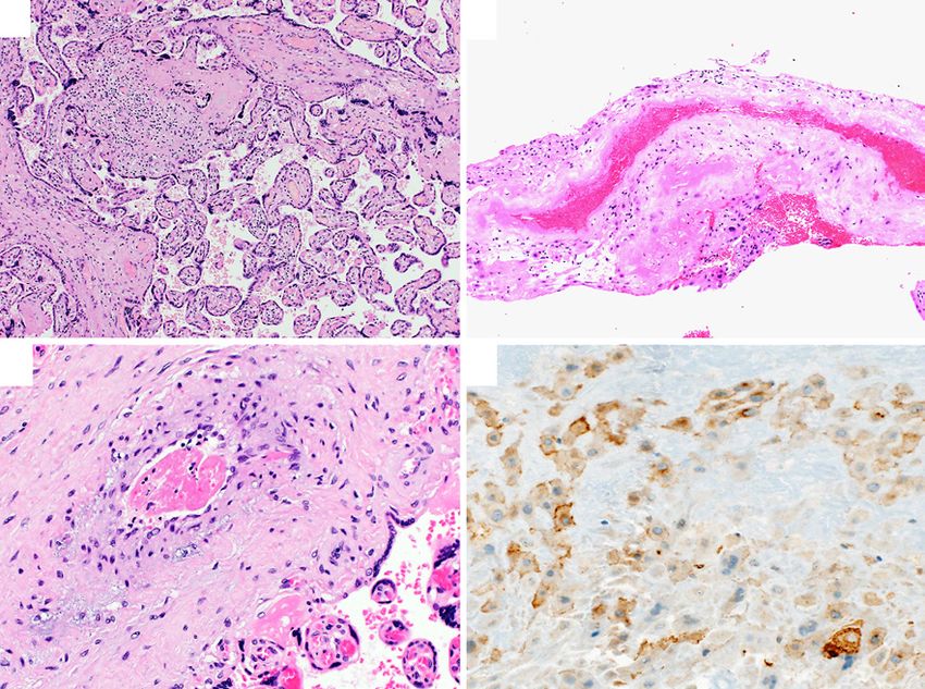
fatalities are primarily seen in elderly patients with rele-    pregnancy outcome in women infected by MERS did not
vant comorbidities, there are also younger patients show-       show a risk for miscarriage, but a higher risk for preterm
ing adverse disease outcome [1, 2]. We cannot know              delivery and preeclampsia. None of the studies dealing
whether pregnant women are at higher risk of developing         with SARS or MERS showed signs of vertical transmis-
more severe complications than the general population as        sion during the follow-up period [13]. So far, no morpho-
the absence of comparisons with appropriate controls is         logic evaluations of placentas of MERS-positive patients
still lacking. Several studies like the national cohort stud-   have been published [14, 15].
ies from France or the UK concluded that the severity of            Several studies have already analysed the histomor-
COVID -19 in pregnant women depends on comorbidi-               phology of placentas in COVID-19 and could describe
ties like age over 35, body mass index (BMI) over 35, ges-      primarily microvascular changes, while an inflammatory
tational diabetes, and hypertension [3, 4]. Since its out-      response was rarely encountered [16–18]. Not all studies
break, there has also been a debate whether the disease –       tested the placental tissue for the presence of the virus or,
as other viral infections – might influence foetal growth       if so, have not morphologically analysed the tissue; there-
and be vertically transmittable through the placenta [5,        fore, it is still difficult to present a comprehensive over-
6].                                                             view of the interaction between SARS-CoV-2, COVID-
    The placenta is an immunoprivileged organ with at-          19-related complications, and the placenta. Furthermore,
tenuated immune response and a target of several viral          little is known about the time course of SARS-CoV-2 in-
infections [7]. Viruses such as cytomegaly virus (CMV),         fection and morphologic alterations of the placenta.
herpes simplex virus 1 and 2 (HSV), Rubella virus, human            Here we present a case series of five placentas of SARS-
immunodeficiency virus (HIV), and lately also Zika virus        CoV-2-positive women who had been diagnosed with vi-
have been shown to be able to cross the placental barrier       rus infection before birth or at the day of birth. We pro-
and may be associated with severe foetal malformations          vide a detailed histopathologic description of morpho-
[8, 9]. The morphological response to viral infections var-     logical changes accompanied by an analysis of
ies: while CMV and HSV classically induce chronic lym-          inflammatory infiltrates, presence of SARS-CoV-2 RNA
phoplasmacytic villitis, in Zika virus infection, there is no   in placental tissue, and expression of the angiotensin-
inflammatory response, but a proliferation of Hofbauer          converting enzyme 2 (ACE2).
cells (specialised placental macrophages) can be observed
[10].
    Pregnant women infected by other coronaviruses such            Materials and Methods
as severe acute respiratory syndrome (SARS) and Middle
East respiratory syndrome (MERS) have already been in-              We included three women who had been tested antenatally for
vestigated in small case series. The rates of ICU admission     SARS-CoV-2 by nasal swabs because of symptoms and two asymp-
                                                                tomatic pregnant women detected by universal screening when
and maternal death were significantly higher for pregnant       entering for delivery at the University Women’s Hospital Basel,
women infected by SARS than in the general population           Switzerland since March 2020. The lockdown was imposed on
and independent of the trimester of infection. Foetal out-      March 15, 2020 in Switzerland. Altogether, five respective placen-
come was characterized by a higher rate of miscarriage,         ta specimens have since been sent for histologic examination to
intrauterine foetal death, and preterm deliveries [11]. Pla-    our institute.
                                                                    After fixation for at least 48 h to reduce infectiousness, placen-
cental weight was below the 5th percentile in a series of       tas were processed according to standard procedures, which in-
seven patients, of which two had abnormal pathology re-         clude histologic examination of one block of the umbilical cord,
sults (thrombotic vasculopathy with avascular fibrotic          one block of the chorionic membranes as well as three blocks of
villi and/or placental infarct) [12]. When infection oc-        placenta tissue [19]. Additionally, macroscopically evident chang-
curred during the week before birth, no foetal growth re-       es such as infarctions of hematomas were embedded. All slides
                                                                were stained by haematoxylin and eosin. Pathological findings
striction was noted. When the infection occurred 1 month        were classified according to the current Amsterdam Placental
or more before birth, two foetuses (2/3, 33%) had foetal        Workshop Group Consensus Statement [20].
growth restriction with oligohydramnios, related to the             In addition, one representative block of each placenta as well as
abnormal placentas presented above. In the group of pa-         of control placentas (5 cases with presence of lymphohistiocytic
tients delivering during the acute phase of illness, placen-    villitis and five cases without evidence of inflammatory infiltrates
                                                                before the outbreak of COVID-19) were stained for fibrin (Dako,
tas showed pronounced features of foetal and maternal           Glostrup, Denmark, A0080). Furthermore, both, the cohort of
malperfusion, but no increased inflammatory infiltrates         SARS-CoV-2 placentas and the control cohort, were stained for
or features of villitis were seen. Pooled information on        ACE2 (polyclonal antibody, Abcam ab15348, Cambridge, UK).

70                   Pathobiology 2021;88:69–77                                         Menter et al.
                     DOI: 10.1159/000511324
Table 1. Clinical findings
Patient 1 Patient 2 Patient 3 Patient 4 Patient 5
Age at birth, years 35.0 39.4 30.3 39.7 27.6
Gestational week 39+0 39+0 39+6 40+4 40+5
Type of birth CS CS VD VD VD
Gravida/para G4P3 G3P1 G1P1 G1P1 G1P1
BMI at time of birth 26 35 32 31 35
SARS-CoV-2 test positive prior to birth, days 5 4 18 –1 35
Gestational week of SARS-CoV-2 infection 38+2 38+3 37+2 40+5 35+5
SARS-CoV-2 test negative prior to birth, days Negative at the Negative 25 days 4 Negative 3 days 18
day of delivery after delivery after delivery
COVID-19-related symptoms None, detected Cough, Mild cough None, detected Mild cough
by screening dyspnoea by screening
Past medical history None Preeclampsia Bariatric surgery
Pregnancy-related complications None Insulin-depen- Smoking Postpartal Pollinosis
dent gestational HELLP
diabetes mellitus syndrome
TORCH serology Negative Negative Negative Negative Negative
Characteristics of neonates
Weight, g (Pc) 3,270 (43) 3,270 (43) 3,180 (17) 2,790 (4) 3,500 (32)
Length, cm (Pc) 48.0 (8) 49.0 (17) 50.0 (15) 51.0 (33) 51.0 (20)
Head circumference, cm (Pc) 35.0 (62) 34.0 (32) 33.5 (6) 33.0 (6) 35.0 (26)
Apgar score 9/10/10 9/10/10 9/10/10 8/9/9 9/10/10
Clinical course NAD Transient hypo- NAD NAD NAD
thermia (35.9 °C)
Type of feeding BM only BM and FM BM and FM BM and FM BM only
BM, breast milk; CS, caesarean section; FM, formula milk; HELLP, haemolysis, elevated liver enzymes, low platelet count syndrome; NAD, no abnor-
malities detected in general appearance, heart rate, respiratory rate, or body temperature; Pc, percentile; VD, vaginal delivery.
A quantitative reverse transcription polymerase chain reac- ISH probes was performed the next day according to manufac-
tion (RT-qPCR) assay to detect the presence of SARS-CoV-2 turer’s protocol (322350, Bio-Techne). Presence of viral RNA
RNA was performed on all samples as previously described [21]. could be confirmed using a second probe (845701 RNAscope
In order to verify also the presence of SARS-CoV-2 RNA in foetal probe – V-nCoV2019-S-sense, Advanced Cell Diagnostics, Hay-
tissue, the umbilical cord was examined in addition to placental ward, CA, USA). This staining was performed on the Leica Biosys-
parenchyma. tems BOND III autostainer (Muttenz, Switzerland) according to
For in situ hybridization, heat-induced epitope retrieval (HIER) the manufacturer’s protocol.
was performed in a Lab VisionTM PT module (Thermo Fisher) with
Dako Target Retrieval Solution, pH 9 (S236784-2, DAKO Agilent,
Santa Clara, CA, USA), for 10 min at 97 ° C before cooling down to
65 ° C and left to reach room temperature. Slides were then washed Results
twice with Milli-Q water (Millipore Sigma, Burlington, MA, USA)
before a 10-min protease digestion at 40 ° C with 1:20 diluted Pro- All placentas were term deliveries (40th and 41st ges-
tease III (322337, Bio-Techne, Minneapolis, MN, USA) in 1× PBS. tational week). The mothers’ age ranged between 27 and
Slides were then washed for 2 × 2 min with Milli-Q water before a 39 years and the BMI between 26 and 35 at the time of
15-min block at 40 ° C (322335, Bio-Techne). Slides were then
washed for 2 × 2 min with Milli-Q water before an overnight hy- birth. One patient (patient 2) presented with persistent
bridization at 40 ° C with probes against SARS-CoV-2 Spike mRNA cough and dyspnoea and was diagnosed with COVID-19
(V-nCoV2019-S-sense, 848561, Bio-Techne). Amplification of the infection 4 days before delivery. We had to perform the
Placental Pathology in the Context of Pathobiology 2021;88:69–77 71
SARS-CoV-2 DOI: 10.1159/000511324caesarean section due to spontaneous rupture of mem- tests for SARS-CoV-2 of umbilical cord blood, breast
branes and transverse lie. The other pregnant women milk, and amniotic fluid were negative.
were all asymptomatic at the time of delivery. We could Clinical findings of all patients and their neonates are
postpone one repeated caesarean section in a SARS- summarized in Table 1. Histomorphological findings are
CoV-2 positive, asymptomatic woman, detected by uni- summarized in Table 2. No patient had a previous medi-
versal screening (patient 1). The positive swab result of cal history of immunodeficiency or autoimmune disease.
the other pregnant woman screened for SARS-CoV-2 was The placenta of patient 1 (SARS-CoV-2 swab negative
obtained after delivery (patient 4). SARS-CoV-2 tests at the day of delivery) showed signs of maternal (increase
were negative at the time of delivery in 3/5 women. All of intervillous fibrin, decidual vasculopathy) and foetal
Table 2. Histomorphological findings
Patient 1 Patient 2 Patient 3 Patient 4 Patient 5 Proportion
Placental weight, g 526 649 549 598 544 n.a.
Percentile 60 >90 60 65 60 n.a.
Neonatal weight, g 3,270 3,270 3,180 2,790 3,500
Percentile 40 45 35a b
c d
e f
g h
1
Placental Pathology in the Context of Pathobiology 2021;88:69–77 73
SARS-CoV-2 DOI: 10.1159/000511324malperfusion (small thrombi in foetal vessels, delayed vil- id chorioamnionitis as well as focal chronic villitis (much
lous maturation, and chorangiosis) as well as mild chron- less extensive than patient 2). No microbiological inves-
ic deciduitis and subchorionitis. There was also a mar- tigation for the analysis of the speculated ascending infec-
ginal insertion of the umbilical cord and hypercoiling of tion was performed. There was also mild vasculitis of the
the umbilical cord. decidua. In addition, it showed signs of maternal malper-
The placenta of patient 2, who presented with COVID- fusion (increase of intervillous fibrin, perivillous fibrin
19-related symptoms at the time of delivery, showed a deposition [gitterinfarcts], decidual vasculopathy).
prominent lymphohistiocytic inflammatory infiltrate re- Findings of the four patients without COVID-19
sulting in chronic lymphohistiocytic villitis and intervil- symptoms at the time of delivery are shown in Figure
lositis accompanied by vasculitis of foetal vessels and fo- 2a–c.
cal thrombosis. The inflammatory infiltrate was predom- Fibrin staining showed a small thrombus in a foetal
inantly composed of CD8-positive cytotoxic T-cells vessel of patient 2. All other placentas of the SARAS-
accompanied by mildly increased CD68-positive macro- CoV-2 cohort and the control group did not show throm-
phages and scarce CD4-positive T-cells and plasma cells. bi in foetal vessels. Analysis of expression of ACE2 showed
There was no increase in neutrophils. The placenta fur- weak expression in cells of the invasive extravillous tro-
thermore showed signs of maternal (infarctions occupy- phoblast in 4/5 cases of our cohort as well as 8/10 control
ing 20% of the placental parenchyma, increase of intervil- cases (Fig. 2d). In decidual stroma cells and decidualized
lous fibrin, intervillous thrombosis) and foetal malperfu- endometrial glands, focal very weak ACE2 expression
sion (thrombi in chorionic vessels with vasculitis, delayed was noted.
villous maturation, and chorangiosis). There was also fo-
cal amnial phagocytosis of meconium and features of pla-
centa accreta. SARS-CoV-2 RNA was detectable at very Discussion
low levels; however, it could be verified independently in
different tissue blocks of umbilical cord and placental pa- In accordance with other studies, we could show that
renchyma. Furthermore, presence of SARS-CoV-2 was microvasculopathy, which manifests as signs of maternal
shown in the decidua by in situ hybridization. A nasal malperfusion and to a lesser degree as foetal malperfu-
swab of the neonate was negative for SARS-CoV-2. A sion, is a common finding in placentas of SARS-CoV-
swab of the placental surface for SARS-CoV-2 was posi- 2-positive women occurring in all and 2/5 patients of our
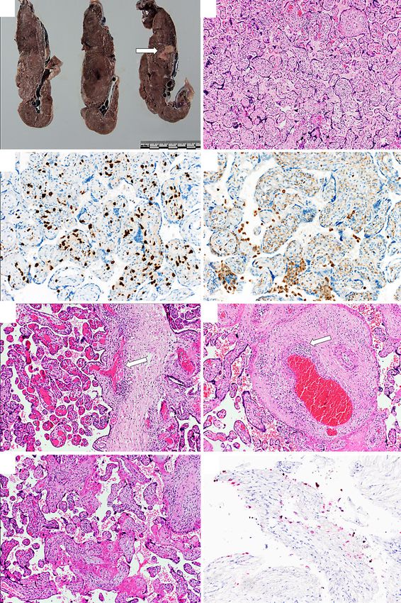
tive at the time of birth. Representative images of the pla- cohort, respectively. In our case of acute COVID-19 (pa-
cental findings are shown in Figure 1. tient 2), prominent lymphohistiocytic villitis occurred,
The placenta of patient 3 (SARS-CoV-2 swab negative which might be potentially attributable to SARS-CoV-2
4 days prior to delivery) showed signs of maternal malp- infection of the placenta. We could document different
erfusion (infarction occupying 10% of the placental pa- time points in regard to SARS-CoV-2 infection ranging
renchyma, increase of intervillous fibrin, perivillous fi- from clearance of the virus for several weeks to both acute
brin deposition [gitterinfarcts], decidual vasculopathy) symptomatic and asymptomatic infections. This feature
and hypercoiling of the umbilical cord. There were no has not been documented systematically in the literature
increased inflammatory infiltrates. This patient was a yet. Furthermore, presence of SARS-CoV-2 RNA in pla-
smoker during pregnancy and had had bariatric surgery. cental tissue is still a rarity.
The placenta of patient 4 (SARS-CoV-2 swab positive In our cohort, we could see that findings differ in rela-
at the day of delivery) also showed signs of maternal mal- tion to the time of detection and clearance of SARS-
perfusion (Tenney-Parker changes) and hypercoiling of CoV-2. Our case with manifest COVID-19 disease
the umbilical cord. As in patient 3, there were no in- showed the most prominent inflammatory response as
creased inflammatory infiltrates. Foetal thoracic cystic well as presence of viral RNA in both the placenta and the
malformation had been suspected at 22 weeks of gesta- umbilical cord and presence of SARS-CoV-2 in the de-
tion and several weeks before the COVID-19 infection. cidua. These findings support the hypothesis that SARS-
Postnatally, chest X-ray and thoracic ultrasound studies CoV-2 can invade the placenta, cause an inflammatory
as well as echocardiography and cranial ultrasound of this response, and is potentially transmittable to the child.
child were unremarkable. They also corroborate findings of Patanè et al. [6] and
The placenta of patient 5 (SARS-CoV-2 swabs persis- other studies looking at the presence of the virus or anti-
tently negative for 18 days prior to delivery) showed flor- SARS-CoV-2 antibodies in neonates [22–24]. In contrast
74 Pathobiology 2021;88:69–77 Menter et al.
DOI: 10.1159/000511324a b
c d
Fig. 2. Findings of placentas with no presence of SARS-CoV-2 at eclampsia or gestation-related hypertension. c Foetal vessel in pa-
the time of delivery and expression of ACE2. a Features of subtle tient 1 showing a small thrombus. In contrast to patient 2, the
chronic villitis in patient 5 showing acute chorioamnionitis as ma- thrombus was not wall-adherent and there were no signs of vascu-
jor finding. The lymphohistiocytic infiltrate was sparse in contrast litis (H&E, 200×). c, d Immunohistochemistry for ACE2 showing
to patient 2 (H&E, 100×). b Decidual arteriopathy in patient 1: weak expression in the extravillous invasive trophoblast (immuno-
decidual artery showing complete necrosis of the arterial wall and histochemistry, 40×).
intraluminal fibrosis. The patient did not show evidence of pre-
to these case reports, intervillositis was not as pronounced; sient hypothermia of one neonate (child to patient No. 2),
however, we could document foci of vasculitis. Charac- which was considered to be due to environmental factors.
terizing the cellular composition of the inflammatory in- All children received breast milk.
filtrate, it did not differ from cases of CMV placentitis The largest series published so far, consisting of 20 pla-
(except for lack of increase of plasma cells) or chronic vil- centas, reports chronic villitis in four patients, yet those
litis of unknown aetiology. Our patient also did not have did not seem to show symptoms of COVID-19 [17]. On
a medical history of autoimmune diseases, which might the other hand, three patients with symptoms of CO
be one of the causes of villitis of unknown aetiology. VID-19 did not show chronic villitis, with one patient
However, she suffered from insulin-dependent gestation- showing signs of acute ascending infection. Shanes et al.
al diabetes and had a history of preeclampsia in her first [16] investigated 16 placentas of SARS-CoV-2-positive
pregnancy. She was under aspirin for prevention of pre- patients and compared their results to large historical
eclampsia until week 36. TORCH serology as well as im- pre-COVID-19 control groups. They could find chronic
munohistochemical exams of the placenta tissue for in- villitis in two cases, one of these patients was in need of
fectious agents (adenovirus, CMV, HSV, toxoplasmosis) oxygen. Compared to their control groups, chronic villitis
had been negative. Postnatal transition and clinical course in general was not more common in SARS-CoV-2-posi-
of all five neonates were unremarkable apart from tran- tive patients in contrast to signs of maternal malperfu-
Placental Pathology in the Context of Pathobiology 2021;88:69–77 75
SARS-CoV-2 DOI: 10.1159/000511324sion. Unfortunately, the authors of these studies did not lial cells of the decidua, which showed very week expres-
report whether the women were tested again at the time sion of ACE2. Studies on animal models have described
of delivery after SARS-CoV-2 had been detected earlier in weak expression of ACE2 in endometrial cells of pregnant
pregnancy. The placental tissue had not been tested for rats [29]. Expression of ACE2 in human endometrial cells
presence of SARS-CoV-2 RNA in these studies. in pregnancy is not described yet; however, first reports
SARS-CoV-2 has an impact on coagulation, yet coagu- on ACE2 expression in the endometrium are currently
lation is already altered by pregnancy itself [25]. Whether being published [30, 31].
SARS-CoV-2 can be attributable to the vascular and cir- To conclude, our study shows the variegated spectrum
culatory pathologies still needs further studies. It is im- of findings in women infected with SARS-CoV-2. Cau-
portant to distinguish between acute and chronic chang- tion has to be taken to consider pregnant women as a ho-
es of the placenta as features such as infarctions, delayed mogenous group as outcomes might depend on the cur-
villous maturation, avascular fibrosed villi, or chorangio- rent state of the infection considering manifest CO
sis need time to evolve. On the other hand, the patients of VID-19 as well as a state after which virus clearance has
our cohort did not have pregnancy-related hypertension, already been achieved. We could show that in acute CO-
which is – in contrast to obesity and gestational diabetes VID-19, prominent lymphohistiocytic villitis may occur
mellitus – a well-established risk factor for maternal mal- and might be attributable to SARS-CoV-2 infection of the
perfusion of the placenta [26]. In the only available cohort placenta. Furthermore, there are pathological findings of
of seven SARS-patients, those having had SARS earlier in maternal and foetal malperfusion which might have a re-
pregnancy showed normal placental histology [12]. Our lationship to an altered coagulative state induced by
patient with manifest COVID-19 showed no abnormali- SARS-CoV-2, yet this cannot be consistently proven con-
ties of her coagulation parameters and there was no doc- sidering the plethora of confounding factors.
umentation of thromboembolic events. Three weeks after
delivery, the patient suffered from an episode of acute
cholecystitis due to cholecystolithiasis and the gall blad- Statement of Ethics
der was removed. This specimen also did not show evi-
dence of current or organized thrombosis. The foetal This study has received permission by the local ethics commit-
tee of central and Northwestern Switzerland (EKNZ, ID 2020-
thrombi were associated with those vessels showing vas- 00629).
culitis, so here the link of thrombi and infection/inflam-
mation is more likely. Mulvey et al. [18] investigated five
cases of the largest series published with signs of foetal Conflict of Interest Statement
malperfusion for activation of the complement pathway,
yet these investigations turned out negative. Additionally, The authors declare to have no competing interests.
these five cases did not show evidence of villitis. In three
of our cases, the diagnosis of presence of SARS-CoV-2
was made less than 1 week prior to delivery and less than Funding Sources
4 weeks since the first confirmed case in Switzerland in
This work is supported by the Botnar Research Centre for Child
two of these cases. The longest interval of our cohort be- Health, BRCCH. S.J. is supported by the Leukemia & Lymphoma
tween detection of SARS-CoV-2 and delivery was 5 weeks Society Career Development Program, a Fast Grant Funding for
and nasal swabs had been consistently negative for 3 COVID-19 Science and the Bill & Melinda Gates Foundation
weeks prior to delivery. This patient showed discrete de- OPP1113682.
cidual vasculopathy and only mild increase of intervillous
fibrin. There was also discrete focal chronic villitis; how-
ever, this was much less pronounced than in patient 2 Author Contributions
having manifest COVID-19.
T.M., I.H., and E.B. designed the study. T.M. wrote the manu-
In accordance with previous studies on ACE2 [27, 28], script. K.D.M. performed qPCR analysis. S.J. and H.C. performed
there was only weak immunohistochemical expression of in situ hybridization for SARS-CoV-2. A.T. analysed expression of
ACE2 in the invasive extravillous trophoblast of both our ACE2. S.W., S.M.S., C.M., and I.H. provided clinical data. All au-
SARS-CoV-2 cohort and control cases. Expression of thors critically read and approved the manuscript.
ACE2 in other cell types was scarce to absent. SARS-
CoV-2 RNA was exclusively seen in transformed epithe-
76 Pathobiology 2021;88:69–77 Menter et al.
DOI: 10.1159/000511324References 1 Petrilli CM, Jones SA, Yang J, Rajagopalan H, pathology of fetuses with congenital Zika vi- and other organs suggesting vascular dys- O’Donnell L, Chernyak Y, et al. Factors asso- rus infection. Arch Gynecol Obstet. 2017 Jun; function. Histopathology. 2020 May; 77(2): ciated with hospital admission and critical ill- 295(6):1361–8. 198–209. ness among 5279 people with coronavirus 11 Lambelet V, Vouga M, Pomar L, Favre G, 22 Egloff C, Vauloup-Fellous C, Picone O, Man- disease 2019 in New York City: prospective Gerbier E, Panchaud A, et al. SARS-CoV-2 in delbrot L, Roques P. Evidence and possible cohort study. BMJ. 2020 May;369:m1966. the context of past coronaviruses epidemics: mechanisms of rare maternal-fetal transmis- 2 Docherty AB, Harrison EM, Green CA, Hard- consideration for prenatal care. Prenat Diagn. sion of SARS-CoV-2. J Clin Virol. 2020 Jul; wick HE, Pius R, Norman L, et al.; ISARIC4C 2020 May;pd.5759. 128:104447. investigators. Features of 20 133 UK patients 12 Ng WF, Wong SF, Lam A, Mak YF, Yao H, Lee 23 Hosier H, Farhadian SF, Morotti RA, in hospital with covid-19 using the ISARIC KC, et al. The placentas of patients with severe Deshmukh U, Lu-Culligan A, Campbell KH, WHO Clinical Characterisation Protocol: acute respiratory syndrome: a pathophysio- et al. SARS-CoV-2 infection of the placenta. J prospective observational cohort study. BMJ. logical evaluation. Pathology. 2006 Jun;38(3): Clin Invest. 2020 Sep;130(9):4947–53. 2020 May;369:m1985. 210–8. 24 Vivanti AJ, Vauloup-Fellous C, Prevot S, Zu- 3 Kayem G, Lecarpentier E, Deruelle P, Bretelle 13 Di Mascio D, Khalil A, Saccone G, Rizzo G, pan V, Suffee C, Do Cao J, et al. Transplacen- F, Azria E, Blanc J, et al. A snapshot of the Co- Buca D, Liberati M, et al. Outcome of corona- tal transmission of SARS-CoV-2 infection. vid-19 pandemic among pregnant women in virus spectrum infections (SARS, MERS, CO- Nat Commun. 2020 Jul;11(1):3572. France. J Gynecol Obstet Hum Reprod. 2020 VID-19) during pregnancy: a systematic re- 25 Benhamou D, Keita H, Ducloy-Bouthors AS; Sep;49(7):101826. view and meta-analysis. Am J Obstet Gynecol CARO working group. Coagulation changes 4 Knight M, Bunch K, Vousden N, Morris E, MFM. 2020 May;2(2):100107. and thromboembolic risk in COVID-19 ob- Simpson N, Gale C, et al.; UK Obstetric Sur- 14 Schwartz DA, Graham AL. Potential Mater- stetric patients. Anaesth Crit Care Pain Med veillance System SARS-CoV-2 Infection in nal and Infant Outcomes from (Wuhan) 2020;39(3):351–353. Pregnancy Collaborative Group. Characteris- Coronavirus 2019-nCoV Infecting Pregnant 26 Bustamante Helfrich B, Chilukuri N, He H, tics and outcomes of pregnant women admit- Women: Lessons from SARS, MERS, and Cerda SR, Hong X, Wang G, et al. Maternal ted to hospital with confirmed SARS-CoV-2 Other Human Coronavirus Infections. Virus- vascular malperfusion of the placental bed as- infection in UK: national population based es. 2020 Feb;12(2):E194. sociated with hypertensive disorders in the cohort study. BMJ. 2020 Jun;369:m2107. 15 Liu W, Wang J, Li W, Zhou Z, Liu S, Rong Z. Boston Birth Cohort. Placenta. 2017 Apr; 52: 5 Penfield CA, Brubaker SG, Limaye MA, Clinical characteristics of 19 neonates born to 106–13. Lighter J, Ratner AJ, Thomas KM, et al. Detec- mothers with COVID-19. Front Med. 2020 27 Pringle KG, Tadros MA, Callister RJ, Lum- tion of SARS-COV-2 in Placental and Fetal Apr;14(2):193–8. bers ER. The expression and localization of Membrane Samples. Am J Obstet Gynecol 16 Shanes ED, Mithal LB, Otero S, Azad HA, the human placental prorenin/renin-angio- MFM. 2020;2(3):100133. Miller ES, Goldstein JA. Placental Pathology tensin system throughout pregnancy: roles in 6 Patanè L, Morotti D, Giunta MR, Sigismondi in COVID-19. Am J Clin Pathol. 2020 Jun; trophoblast invasion and angiogenesis? Pla- C, Piccoli MG, Frigerio L, et al. Vertical 154(1):23–32. centa. 2011 Dec;32(12):956–62. transmission of coronavirus disease 2019: se- 17 Baergen RN, Heller DS. Placental Pathology 28 Valdés G, Neves LA, Anton L, Corthorn J, vere acute respiratory syndrome coronavirus in Covid-19 Positive Mothers: preliminary Chacón C, Germain AM, et al. Distribution of 2 RNA on the fetal side of the placenta in Findings. Pediatr Dev Pathol. 2020 May-Jun; angiotensin-(1-7) and ACE2 in human pla- pregnancies with coronavirus disease 23(3):177–80. centas of normal and pathological pregnan- 2019-positive mothers and neonates at birth. 18 Mulvey JJ, Magro CM, Ma LX, Nuovo GJ, cies. Placenta. 2006 Feb-Mar;27(2-3):200–7. Am J Obstet Gynecol MFM. 2020 Aug; 2(3): Baergen RN. Analysis of complement deposi- 29 Neves LA, Stovall K, Joyner J, Valdés G, Gal- 100145. tion and viral RNA in placentas of COVID-19 lagher PE, Ferrario CM, et al. ACE2 and 7 Racicot K, Mor G. Risks associated with viral patients. Ann Diagn Pathol. 2020 Jun; 46: ANG-(1-7) in the rat uterus during early and infections during pregnancy. J Clin Invest. 151530. late gestation. Am J Physiol Regul Integr 2017 May;127(5):1591–9. 19 sgpath.ch/docs/QRL/QR_SGPath_DE_2011. Comp Physiol. 2008 Jan;294(1):R151–61. 8 Lee JK, Oh SJ, Park H, Shin OS. Recent Up- pdf. 30 Henarejos-Castillo I, Sebastian-Leon P, Deve- dates on Research Models and Tools to Study 20 Khong TY, Mooney EE, Ariel I, Balmus NC, sa-Peiro A, Pellicer A, Diaz-Gimeno P. SARS- Virus-Host Interactions at the Placenta. Vi- Boyd TK, Brundler MA, et al. Sampling and CoV-2 infection risk assessment in the endo- ruses. 2019 Dec;12(1):E5. Definitions of Placental Lesions: Amsterdam metrium: viral infection-related gene expres- 9 Antoniou E, Orovou E, Sarella A, Iliadou M, Placental Workshop Group Consensus State- sion across the menstrual cycle. Fertil Steril. Rigas N, Palaska E, et al. Zika Virus and the ment. Arch Pathol Lab Med. 2016 Jul;140(7): 2020 Aug;114(2):223–32. Risk of Developing Microcephaly in Infants: 698–713. 31 Chadchan SB, Maurya VK, Popli P, Kom- A Systematic Review. Int J Environ Res Public 21 Menter T, Haslbauer JD, Nienhold R, Savic S, magani R. The SARS-CoV-2 receptor, Angio- Health. 2020 May;17(11):E3806. Hopfer H, Deigendesch N, et al. Postmortem tensin converting enzyme 2 (ACE2) is re- 10 Schwartz DA. Viral infection, proliferation, examination of COVID-19 patients reveals quired for human endometrial stromal cell and hyperplasia of Hofbauer cells and absence diffuse alveolar damage with severe capillary decidualization. bioRxiv. 2020. https://doi. of inflammation characterize the placental congestion and variegated findings in lungs org/10.1101/2020.06.23.168252. Placental Pathology in the Context of Pathobiology 2021;88:69–77 77 SARS-CoV-2 DOI: 10.1159/000511324
You can also read