Lipoedema: from clinical presentation to therapy. A review of the literature
←
→
Page content transcription
If your browser does not render page correctly, please read the page content below
R E V I E W A RT I C L E                                                                              BJD      British Journal of Dermatology

Lipoedema: from clinical presentation to therapy. A review
of the literature
S.I. Langendoen, L. Habbema, T.E.C. Nijsten and H.A.M. Neumann
Department of Dermatology, Erasmus Medical Center, Postbus 2040, 3000 CA Rotterdam, the Netherlands

                                                     Summary

Correspondence                                       Lipoedema is an infrequently recognized disorder in women. Lipoedema is char-
Sterre Langendoen.                                   acterized by bilateral enlargement of the legs due to abnormal depositions of
E-mail: s.langendoen@erasmusmc.nl                    subcutaneous fat associated with often mild oedema. There is substantial variabil-
                                                     ity in disease severity. The diagnosis should be made as early as possible to pre-
Accepted for publication
9 July 2009
                                                     vent complications of the disorder, which is associated with increasing functional
                                                     and cosmetic morbidity. This review describes clinical manifestations, pathogene-
Key words                                            sis, technical investigations, management and therapies of lipoedema, with the
lipoedema, liposuction, pathophysiology, treatment   aim of optimizing management and care of patients with lipoedema.
Conflicts of interest
None declared.

DOI 10.1111/j.1365-2133.2009.09413.x

Lipoedema is an infrequently recognized disorder, first                            diagnosed with lipoedema.8 Although the incidence of lipo-
described by Allen and Hines in 1940. Lipoedema is character-                      edema in the general population is not known, these two stud-
ized by bilateral enlargement of the legs due to abnormal                          ies from specialty clinics support our suspicion that lipoedema
depositions of subcutaneous fat associated with often mild                         is more common than most physicians realize. Epidemiological
oedema.1 Different synonyms are found in the literature                            studies estimating the occurrence of lipoedema are warranted.
(Table 1), but because of the lack of a clear definition it is                     Lipoedema occurs almost exclusively in women. To our
uncertain whether all publications in fact discuss lipoedema.                      knowledge, only two men with lipoedema have been reported
This abundance of terminology and unclear definitions has                          in the literature.9,10 Lipoedema most commonly starts during
resulted in confusion about lipoedema, underdiagnosis and                          or soon after puberty. It has been suggested that in all women
mistreatment. For example, patients with lipoedema are often                       with increased fat deposits of the lower extremities, 60% is
diagnosed and treated as if they had primary lymphoedema.                          caused by obesity, 20% by lipoedema and 20% by a combin-
Lipoedema is a chronic, progressive condition that may be as-                      ation of both.7 Although the proportion of women with a self-
sociated with considerable morbidity. Initially, patients experi-                  reported positive family history of lipoedema varies between
ence discomfort, easy bruising and tenderness of the                               16% and 64%,1,5,8 lipoedema is not included in the OMIM
disproportionately enlarged legs, which may progress to high-                      database for hereditary diseases (Online Mendelian Inheritance
intensity pain and limited mobility. In addition to physical                       in Man, http://www.ncbi.nlm.nih.gov/omim/).
problems, lipoedema may be associated with psychological
morbidity. Therefore, it is important that lipoedema is recog-
                                                                                   Clinical presentation
nized as early as possible and that patients receive optimal
information and care.                                                              Allen and Hines1 described lipoedema as a clinical syndrome,
   The objective of this review is to increase the awareness of                    characterized by the combination of subcutaneous deposition
physicians about lipoedema and to describe its clinical presen-                    of fat in the buttocks and lower extremities and the accumula-
tation, pathogenesis, management and therapies.                                    tion of fluid in the legs. In 1951, Wold et al.9 suggested the
                                                                                   following diagnostic criteria: (i) occurrence almost exclusively
                                                                                   in women; (ii) a bilateral and symmetrical nature with mini-
Epidemiology
                                                                                   mal involvement of the feet; (iii) minimal pitting oedema;
Although some authors state that lipoedema is a rare disease,2                     (iv) pain, tenderness and easy bruising; and (v) persistent
the majority feels that it is more frequent than expected and is                   enlargement after elevation of the extremities or weight loss.
often missed.3–6 In one specialized lymphology clinic lipo-                           Patients complain of sensations of heaviness and discomfort
edema was diagnosed in 15% of the patients referred,7 and                          of the legs, with moderate to severe sensitivity to digital pres-
10–15% of the patients of another oedema clinic were                               sure. The swelling and aching of lipoedema gets worse during

                                                                                                                                     ! 2009 The Authors
980                                  Journal Compilation ! 2009 British Association of Dermatologists • British Journal of Dermatology 2009 161, pp980–986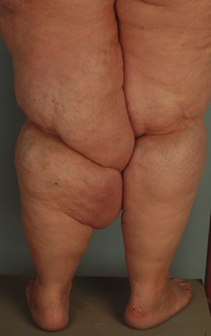
Lipoedema, S.I. Langendoen et al. 981

Table 1 Different synonyms of lipoedema used in the literature.
Because a clear definition is lacking, it is usually uncertain whether
authors mean lipoedema, lipohypertrophy or obesity when using
these terms

   Focused on fat
     Adipocyanosis
     Adiposalgie
     Adipose segmentaire
     Adipositas (or adiposis) dolorosa
     Adipositas (or adiposis) oedematosa
     Adipositas spongiosa
     Fettbein
     Obésité inférieure
     Painful fat syndrome
     Zonale Adipositas
   Focused on oedema
     Oedema cyanotique des jambes                                           Fig 1. Filling of the retromalleolar sulcus.
     Schmerzhaftes Lipödem
   Focused on morphology ⁄ descriptive
     Derkumkrankheit                                                        apparent cuff sign. In a number of patients, similar fat depos-
     Lipohypertrophia corporis inferioris                                   its exist in the upper extremities that end abruptly above the
     Rot-Dick-Schenkel                                                      wrists, sparing the hands.
     Säulenbein
                                                                               The course of lipoedema is variable. Some women develop
     Stovepipe legs
                                                                            minor lipoedema, which stabilizes and does not show pro-
     Sülzbein
     Zonaler Riesenwuchs                                                    gression over time. Other women show a gradual progression
                                                                            of the lipoedema, whereas in other patients an exacerbation is
                                                                            sometimes provoked by a stress situation such as pregnancy or
exercise and warm weather and does not recede with elevation                surgery.11,12
of the legs, although a (usually mild) pitting oedema com-
ponent will benefit from elevation. The oedema component                    Historical classifications
becomes more prominent in time and is nonpitting but may
become pitting to some extent at the end of the day.                        Different phenotypes of lipoedema have been recognized. In
   The medical history of most patients shows that the major-               addition to the first presentation described by Allen and
ity has a history of dieting and exercising without any consid-             Hines,1,9 Moncorps et al.13 described a subtype of lipoedema
erable benefit on the contours and complaints of the lower                  named the ‘typus rusticanus’ in 1940, nowadays known as
extremities. This adds to the psychological burden and low                  ‘type rusticanus Moncorps’. The difference between these
self-esteem of patients, which are risk factors for (increasing)            groups is made clinically and is important because patients
obesity. Also, limited mobility due to embarrassment and ⁄or                with the ‘type rusticanus Moncorps’ experience more serious
discomfort, pain or mechanical hindrance may contribute to                  complaints at a younger age, especially a dull spontaneous
obesity in time. Limited mobility due to mechanical friction                pain in the legs that is most prominent at the end of the
and skin irritation are well-known sequelae, even leading to                day, which may resemble symptoms of chronic venous
inability to work. For many patients, it is impossible to wear
ready-made clothes.
   The symmetrical, bilateral fat deposits on the lower extrem-
ities develop slowly and gradually. Although about half of the
patients may also be obese,7,9 the enlargement of the lower
extremities is disproportionate in relation to the trunk and
upper extremities, face and neck. In early-stage lipoedema, the
only sign can be the disappearance of the concave spaces on
both sides of the Achilles tendon (filling of the retromalleolar
sulcus) (Fig. 1). As the lipoedema progresses, the disorder
becomes easier to recognize. Characteristically, the feet are
spared and the fat deposits begin abruptly above the malleoli
causing a sharp demarcation between normal and abnormal
tissue at the ankle (cuff sign of lipoedema) (Fig. 2). The con-
tours of the lower legs are described as ‘stovepipe legs’.
Patients in whom only the thigh is affected may show a less                 Fig 2. ‘Cuff sign’ of lipoedema.

! 2009 The Authors
Journal Compilation ! 2009 British Association of Dermatologists • British Journal of Dermatology 2009 161, pp980–986
982 Lipoedema, S.I. Langendoen et al.
insufficiency in the absence of varicose veins.14 The ‘type rus-
ticanus Moncorps’ is associated with erythrocyanosis crurum
puellarum (i.e. cyanotic, bluish discoloration of the lower
third of the legs), cinnabar spots (i.e. bright red hyperaemic
spots) and follicular hyperkeratosis. It has been hypothe-
sized14,15 that there is an intrinsic connective tissue defect of
the skin of these patients, clinically visualized as stretch marks,
after a skin elasticity deficit of the skin of the calf was
observed. Furthermore, the frequent presence of knock knees,
flat feet and moderately impaired calf muscle pump function
(venous return), that may be due to musculofascial insuffi-
ciency (i.e. relatively weak connective tissue of the fascial
compartment), might even point to a more generalized con-
nective tissue defect.14 Nowadays, the ‘type rusticanus Mon-
corps’ is called erythrocyanosis crurum puellarum, as
described by Klingmüller in 1921, and is seen as a variation
of acrocyanosis in combination with fat legs, but without
oedema.16 Schmitz17 described three variants of lipoedema
including the type rusticanus Moncorps and two patient
groups with varicose veins (i.e. obese women with dispropor-
tionate fat deposits on the lower extremities and varicose
veins, and slim women with disproportionately fat legs who Fig 3. Lipoedema stage II.
also frequently have varicose veins). Three different shapes in
the legs (upper leg type, lower leg type, ankle type) and two
shapes in the arms (upper and lower arm type) were men- subcutaneous fat deposits, mostly on the legs and arms in
tioned by Herpertz.7 Meier-Vollrath and Schmeller18 described women. According to Herpertz,7 lipoedema is always pre-
three stages of lipoedema. In stage I, the skin looks flat, but ceded by lipohypertrophy. The difference between lipohyper-
the subcutis is enlarged and, when palpating the skin, feels trophy and lipoedema is the absence of oedema and pain in
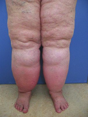
like ‘styrofoam balls in a plastic bag’. In stage II (Fig. 3), wal- lipohypertrophy. The term ‘lipodystrophy’ is reserved for local
nut- to apple-like indurations may develop and the overlying
skin has an irregular appearance (‘mattress phenomenon’).
Stage III shows bigger indurations and deforming fat deposits
(Fig. 4). The authors also suggested a classification according
to the location of the fat deposits: mainly buttocks (type I),
buttocks to knees (type II), buttocks to malleoli (type III),
mainly arms (type IV) and mainly lower legs (type V).18
These regional classifications are developed mainly for thera-
peutic follow-up purposes and are not based on pathophysio-
logical knowledge.
Differential diagnosis
Lipoedema has a distinct clinical presentation, and for phys-
icians familiar with the disease the diagnosis can be made by
patient history and clinical examination. There is no patho-
gnomonic diagnostic test for lipoedema. The most relevant
differential diagnosis of lipoedema (Table 2) includes obesity,
lipohypertrophy and lymphoedema. Further elaboration of the
causes of oedema of the lower legs (chronic venous insuffi-
ciency, idiopathic cyclic oedema, oedema due to internal dis-
ease, and orthostatic oedema) which usually concerns pitting
oedema, is beyond the scope of this review.
In obesity, the increase of subcutaneous fat deposits is gen-
eralized and not disproportionate. Furthermore, the typical
sparing of the feet and the pain of lipoedema are lacking. Her-
pertz7 described lipohypertrophy as increased symmetrical Fig 4. Lipoedema stage III.
! 2009 The Authors
Journal Compilation ! 2009 British Association of Dermatologists • British Journal of Dermatology 2009 161, pp980–986Lipoedema, S.I. Langendoen et al. 983
Table 2 Differential diagnosis of lipoedema
Lipoedema Obesity Lipohypertropy Lymphoedema
Patient history
Sex Female Male and female Female Male and female
Family history positive Common Common Possible Present in primary
lymphoedema
Proven hereditary factor Absent Absent Absent Present in primary
lymphoedema
History of erysipelas Absent Absent Absent Usually present
Progression Involved areas All over body, Not Proximally, from distal
although in most progressive portion of limb
men (and some women)
limited to trunk
Response to diet None Excellent None None
Effect of elevation Minimal (limited to pitting Ineffective Ineffective Effective initially
(oedema reduction) oedema component)
Physical examination
Bilateral involvement Always Always Always Primary: often;
secondary: rare
Foot involvement Absent Common Absent Common
Malleolar fat pad Present Absent Absent Absent
Consistency on Soft-firm Soft Soft Firm
palpation
Pitting oedema Minimal Absent Absent Always present in
variable severity
Pain on pressure Common Absent Absent Absent
damaged subcutaneous fat (e.g. insulin injections in patients oedematous state of the fat tissue. In healthy fat, the amount
with diabetes, or local trauma). of interstitial space is much less than in many other tissues
Lipoedema can be differentiated from chronic venous insuf- (about 10% of its total volume, whereas in many tissues it
ficiency because chronic venous insufficiency is associated exceeds 20%).21 In lipoedema, the anatomy of the lymphatic
with hyperpigmentation, ankle flare and pitting oedema that vessel system has been found to be normal, as far as the large
is minimal after bed rest. Moreover, in lipoedema the symp- lymph vessels are concerned. However, the increased inter-
toms increase with exercise, in contrast to chronic venous cellular pressure due to expanding fat tissue (because of dis-
insufficiency [provided that medical elastic compression stock- proportionate enlargement of the adipocytes) may cause slight
ings (MECS) are worn during exercise]. In contrast to lym- mechanical obstruction of the small lymphatic vessels in the
phoedema, Stemmer’s sign (i.e. the inability to pinch a fold septa, which results in mild lymphostasis and oedema of the
of skin at the base of the second toe due to thickening of the subcutaneous tissue. Photoplethysmography and quantitative
skin and subcutaneous tissues) is negative, and lipoedema is lymphoscintigraphic studies showed that mild venous and
always symmetrical. lymphatic insufficiency, respectively, may be present, as com-
pared with healthy controls. However, the degree of insuffi-
ciency never reached the level of true chronic venous
Pathophysiology
insufficiency or lymphoedema, and large lymph vessels were
The aetiology of lipoedema is still unknown, and some normal and sufficient.4,5,22–24 Others found that in lipoedema
authors consider it a physiological variant. The two main in the early stages the lymph flow in some cases was reduced,
components of lipoedema, consisting of enlargement of the but in others increased. It was found that lymph transport in
subcutaneous fatty tissue in combination with the formation lipoedema decreases as the body ages and the fibrosis
of oedema, have been studied but are still not well under- increases.25,26 In long-standing lipoedema, small alterations of
stood. Histopathology of biopsies and liposuction aspirate does the lymphatic tissue may appear, which can be visualized by
not show any abnormalities, except for oedema of the fat cells indirect lymphography. In lipoedema, the injection depots
and ⁄or interstitium.4,7,8,10,19,20 look flame-like, unlike the usually visualized round deposits.
Fat tissue consists of fat lobes (i.e. collection of fat cells), The ‘tongues of flame’ are likely to represent distended pre-
surrounded by connective tissue septa in which free nerve lymphatic spaces.7,22,27–31 Some investigators found enlarged
fibres, arterioles, venules and lymphatic vessels are located. and obliterated lymphatic microvessels,29 lymphatic collectors
Water may collect in fat tissue by swelling of the fat cells following a tortuous course through the fatty subcutaneous
and ⁄or collecting interstitial fluid, thereby producing an tissue22,29 and multiple microlymphatic aneurysms of lympha-
! 2009 The Authors
Journal Compilation ! 2009 British Association of Dermatologists • British Journal of Dermatology 2009 161, pp980–986984 Lipoedema, S.I. Langendoen et al.
tic capillaries in patients with lipoedema,30 of which the patho- of the disproportionate enlargement of the lower extremities,
physiological role remains to be established. the body mass index and total body weight are suboptimal
The relationship between lymphatics and adipose tissue obesity parameters in patients with lipoedema. We advise our
remains controversial. Because a relationship between the size patients to use their waist circumference as an indicator for
of fat lobules and the relative richness of blood supply and their ‘healthy’ weight. Cut-off points for categorization of waist
lymphatic drainage was found (a slow flow rate and stagna- circumference are ethnicity dependent.43 Based on the recom-
tion of lymphatic drainage enhance the deposition of fat),21,32 mendations of the World Health Organisation and Health
the mechanism of lipoedema might be a continuing deteriora- Council of the Netherlands,44 women with a waist circumfer-
tion in which the growing adipocytes keep slowing the lym- ence of < 80 cm are categorized as normal, 80–87Æ9 cm as
phatic drainage, while it is still unclear whether the primary overweight and > 88 cm as obese. Diet recommendations are
factor is the growing adipocyte or an intrinsic problem in the therefore focused on achieving the desired waist circumference
interstitial space or microlymphatic pathway. Compression of and avoiding daily variation in calorie intake.
the nerve fibres in the septa may also explain the complaints
of discomfort and, in later stages, pain. Although this is an
Conservative therapy
attractive theory, it cannot fully explain the pathogenesis of
lipoedema. Moreover, in other conditions with comparable or A combination of manual lymphatic drainage therapy and
even more enlarged fat deposits, such as lipohypertrophy or compression therapy (‘complex physical decongestion ther-
adiposis, there is no reduction of the lymphatic flow. apy’)7,11,14,41,45 is widely accepted as standard therapy. How-
ever, good clinical evidence is lacking. Compression therapy
may improve, in part, the symptoms of lipoedema and pre-
Investigations
vent the progression of the lymphatic component of lipo-
As varicose veins are invisible in the thick adipose tissue and edema. Patients with concomitant chronic venous insufficiency
lipoedema can be present concomitantly with venous disease, and ⁄or lymphoedema have an additional indication for com-
duplex ultrasound is indicated if complaints cannot fully be pression therapy. Patients with the ‘type rusticanus Moncorps’
explained by lipoedema. Dynamic lymphoscintigraphy is a may also benefit from compression therapy, probably because
good tool to rule out true lymphoedema. Imaging studies of correction of the moderately impaired calf muscle pump
using computed tomography, magnetic resonance imaging function. For some patients with lipoedema, wearing MECS is
and ⁄or ultrasound showed that the oedema is minimal and too painful.7 Diuretics7,20,46 and limb elevation have unsatis-
that limb swelling is due entirely to bilateral homogeneous factory results in patients with lipoedema,5,7 although the
enlargement of the subcutaneous compartment in the early oedema component will benefit from elevation. Disturbed
stages of lipoedema.33–38 Because there are no diagnostic and walking patterns caused by orthopaedic problems (such as flat
therapeutic implications of these investigations, we consider feet) or mechanical hindrance by the enlarged fat deposits
them not indicated in patients with lipoedema. (mainly on the medial thighs) should be treated if possible.
Therapy Surgery
Surgical debulking procedures or liposuction under general
Patient education
anaesthesia without massive subcutaneous infiltration of the
Most patients are relieved by the fact that their complaints and excessive fat depots19,45 are contraindicated because of the risk
appearance are caused by a disease instead of a lack of self- of iatrogenic damage to the lymphatics.40 For some authors,
control. This is, however, followed by disappointment because lipoedema is a (relative) contraindication for surgery of vari-
there is no easy solution to their problems. Acknowledgment cose veins because it may worsen lipoedema and may be asso-
of the severity of the complaints and optimal information and ciated with complications such as delayed wound healing and
education about lipoedema are essential in the management of postoperative swelling.11,42,47 It might be advisable not to risk
patients with lipoedema. Psychological counselling may be exacerbation of lipoedema by performing surgery on these
indicated for some patients. patients, but instead prevent progression of chronic venous in-
sufficiency and ⁄or lymphoedema by adequate compression
therapy.
Weight control
The introduction of tumescent local anaesthesia in the
People with lipoedema do not inherently have obesity, but 1980s has greatly changed the therapeutic options for lipo-
about 50% are overweight.39 Prevention of obesity is impor- edema.48 In tumescent local anaesthesia, large amounts of
tant, because the additional weight gain of the body areas fluid (containing saline, lidocaine, sodium bicarbonate and
affected by lipoedema is very resistant to dieting and exercise. adrenaline) are infiltrated in the subcutaneous tissues. Tumes-
Therefore, weight loss will not improve patients’ discomfort cent liposuction is at least as effective as the conventional
and appearance, resulting in frustration and demotivation, (‘dry’) liposuction and the so-called ‘wet’ liposuction in
which may enhance the development of obesity.40–42 Because removing adipose aspirates, but has the advantage that it is
! 2009 The Authors
Journal Compilation ! 2009 British Association of Dermatologists • British Journal of Dermatology 2009 161, pp980–986Lipoedema, S.I. Langendoen et al. 985
significantly less likely to damage the lymphatic vessels.12 The 9 Wold LE, Hines EA Jr, Allen EV. Lipedema of the legs; a syndrome
use of vibrating microcannulas further improved the results in characterized by fat legs and edema. Ann Intern Med 1951; 34:1243–
patients with lipoedema. 50.
10 Chen SG, Hsu SD, Chen TM et al. Painful fat syndrome in a male
Although tumescent liposuction cannot cure lipoedema,
patient. Br J Plast Surg 2004; 57:282–6.
results are promising: especially an impressive improvement of 11 Földi M, Idiazabal G. The role of operative management of varicose
pain is reported by patients with lipoedema.49 Furthermore, veins in patients with lymphedema and ⁄ or lipedema of the legs.
functional improvement in mobility is noted. A recent case ser- Lymphology 2000; 33:167–71.
ies of patients with lipoedema demonstrated that this technique 12 Hoffmann JN, Fertmann JP, Baumeister RGH et al. Tumescent and
improved appearance and quality of life and reduced symptoms dry liposuction of lower extremities: differences in lymph vessel
such as tendency to swelling and pain.2,18 Another group injury. Plast Reconstr Surg 2003; 113:718–24.
13 Moncorps C, Brinkhaus G, Herfeld-Münster F. [Experimental inves-
reported follow-up periods of more than 8 years without com-
tigations in acrocyanosis]. Arch Dermatol Syph 1940; 180:209–15.
plications or negative results.50 Because often extensive amounts 14 Jagtman BA, Kuiper JP, Brakkee AJ. [Lipedema of the legs]. Ned
of adipose tissue have to be removed, multiple sessions are Tijdschr Geneeskd 1987; 131:345–8.
necessary, thereby making it a time-consuming method. Ideally 15 Jagtman BA, Kuiper JP, Brakkee AJM. [Measurements of skin elas-
it is performed relatively early to prevent progression of the ticity in patients with lipedema of the Moncorps ‘rusticanus’ type].
disorder.51 The German Phlebological Society recommends Phlebologie 1984; 37:315–19.
liposuction as part of the therapeutic armamentarium in the 16 Braun-Falco O, Wolff HH, Plewig G et al. Erythrocyanosis crurum
puellarum. In: Dermatologie und Venerologie (Braun-Falco O, Wolff HH,
management of lipoedema.52 Unfortunately, this therapy is
Plewig G, Landthaler M, Burgdorf WHC, eds), 5th edn. Heidel-
usually not supported by health insurance companies. berg: Springer Medizin Verlag, 2005; 773.
17 Schmitz R. [Lipedema from the differential diagnostic and thera-
peutic viewpoint]. Z Hautkr 1987; 62:146–57.
Conclusion 18 Meier-Vollrath I, Schmeller W. [Lipoedema—current status, new
Because lipoedema is a relatively frequent, chronic disorder perspectives]. J Dtsch Dermatol Ges 2004; 2:181–6.
with considerable morbidity, the diagnosis should be made as 19 Stallworth JM, Hennigar GR, Jonsson HT et al. The chronically
swollen painful extremity. JAMA 1974; 228:1656–9.
early as possible to inform patients and, hopefully, prevent
20 Rudkin GH, Miller TA. Lipedema: a clinical entity distinct from
progression. The diagnosis is based on patient history and lymphedema. Plast Reconstr Surg 1994; 94:841–7.
physical examination. Duplex ultrasound and ⁄or dynamic 21 Ryan T. Lymphatics and adipose tissue. Clin Dermatol 1995; 13:493–
lymphoscintigraphy are indicated if complaints cannot fully be 8.
explained by lipoedema. The aim of our treatment is to pre- 22 Weissleder H, Brauer JW, Schuchhardt C et al. [Value of functional
vent or reduce obesity, to optimize patient mobility, reduce lymphoscintigraphy and indirect lymphangiography in lipedema
complaints and to improve overall quality of life. The impor- syndrome]. Z Lymphol 1995; 19:38–41.
23 Boursier V, Pecking A, Vignes S. [Comparative analysis of lympho-
tance of weight control is emphasized in all patients. Conser-
scintigraphy between lipedema and lower limb lymphedema].
vative therapy such as manual lymphatic drainage, MECS and J Mal Vasc 2004; 29:257–61.
orthopaedic or psychological counselling is started on indica- 24 van Geest AJ, Esten SCAM, Cambier J-PRA et al. Lymphatic disturb-
tion. For patients with acceptable waist circumference, the ances in lipoedema. Phlebologie 2003; 32:138–42.
treatment of choice is tumescent liposuction, which is at this 25 Brauer WJ, Brauer VS. Altersabhängigkeit des Lymphtransportes
moment the only available technique to correct the abnormal beim Lipödem und Lipolymphödem. LymphForsch 2005; 9:6–9.
adipose tissue. 26 Brauer WJ, Weissleder H. Methodik und Ergebnisse der Funk-
tionslymphszintigraphie: Erfahrungen bei 924 Patienten. Phlebologie
2002; 31:118–25.
References 27 Partsch H, Stoberl C, Urbanek A et al. Clinical use of indirect lym-
phography in different forms of leg edema. Lymphology 1988;
1 Allen EV, Hines EA. Lipedema of the legs: a syndrome character- 21:152–60.
ized by fat legs and orthostatic edema. Proc Staff Meet Mayo Clin 28 Tiedjen KU, Heimann KD, Tiedjen-Kraft U et al. Indirect xero-lym-
1940; 15:184–7. phography in lymphedema, lipedema and venous insufficiency.
2 Schmeller W, Meier-Vollrath I. Tumescent liposuction: a new Phlebologie 1992; 92:396–8.
and successful therapy for lipedema. J Cutan Med Surg 2006; 10: 29 Bollinger A. Microlymphatics of human skin. Int J Microcirc Clin Exp
7–10. 1993; 12:1–15.
3 Beninson J, Edelglass JW. Lipedema – the non-lymphatic masquer- 30 Amann-Vesti BR, Franzeck UK, Bollinger A. Microlymphatic aneu-
ader. Angiology 1984; 35:506–10. rysms in patients with lipedema. Lymphology 2001; 34:170–5.
4 Bilancini S, Lucchi M, Tucci S et al. Functional lymphatic alterations 31 Stoberl C, Partsch H, Wruhs M. [Diagnostic value and assessment
in patients suffering from lipedema. Angiology 1995; 46:333–9. criteria of indirect lymphography in lymphedema]. Vasa 1990;
5 Harwood CA, Bull RH, Evans J, Mortimer PS. Lymphatic and ven- 19:212–17.
ous function in lipoedema. Br J Dermatol 1996; 134:1–6. 32 Földi E, Földi M, Tischendorf F. [Adipositas, lipedema and lym-
6 Fonder MA, Loveless JW, Lazarus GS. Lipedema, a frequently phostasis]. Med Welt 1983; 34:198–200.
unrecognized problem. J Am Acad Dermatol 2007; 57:S1–3. 33 Hadjis NS, Carr DH, Banks L et al. The role of CT in the diagnosis
7 Herpertz U. [Lipedema]. Z Lymphol 1995; 19: 1–11. of primary lymphedema of the lower limb. AJR Am J Roentgenol
8 Gregl A. [Lipedema]. Z Lymphol 1987; 11: 41–3. 1985; 144:361–4.
! 2009 The Authors
Journal Compilation ! 2009 British Association of Dermatologists • British Journal of Dermatology 2009 161, pp980–986986 Lipoedema, S.I. Langendoen et al.
34 Vaughan BF. CT of swollen legs. Clin Radiol 1990; 41:24–30. 43 Zhu S, Heshka S, Wang Z et al. Combination of BMI and waist cir-
35 Monnin-Delhom ED, Gallix BP, Achard C et al. High resolution un- cumference for identifying cardiovascular risk factors in whites.
enhanced computed tomography in patients with swollen legs. Obes Res 2004; 12:633–45.
Lymphology 2002; 35:121–8. 44 Health Council of the Netherlands. [Advisory Report on Excess Weight and
36 Duewell S, Hagspiel KD, Zuber J et al. Swollen lower extremity: Obesity]. The Hague: Health Council of the Netherlands, 2003.
role of MR imaging. Radiology 1992; 184:227–31. 45 Rank BK, Wong GSC. Lipoedema. Aust NZ J Surg 1966; 35:165–9.
37 Werner GT, Rodiek SO. [Value of nuclear magnetic resonance 46 Wienert V, Leeman S. [Lipedema]. Hautarzt 1991; 42:484–6.
tomography in leg edema of unknown origin. Preliminary report]. 47 Pereira de Godoy JM, de Fatima Guerreiro Godoy M, Hayashida M.
Z Lymphol 1993; 17:2–5. Lipoedema and varicose vein surgery: a worse prognosis? Acta Angiol
38 Dimakakos PB, Stefanopoulos T, Antoniades P et al. MRI and ultra- 2005; 11:186–7.
sonographic findings in the investigation of lymphedema and lipe- 48 Klein JA. The tumescent technique: anesthesia and modified lipo-
dema. Int Surg 1997; 82:411–16. suction technique. Dermatol Clin 1990; 8:425–37.
39 Greer KE. Lipedema of the legs. Cutis 1974; 14:89. 49 Schmeller W, Meier-Vollrath I. [Successful surgical therapy of lipe-
40 Stiefelhagen P. [No lymphedema, no obesity. How can lipedema dema by liposuction]. Phlebologie 2004; 33:23–9.
be treated?]. MMW Fortschr Med 2001; 143:15. 50 Rapprich S, Loehnert M, Hagedorn M. Therapy of lipoedema syn-
41 Macdonald JM, Sims N, Mayrovitz HN. Lymphedema, lipedema drome by liposuction under tumescent local anaesthesia. Ann Derma-
and the open wound: the role of compression therapy. Surg Clin tol Venereol 2002; 129:1S711.
North Am 2003; 83:639–58. 51 Sattler G, Bergfeld D, Sommer B. [Liposuction]. Hautarzt 2004;
42 Brunner U. [Vascular diseases in lipedema of the legs. Special 55:599–604.
symptoms, common therapeutic results, viewpoint on vascular sur- 52 Wienert V, Földi E, Schmeller W et al. [Guideline: lipedema of the
gery]. Schweiz Med Wochenschr 1982; 112:1130–7. legs]. Phlebologie 2005; 34:42–4.
! 2009 The Authors
Journal Compilation ! 2009 British Association of Dermatologists • British Journal of Dermatology 2009 161, pp980–986You can also read


