Molecular mechanism of the anti inflammatory effects of Sophorae Flavescentis Aiton identified by network pharmacology - Nature
←
→
Page content transcription
If your browser does not render page correctly, please read the page content below
www.nature.com/scientificreports

                OPEN             Molecular mechanism
                                 of the anti‑inflammatory
                                 effects of Sophorae Flavescentis
                                 Aiton identified by network
                                 pharmacology
                                 Naiqiang Zhu & Jingyi Hou*

                                 Inflammation, a protective response against infection and injury, involves a variety of biological
                                 processes. Sophorae Flavescentis (Kushen) is a promising Traditional Chinese Medicine (TCM) for
                                 treating inflammation, but the pharmacological mechanism of Kushen’s anti-inflammatory effect
                                 has not been fully elucidated. The bioactive compounds, predicted targets, and inflammation-related
                                 targets of Kushen were obtained from open source databases. The “Component-Target” network
                                 and protein–protein interaction (PPI) network were constructed, and hub genes were screened out
                                 by topological analysis. Gene ontology (GO) and Kyoto Encyclopedia of Genes and Genomes (KEGG)
                                 enrichment analyses were performed on genes in the PPI network. Furthermore, nitric oxide (NO)
                                 production analysis, RT-PCR, and western blot were performed to detect the mRNA and protein
                                 expression of hub genes in LPS-induced RAW264.7 cells. An immunofluorescence assay found
                                 that NF-κB p65 is translocated. A total of 24 bioactive compounds, 465 predicted targets, and 433
                                 inflammation-related targets were identified and used to construct “Component-Targets” and PPI
                                 networks. Then, the five hub genes with the highest values-IL-6, IL-1β, VEGFA, TNF-α, and PTGS2
                                 (COX-2)- were screened out. Enrichment analysis results suggested mainly involved in the NF-κB
                                 signaling pathway. Moreover, experiments were performed to verify the predicted results. Kushen
                                 may mediate inflammation mainly through the IL-6, IL-1β, VEGFA, TNF-α, and PTGS2 (COX-2), and
                                 the NF-κB signaling pathways. This finding will provide clinical guidance for further research on the
                                 use of Kushen to treat inflammation.

                                 Abbreviations
                                 ANOVA	Analysis of variance
                                 BP	Biological process
                                 CC	Cell component
                                 DL	Drug-likeness
                                 DMEM	Dulbecco’s modified Eagle’s medium–high glucose
                                 ETCM	Encyclopedia of Traditional Chinese Medicine
                                 GO	Gene ontology
                                 HI-FBS	Heat inactivated fetal bovine serum
                                 IL-1β	Interleukin-1β
                                 IRA	Inflammatory receptor agonist
                                 KEGG	Kyoto Encyclopedia of Genes and Genomes
                                 LPS	Lipopolysaccharide
                                 MF	Molecular function
                                 NSAIDs	Non-steroidal anti-inflammatory drugs
                                 OB	Oral bioavailability
                                 PGE2	Prostaglandin E2

                                 Department of Minimally Invasive Spinal Surgery, the Affiliated Hospital of Chengde Medical College,
                                 Chengde 067000, China. *email: hjy_2016@126.com

Scientific Reports |   (2021) 11:1005              | https://doi.org/10.1038/s41598-020-80297-y                                          1

                                                                                                                                 Vol.:(0123456789)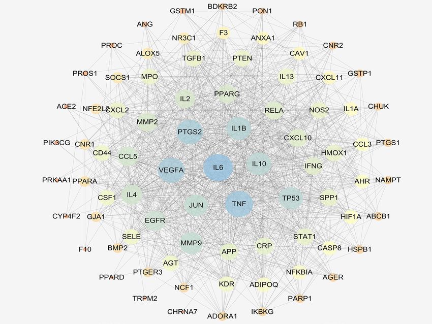
www.nature.com/scientificreports/
PPI Protein–protein interaction
SEA Similarity ensemble approach
TNF-α Tumor necrosis factor-α
TCM Traditional Chinese Medicine
Inflammation can be discovered and diagnosed early, and it is associated with huge risks in a variety of dis-
eases, including a rthritis1, atherosclerosis2, Alzheimer’s d
iseases3, inflammatory bowel syndrome4, and i njury5.
Cytokines are involved in both inflammation and anti-inflammatory effects via multifunctional molecules
depending on the manner of the inflammatory response6. Thus, pro-inflammatory cytokines IL-6, tumor
necrosis factor-α (TNF-α), and IL-18 initiate and amplify inflammatory processes, whereas anti-inflammatory
cytokines, such as IL-10, inflammatory receptor agonist (IRA), and transforming growth factor-β (TGF-β)
negatively regulate these p rocesses7. Almost all acute and chronic disease are driven or regulated by inflamma-
tion, and inflammatory response are fairly complicated processes that involve the synthesis and release of a large
number of inflammatory factors. Currently, the anti-inflammatory drugs approved for clinical use include (a)
non-steroidal anti-inflammatory drugs (NSAIDs)8, such as aspirin, paracetamol, anti-inflammatory drugs, and
ibuprofen, and (b) corticosteroid hormones, such as prednisone and steroidal anti-inflammatory drugs such as
adrenocorticosteroids and androgens)9. However, the long-term use of anti-inflammatory drugs is associated
with many side effects and poor prognosis, resulting in serious the gastrointestinal reactions and causing specific
damage to certain parts of the cardiovascular system.
Kushen (Sophorae Flavescentis) is a dry stem of the medicinal plant of Sophora flavescens, which is used to
clear heat and dampness from the body and kill insects10. Kushen products are abundant in China, and they
are produced in a number of provinces, including the Hebei, Henan, Shandong, Anhui, Hubei, and Xinjiang
provinces11. Clinical trials have shown that Kushen has low toxicity and side effects. Moreover, Kushen exerts a
variety of therapeutic effects, such as swelling reduciton, immunostimulation, and anti-arrhythmic, anti-tumor,
and anti-bacterial effects12. Kushen can act on many key pathways and links in the anti-inflammatory process;
however, the specific mechanisms have not been fully explained.
Network pharmacology is based on the theory of receptors and biological network technology. It analyzes
multi-component, multi-target, and multi-pathway synergistic relationships between drugs, targets, and diseases,
and then use them to explain drug action13. Traditional Chinese Medicine (TCM) operates systematically and
holistically, and network pharmacology has allowed TCM research to go beyond focusing on a single ingredient,
target, and disease and to study the efficacy of TCM and its mechanisms in d epth14. In this study, the network
pharmacology method was used to predict the effective components and potential targets of Kushen and its
hub genes involved in the treatment of inflammation. This network pharmacology approach in combination
with molecular biology experiments for further verification provided a basis for future research on the anti-
inflammatory mechanism of Kushen (Fig. 1).
Results
Identification of bioactive compounds and targets. The TCMSP, ETCM, and SymMap databases were
used to determine the bioactive compounds in Kushen. A total of 19 bioactive compounds were identified based
on the parameters oral bioavailabilty (OB) ≥ 30% and drug-likeness (DL) ≥ 0.18. Further, several bioactive com-
pounds, whose OB and DL values were below the screening thresholds were excluded. According to the studies,
kurarionl, dehydromiltirone, kushenol B, kushenol F, and kushenonl E were also included (Table 1). Next, we
found 465 targets corresponding to the bioactive compounds in Kushen (Supplementary Excel 1). Furthermore,
OMIM, Genecards, and PubMed-gene were screened using the search terms as anti-inflammatory, inflammation,
and anti-inflammation, and 433 targets were found in all three databases (Supplementary Excel 2).
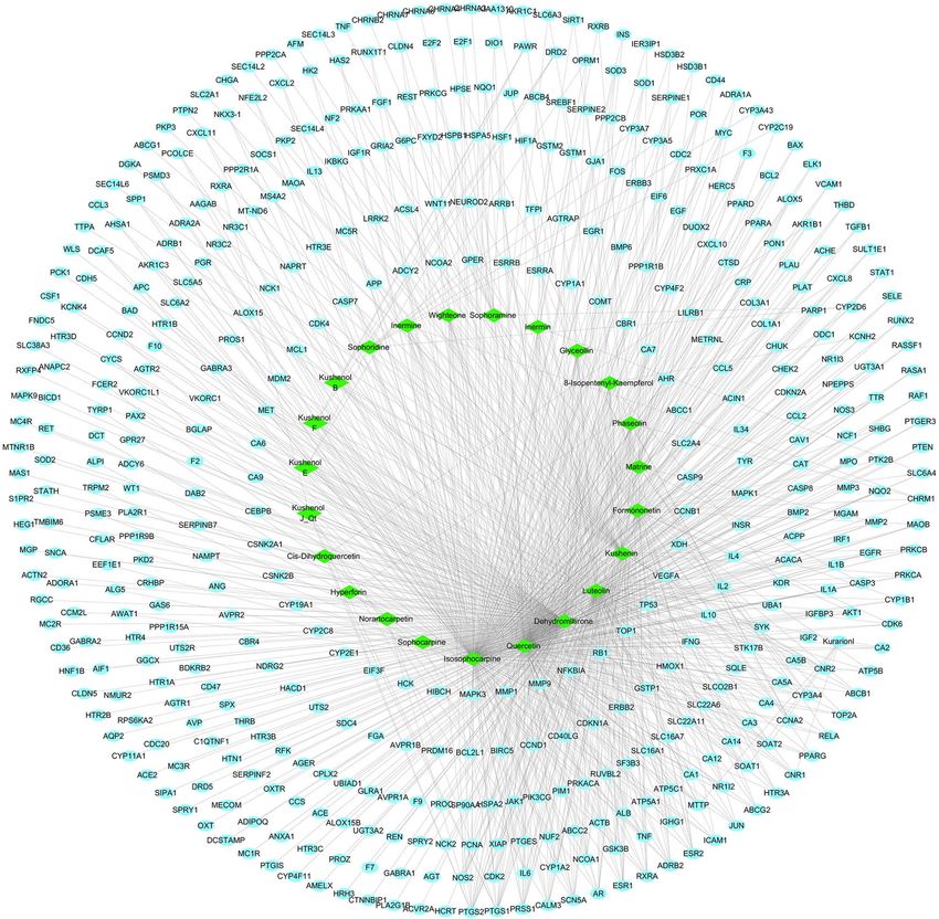
Compound‑target network construction and analysis. As shown in Fig. 2, the compound-target
network consists of 489 nodes (24 bioactive ingredients and 465 targets) and 974 interaction edges. In descend-
ing order of degree, the top four bioactive ingredients were isosophocarpine (degree 218), quercetin (degree
207), dehydromiltirone (degree 166), and luteolin (degree 129) (Table 2), suggesting these bioactive ingredients
are crucial to the pharmacological action of Kushen.
PPI network for Kushen in the treatment of inflammation and hub genes analyses. In order
to clarify Kushen’s potential anti-inflammatory mechanisms, the obtained anti-inflammatory targets of the bio-
active ingredients in Kushen were introduced into the STRING online database (PPI combined score > 0.7) to
construct a PPI network, consisting of 81 nodes and 1088 interaction edges (Fig. 3). Based on the plug-in cyto-
Hubba, the following hub genes were screened out in the PPI network according to the top five values of degree:
IL-6, TNF-α, IL-1β, VEGFA, and PTGS2 (COX-2). Table 2 shows that these hub genes are mainly involved in
enzymes, and signaling.
Pathway enrichment analysis. As shown in Fig. 4, GO enrichment analysis of the targets in the PPI
network was performed using ClusterProfiler in R. The biological process (BP) results suggest that these targets
respond to lipopolysaccharides, molecules of bacterial origin, oxidative stresses, nutrient levels, peptides, oxygen
levels, decreased oxygen levels, reactive oxygen species, regulation of smooth muscles, and smooth muscle, mus-
cle cell proliferation. Component cellular (CC) results included the vesicle, endoplasmic reticulum, secretory
granule, Golgi, and platelet alpha granule lumen. For molecular function (MF), the targets mostly involved the
binding of the cytokine receptor, G protein-coupled receptor, promoter sequence-specific DNA, growth factor
receptor, and chemokine receptor. Furthermore, KEGG enrichment analysis has suggested that targets were
Scientific Reports | (2021) 11:1005 | https://doi.org/10.1038/s41598-020-80297-y 2
Vol:.(1234567890)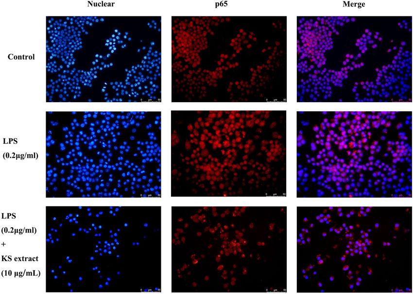
www.nature.com/scientificreports/
Figure 1. The framework of Kushen extract for the treatment of inflammation.
mainly associated with the TNF signaling pathway, NF-kappa B signaling pathway, cytokine-cytokine receptor
interaction, chemokine signaling pathway, and PI3K-Akt signaling pathway (Table 3).
Effects of Kushen on cell viability and NO production. Previous studies have reported that the main
components of Kushen, such as M atrine15, Oxymatrine16, Sophoridine17, Kushenol B18, own excellent anti-
inflammatory effects. In this study, LPS-induced RAW264.7 cells inflammatory model was used cell to inves-
tigate the mechanism underlying Kushen’s anti-inflammatory effect on macrophagocytes. After an incubation
period, the effects of Kushen extract on the viability of RAW264.7 cells were detected by CellTriter-Lumi ™ Plus.
A viability assay showed that Kushen extract does not inhibit cell proliferation at any concentrations up to 10 μg/
mL (Fig. 5A). In addition, the anti-inflammatory effects of Kushen extract on NO production in LPS-treated
cells were detected by Griess reagent. As shown in Fig. 5B, the Kushen extract significantly inhibited NO pro-
duction. Furthermore, laser microscopy showed that Kushen extract is a stronger inhibitor of intracellular NO
production than LPS stimulation alone (Fig. 6).
Suppression of the mRNA and protein expression of the hub genes by Kushen extract. To
investigate the effects of Kushen extract on the predicted hub genes by network pharmacology, the mRNA levels
of IL-6, IL-1β, VEGFA, TNF-α, and PTGS2 (COX-2) were measured by quantitative real-time PCR, whereas the
protein levels of IL-6, IL-1β, VEGFA, TNF-α, and PTGS2 (COX-2) were measured using western blot analysis.
As shown in Fig. 7, the mRNA expression of IL-6, IL-1β, VEGFA, TNF-α, and PTGS2 (COX-2) was significantly
increased after LPS stimulation (0.2 μg/mL). Moreover, the mRNA expression of these genes was significantly
inhibited by all concentrations of Kushen extract in a concentration-dependent manner. In addition, the protein
expression of IL-6, IL-1β, VEGFA, TNF-α, and PTGS2 (COX-2) in cells treated with Kushen extract was signifi-
cantly inhibited compared to their expression in the LPS group alone (0.2 μg/mL) group (Fig. 8). Collectively,
our study suggests that Kushen extract mainly treats inflammation by inhibiting these genes.
Translocation of the NF‑κB p65 subunit. As shown in Table 4, the NF-κB signaling pathway is the key
signaling pathway underlying the anti-inflammatory action of Kushen extract. The translocation of the NF-κB
p65 subunit was determined by immunofluorescence. As shown in Fig. 9, after stimulation by LPS, p65 (red) was
translocated from the cytoplasm to the nucleus; this clearly attenuated by Kushen extract (10 μg/mL), suggesting
that the Kushen extract inhibited NF-κB activation.
Scientific Reports | (2021) 11:1005 | https://doi.org/10.1038/s41598-020-80297-y 3
Vol.:(0123456789)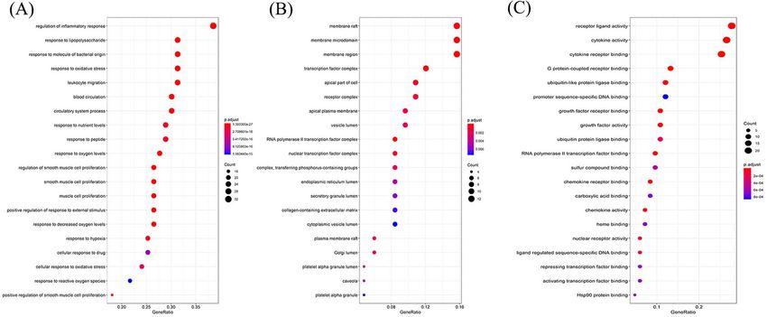
www.nature.com/scientificreports/

                                             Molecular name             OB (%)   DL     Degree   Structure

                                             Luteolin                   36.16    0.25   129

                                             8-Isopentenyl-Kaempferol   38.04    0.39   13

                                             Sophoramine                42.16    0.25   11

                                             Wighteone                  42.80    0.36   11

                                             Hyperforin                 44.03    0.6    4

                                             Quercetin                  46.43    0.28   207

                                             Kushenin                   47.62    0.38   49

                                             Kurarionl                   0.95    0.67   5

                                             Kushenol J_Qt              50.86    0.24   6

                                             Norartocarpetin            54.93    0.24   3

                                             Continued

          Scientific Reports |   (2021) 11:1005 |                  https://doi.org/10.1038/s41598-020-80297-y   4

Vol:.(1234567890)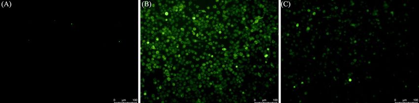
www.nature.com/scientificreports/

                                   Molecular name             OB (%)   DL     Degree   Structure

                                   Sophoridine                60.07    0.25   9

                                   Isosophocarpine            61.57    0.25   218

                                   Matrine                    63.77    0.25   18

                                   Sophocarpine               64.26    0.25   2

                                   Inermin                    65.83    0.54   11

                                   Cis-Dihydroquercetin       66.44    0.27   4

                                   Formononetin               69.67    0.21   48

                                   Dehydromiltirone           24.57    0.26   166

                                   Inermine                   75.18    0.54   11

                                   Phaseolin                  78.20    0.73   16

                                   Continued

Scientific Reports |   (2021) 11:1005 |                   https://doi.org/10.1038/s41598-020-80297-y        5

                                                                                                       Vol.:(0123456789)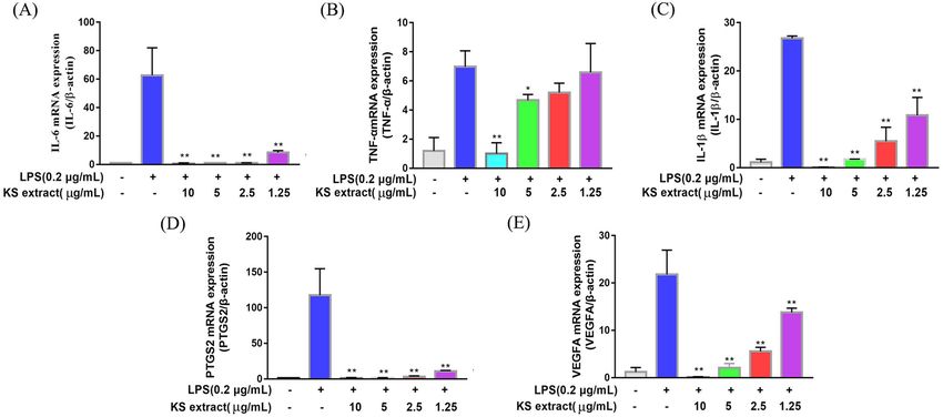
www.nature.com/scientificreports/
Molecular name OB (%) DL Degree Structure
Glyceollin 97.27 0.76 12
Kushenol B 1.21 0.75 7
Kushenol F 18.85 0.61 7
Kushenol E 19.66 0.59 7
Table 1. Information of bioactive compounds of Kushen.
Discussion
Inflammation is a protective response against infection and injury19, which is closely linked to various chronic or
malignant diseases, such as type II d iabetes20, atherosclerosis21, and c ancer22. Inflammation is a complex process
involving multiple genes and signaling p athways23, and TCM is considered to have anti-inflammatory potential
due to its effects via multiple-compounds, multiple-targets, and multiple pathways involved24. Kushen, well-
known for its efficacy in clearing body heat, is a TCM mostly used to treat various syndrome that are caused by
inflammation25 or i nfection26. Therefore, Kushen and its preparation, such as Kushen Lotion, Kushen Injection,
are clinically used as adjuncts to treat inflammation-related diseases27. Through data mining and analysis, net-
work pharmacology systematically interprets the overall relationship between drugs and targets, which perfectly
fits TCM’s strategy of disease management through multiple ingredients, targets, and p athways28. Unlike previ-
ous studies, our study was the first to fully elucidate the anti-inflammatory mechanism of Kushen extract via
network pharmacology methods and experimental validation, laying the foundation for further clinical research.
In this study, we retrieved 19 bioactive compounds of Kushen from the TCMSP, ETCM, and SymMap data-
bases and five bioactive compounds with noteworthy pharmacological effects from literatures and found 465
predicted targets and used them to construct a “Component-Target” network. In the network, important com-
pounds such as isosophocarpine (degree 218), quercetin (degree 207), dehydromiltirone (degree 166), and
luteolin (degree 129) have a high degree value and are associated with many targets. Isosophocarpine, a tetracy-
clic quinolizidine alkaloid, shows anti-cancer effects on different types of cancer by attenuating inflammation29.
Quercetin is a flavonoid with antioxidant, antiviral, and antibacterial effects, and is widely distributed in various
plants and food. Lin et al.30 found that quercetin suppresses inflammation by countering the Azoxymethane/
Dextran sodium sulfate (AOM/DSS)-induced carcinogenesis progression. In addition, Yue et al. have found that
the dehydromiltirone is an anti-inflammatory compound that initiates p38 and the NF-κB signaling pathway
in LPS-induced Kupffer c ells31. Luteolin is a flavonoid commonly found in plants, such as celery, green pepper,
honeysuckle, and chamomile. Previous studies have suggested that luteolin can reduce inflammation via inhibit-
ing inflammation via activating the Nrf2/ARE, NF-κB, and MAPK signaling pathways32. These findings suggested
that Kushen extract exerts its anti-inflammatory effects through multiple components and multiple targets.
A PPI network was constructed with 81 nodes and 1088 interaction edges to elucidate further the mecha-
nisms of Kushen’s anti-inflammatory response. Then, the hub genes, namely IL-6, IL-1β, VEGFA, TNF-α, and
PTGS2 (COX-2) were screened out based on the topological properties’ analysis. LPS-induced RAW264.7 cells
constitute a typical inflammation model, and therefore the present study used them to investigate the anti-
inflammatory effects of Kushen extract. A NO production assay showed that any concentration of Kushen
extract inhibits the production of NO in a concentration-dependent manner, and intracellular NO production
assay intuitively showed that Kushen extract shows maximum anti-inflammatory effect at 10 μg/mL. Moreover,
RT-PCR and western blot further verified that the mRNA and protein levels of IL-6, IL-1β, VEGFA, TNF-α, and
PTGS2 (COX-2) differ significantly between the LPS-induced and Kushen extract groups, suggesting that Kushen
mainly exerts the anti-inflammatory effects via these predicted hub genes. IL-6, IL-1β, and TNF-α are crucial
pro-inflammatory cytokines that coordinate a variety of inflammatory and immunomodulatory pathways that
have broad effects on cells of the immune s ystem31,33. VEGFA, a pleiotropic cytokine, has been considered as the
Scientific Reports | (2021) 11:1005 | https://doi.org/10.1038/s41598-020-80297-y 6
Vol:.(1234567890)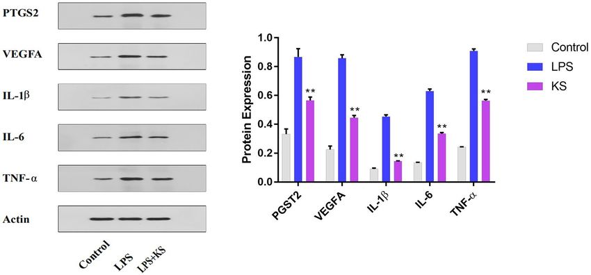
www.nature.com/scientificreports/

                                 Figure 2.  The Compound-Target network of Kushen. Green nodes indicate bioactive compounds, blue nodes
                                 indicate target proteins, and edges indicates interaction between ingredients and targets.

                                   Gene Symbol   Uniprot ID   Target                                  Target Class   Degree
                                   IL6           P05231       Interleukin 6                           None           70
                                   TNF-α         P01375       Tumor necrosis factor                   Signaling      66
                                   VEGFA         P15692       Vascular endothelial growth factor A    Signaling      63
                                   PTGS2         P35354       Prostaglandin-endoperoxide synthase 2   Enzyme         62
                                   IL1β          P01584       Interleukin 1 beta                      None           57

                                  Table 2.  Hub genes and topological properties.

Scientific Reports |   (2021) 11:1005 |               https://doi.org/10.1038/s41598-020-80297-y                                          7

                                                                                                                                   Vol.:(0123456789)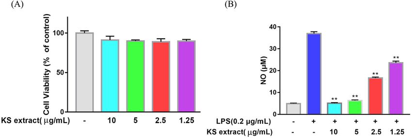
www.nature.com/scientificreports/
Figure 3. Protein–protein interaction (PPI) network analysis of Kushen for the treatment of inflammation
using STRING database.
Figure 4. GO enrichment analysis of the putative targets. (A) The top 20 significant enriched terms in
biological process (BP); (B) The top 20 significant enriched terms in cellular component (CC); (C) The top 20
significant enriched terms in molecular function (MF).
indispensable part of angiogenesis, which mainly leads to cancer-related inflammation34. PTGS2 (COX-2), an
enzyme induced by pro-inflammatory cytokines, releases prostaglandin E2 (PGE2) and promotes the synthesis of
prostaglandins stimulating cancer cell proliferation, development, and metastasis; thus, it serves as a therapeutic
target for anti-inflammatory drugs.
Scientific Reports | (2021) 11:1005 | https://doi.org/10.1038/s41598-020-80297-y 8
Vol:.(1234567890)www.nature.com/scientificreports/
Description Gene ratio Bg Ratio p value p. adjust q value
TNF signaling pathway 18/79 112/8033 1.33E−17 7.30E−16 3.01E−16
NF-kappa B signaling pathway 9/79 104/8033 6.99E−07 3.03E−06 1.25E−06
Toll-like receptor signaling pathway 16/79 104/8033 2.10E−15 7.13E−14 2.95E−14
Inflammatory bowel disease (IBD) 13/79 65/8033 3.15E−14 8.03E−13 3.32E−13
Cytokine-cytokine receptor interaction 18/79 294/8033 3.03E−10 3.25E−09 1.34E−09
Chemokine signaling pathway 14/79 189/8033 3.21E−09 2.52E−08 1.04E−08
T cell receptor signaling pathway 11/79 104/8033 4.35E−09 3.17E−08 1.31E−08
MAPK signaling pathway 15/79 294/8033 1.29E−07 7.31E−07 3.02E−07
PI3K-Akt signaling pathway 16/79 354/8033 2.46E−07 1.25E−06 5.17E−07
VEGF signaling pathway 5/79 59/8033 2.67E−04 6.9E−04 2.8E−04
Table 3. KEGG pathway enrichment analysis.
Figure 5. Effect of Kushen extract on RAW264.7 cells. (A) Cell viability of RAW264.7 cells after being
treated by Kushen extract was detected by the CellTiter-LumiTM Plus, (B) NO production of LPS stimulated
RAW264.7 cells after being treated with Kushen extract was detected by the Griess reaction. Data were presented
as the mean ± SEM (n = 6), *p < 0.05 and **p < 0.01 versus LPS-treated group was considered statistically
significant differences.
Figure 6. Effect of Kushen extract on intracellular NO production in RAW264.7 cells. The NO production of
(A) Control group, (B) LPS-stimulated group, and (C) LPS-stimulated treated with Kushen extract group were
determined by DAF-FM diacetate.
In, addition, the KEGG enrichment analysis of targets in the PPI network showed that Kushen’s anti-inflam-
matory effect is mainly enriched in the NF-κB signaling pathway and the PI3K-Akt signaling pathway. The
PI3K-Akt/NF-κB signaling pathways play different roles in normal physiological responses and inflammatory
processes31,35, including promoting cell proliferation, survival and differentiation. After activation of PI3K-Akt
pathway, Akt enhances the phosphorylation and lowers the phosphorylation of the NF-κB inhibitor protein IκB
kinase. PI3K/Akt/NF-κB signaling pathway server as the intersection of T and B cell inflammatory signaling
pathway, resulting in increased expression of its maker proteins IKK-α, IκB-α, NF-κB p65, PI3K, p-AKT, p-NF-κB
p65 in inflammatory m odel36. Among these, p65/RelA and p50 take important part in the NF-κB signaling
pathway; p65 degrades when NF-κB signaling shuts d own37. The immunofluorescence results also verified that
Kushen extract (10 μg/mL) attenuates p65 from the cytoplasm to the nucleus in LPS-induced RAW264.7 cells,
suggesting that the Kushen plays a crucial role in modulating inflammation via the NF-κB signaling pathway.
Scientific Reports | (2021) 11:1005 | https://doi.org/10.1038/s41598-020-80297-y 9
Vol.:(0123456789)www.nature.com/scientificreports/
Figure 7. Effect of Kushen extract on key mRNA expression levels in RAW264.7 cells. The expression of (A)
IL-6; (B) TNF-α; (C) IL-1β; (D) PTGS2 (COX-2); and (E) VEGFA mRNA levels were determined by RT-PCR.
Data were presented as the mean ± SEM (n = 3), *p < 0.05 and **p < 0.01 versus LPS-treated group was considered
statistically significant differences.
Figure 8. Effect of Kushen extract on key proteins expression levels in RAW264.7 cells. The expression of IL-6,
TNF-α, IL-1β, PTGS2, and VEGFA protein levels were determined by western blot with specific antibodies and
quantification. Data were presented as the mean ± SEM (n = 3), *p < 0.05 and **p < 0.01 versus LPS-treated group
was considered statistically significant differences.
In conclusion, a network pharmacology approach was developed to elucidate the underlying molecular
mechanism of anti-inflammatory effects of Kushen on inflammation. A total of 24 bioactive compounds, 465
Kushen-related targets, and 433 inflammation-related targets were obtained from open source databases. Fur-
thermore, five hub genes were screened out based on a topological property analysis of the PPI network: IL-6,
IL-1β, VEGFA, TNF-α, and PTGS2 (COX-2). Then, an experimental in vitro validation was performed to confirm
the mRNA and protein expression of these hub genes and for enrichment analysis. Considering the complexity
of the inflammatory process and TCM predicted targets, further research and clinical trials are necessary to
confirm our findings.
Scientific Reports | (2021) 11:1005 | https://doi.org/10.1038/s41598-020-80297-y 10
Vol:.(1234567890)www.nature.com/scientificreports/
Gene Primer Sequence (5′–3′)
Forward TGTTACCAACTGGGACGACA
β-actin
Reverse GGGGTGTTGAAGGTCTCAAA
Forward TGAGTACCGCAAACGCTTCTC
COX-2
Reverse TGGACGAGGTTTTCCACCAG
Forward TAGCCAGGAGGGAGAACAGA
TNF-α
Reverse TTTTCTGGAGGGAGATGTGG
Forward CTGGAGCCCACCAAGAACGA
IL-6
Reverse GCCTCCGACTTGTGAAGTGGT
Forward ATGCCACCTTTTGACAGTGATG
IL-1β
Reverse GTTGATGTGCTGCTGCGAGATT
Forward TGAAGTGATCAAGTTCATGGACGT
VEGFA
Reverse TCACCGCCTTGGCTTGTC
Table 4. Primers used for the quantitative RT-PCR.
Figure 9. Effect of Kushen extract on the p65 subunit of NF-κB in RAW264.7 cells. The nuclear localization of
the p65 subunit of NF-κB in (A) Control group, (B) LPS groups, and (C) LPS + Kushen extract were determined
by immunofluorescence.
Materials and methods
Reagents. Bacterial lipopolysaccharide (LPS) was obtained from Sigma. Dulbecco’s modified Eagle’s
medium–high glucose (DMEM-HG) and heat inactivated fetal bovine serum (HI-FBS) were purchased from
Biological Industries. Griess reagent system, DAPI, CellTiter-Lumi Plus Detection Kit, DAF-FM DA Detection
Kit, and Cy3-labeled goat anti-rabbit IgG were obtained from Beyotime. RT-PCR kits were purchased from
Takara Biotechnology Co., Ltd. Affinity provided the following antibodies: NF-κB p65, IL-6, IL-1β, TNF-α,
VEGFA, PTGS2 (COX-2), and β-actin.
Preparation of Kushen extract. Kushen herb was purchased from the Tongren Pharmaceutical Co., Ltd.
(BEIJING, CHINA). Kushen (50 g) was soaked in 1 L of cold water for 30 min and then boiled for 30 min; this
procedure was repeated three times. The combined extracts were concentrated to 1 g/mL (crude herbal dose)
Scientific Reports | (2021) 11:1005 | https://doi.org/10.1038/s41598-020-80297-y 11
Vol.:(0123456789)www.nature.com/scientificreports/
in a vacuum rotary evaporator. Then, the extract (1 mL) was filtered through microporous membrane before
quantification.
Network pharmacology analysis. Screening for active ingredients of Kushen. The bioactive ingredients
of Kushen were screened from the following databases: Traditional Chinese Medicine Systems Pharmacology
(TCMSP, https://tcmspw.com/tcmsp.php)38, the Encyclopedia of Traditional Chinese Medicine (ETCM, http://
www.tcmip.cn/ETCM/index.php/Home/Index/All)39 and the SymMap (https://www.symmap.org/)40. Accord-
ing to the most common criteria of network pharmacology analysis14, the active ingredients with oral bioavail-
ability (OB) ≥ 30% and drug-likeness (DL) ≥ 0.18 were selected for subsequent analysis.
Screening of potential targets for Kushen. Targets related to the candidate bioactive compounds of Kushen were
collected from the ETCM, Search Tool for Interacting Chemicals (STITCH, http://stitch.embl.de/), SymMap,
and Similarity ensemble approach (SEA, https://www.sogou.com/link?url=LeoKdSZoUyC9U6gWDurrLbchw
v7HyEQP) databases41 with the “Homo sapiens” setting (Supplementary Excel 1).
Construction of a bioactive component‑target network. To intuitively understand the mechanisms of Kushen
extract treatment on anti-inflammation, a “Component-Target” network was constructed via Cytoscape 3.7.0
based on the bioactive components and predicted targets. In this network, the green rhombus node, blue round
node, and edges indicate a bioactive component, a predicted target, and the interaction between the bioactive
compounds and targets, respectively. The plug-in “Cytohubba” was applied to calculate the “degree” value of the
node, which suggests the number of edges between the nodes in the network.
Screening for potential targets of inflammation. The keywords “inflammation,” “anti-inflammatory,” or “anti-
inflammation” were used to search disease-related genes on the following databases: the Online Mendelian
Inheritance in Man (OMIM, https://omim.org/), GeneCards (https://www.genecards.org/), and National Center
for Biotechnology Information Gene Database (https://pubmed.ncbi.nlm.nih.gov/29140470/); the genes that
overlapped among these databases were recorded (Supplementary Excel 2).
Protein–protein interaction network construction and hub genes analysis. To further elucidate
the potential mechanism underlying Kushen’s anti-inflammatory effect, a website was used to find overlap-
ping inflammatory-related and predicted targets of Kushen. These overlapping targets were used to construct
a protein–protein interaction (PPI) network on the Search Tool for the Retrieval of Interacting Genes/Proteins
(STRING) database (https://string-db.org/) with the “Homo sapiens” setting. Cytoscape 3.7.0 was used to visual-
ize PPI network, and the plug-in “Network Analysis” was performed to visualize the topological properties of
each node in the network. To further elucidate the mechanism by which Kushen treats inflammation, the hub
genes were screened out based on the topological properties of nodes in the PPI network. The plug-in “cyto-
Hubba” was applied to calculate the value of degree in the PPI network, and the five genes with the highest values
of degree were selected as anti-inflammatory hub genes for Kushen. Further, information on the target type
(protein class) of the hub genes was taken from the DisGeNET database (https://www.disgenet.org).
Gene ontology and KEGG pathway enrichment analyses. Gene ontology (GO) enrichment analy-
sis is a bioinformatics tool for predicting gene function, while the Kyoto Encyclopedia of Genes and Genomes
(KEGG, https://www.kegg.jp/) is a database for identifying the systematic functions and biological relevance of
targets42. In order to analyze the biological pathways of genes in the PPI network, the “clusterProfiler” package
(https://bioconductor.org/packages/release/bioc/html/clusterProfiler.html) in R (version: 3.6.3)43 was applied to
analyze GO enrichment and KEGG pathway enrichment (adjusted to P. < 0.05).
Cell culture. RAW264.7 cells were obtained from Cell Culture Center of the Chinese Academy of Medical
Sciences. Cells were cultured in DMEM supplemented with 10% HI-FBS, 100 units/mL penicillin, and 100 μg/
mL streptomycin at 37 °C in a fully humidified incubator containing 5% C
O2.
Cell viability assay. RAW264.7 cells were cultured in a 96-well plate at of 5 × 10³ cells/well and incubated
overnight at 37 °C in an incubator containing 5% CO2. Subsequently, the cells were incubated with various
concentrations of Kushen extract (10, 5, 2.5, and 1.25 μg/mL) for 24 h. Then, 100 μL of CellTiter-Lumi Plus
detection reagent was added to each well, and the wells were vibrated for 5 min to fully lyse the cells completely.
A luminometer (multifunctional microplate reader) was used to measure the luminescence (RLU) of each well.
The viability of the Kushen extract was calculated as follows: ( RLUcontrol- RLUtreated/RLUcontrol) × 100%.
Nitrite assay. RAW264.7 cells were cultured in 96-well plates at 1 × 10⁴ cells/well and treated as described
above. NO production in the supernatant of the medium was measured by the Griess assay according to the
manufacturer’s instructions and absorbance at 540 nm (OD540) was measured with a multifunctional microplate
reader. In addition, DAF-FM was applied to qualitatively detect the concentration of NO. The cells were cultured
and treated as described above. According to the instructions for the DAF-FM DA Kit, the images were gener-
ated with a Laser microscope (495 nm/515 nm).
Total mRNA extraction and RT‑PCR. Total RNA was isolated from cells treated with Kushen extract
(10, 5, 2.5, and 1.25 μg/mL) and LPS (0.2 μg/mL) using Trizol reagent and reverse-transcribed into cDNA with
Scientific Reports | (2021) 11:1005 | https://doi.org/10.1038/s41598-020-80297-y 12
Vol:.(1234567890)www.nature.com/scientificreports/
PrimeScript II 1st Strand cDNA Synthesis Kit. The real-time PCR detection system and SYBR were applied to
the PCR-amplified hub genes in accordance with the manufacture’s instruction manual. The primers for the
hub genes are described in Table 4. β-Actin served as the internal control. The relative expression of mRNA was
calculated as 2−∆∆CT.
Protein extraction and western blot. Total protein was isolated from RAW264.7 cells treated with
Kushen extract (10 μg/mL), and LPS (0.2 μg/mL) using RIPA lysis buffer: the protein concentration was quanti-
fied with a BCA protein assay kit as described previously44. Then, equal amounts of protein were separated by
SDS-PAGE and transferred to PVDF membranes. After blocking in 0.01% Tween 20 containing 5% skimmed
milk powder for 4 h, the membranes were incubated with a primary antibody (IL-6, IL-1β, PTGS2 (COX-2),
TNF-α, VEGFA and β-actin) at 1:800 dilution overnight. Next, the membranes were incubated with anti-rabbit
IgG secondary antibodies for 1 h. The blot bands were visualized and quantified using Gel Image system.
Immunofluorescence assay. The total nuclear translocation of active p65 from the cytosol was assessed
by immunofluorescence as described p reviously44. Briefly, RAW264.7 cells were stimulated with LPS (0.2 μg/mL)
and treated with Kushen extract (10 μg/mL) for 6 h. Then, the cells were permeabilized with 0.1% Triton X-100
and incubated with anti-NF-κB p65 antibody (1:100 with 2% BSA) overnight at 4 °C. Next, the cells were incu-
bated with Cy3-labeled goat anti-rabbit IgG, followed by DAPI mounting. Micrographs were captured under a
fluorescence microscope.
Statistical analysis. The data were analyzed with one-way analysis of variance (ANOVA) and P val-
ues < 0.05 were considered statistically significant. Statistical tests were performed using the GraphPad.
Received: 23 July 2020; Accepted: 16 December 2020
References
1. Noack, M. & Miossec, P. Selected cytokine pathways in rheumatoid arthritis. Semin. Immunopathol. 39, 365–383. https://doi.
org/10.1007/s00281-017-0619-z (2017).
2. Gistera, A. & Hansson, G. K. The immunology of atherosclerosis. Nat. Rev. Nephrol. 13, 368–380. https://doi.org/10.1038/nrnep
h.2017.51 (2017).
3. Lodygin, D. et al. beta-Synuclein-reactive T cells induce autoimmune CNS grey matter degeneration. Nature 566, 503–508. https
://doi.org/10.1038/s41586-019-0964-2 (2019).
4. van Hemert, S. et al. Migraine associated with gastrointestinal disorders: review of the literature and clinical implications. Front.
Neurol. 5, 241. https://doi.org/10.3389/fneur.2014.00241 (2014).
5. Thomas, M. R. & Storey, R. F. The role of platelets in inflammation. Thromb. Haemost. 114, 449–458. https://doi.org/10.1160/
TH14-12-1067 (2015).
6. Chousterman, B. G., Swirski, F. K. & Weber, G. F. Cytokine storm and sepsis disease pathogenesis. Semin. Immunopathol. 39,
517–528. https://doi.org/10.1007/s00281-017-0639-8 (2017).
7. Yeung, Y. T., Aziz, F., Guerrero-Castilla, A. & Arguelles, S. Signaling pathways in inflammation and anti-inflammatory therapies.
Curr. Pharm. Des. 24, 1449–1484. https://doi.org/10.2174/1381612824666180327165604 (2018).
8. Kohler, O., Krogh, J., Mors, O. & Benros, M. E. Inflammation in depression and the potential for anti-inflammatory treatment.
Curr. Neuropharmacol. 14, 732–742. https://doi.org/10.2174/1570159x14666151208113700 (2016).
9. Wongrakpanich, S., Wongrakpanich, A., Melhado, K. & Rangaswami, J. A comprehensive review of non-steroidal anti-inflammatory
drug use in the elderly. Aging Dis. 9, 143–150. https://doi.org/10.14336/AD.2017.0306 (2018).
10. Ding, P. L., He, C. M., Cheng, Z. H. & Chen, D. F. Flavonoids rather than alkaloids as the diagnostic constituents to distinguish
Sophorae Flavescentis Radix from Sophorae Tonkinensis Radix et Rhizoma: an HPLC fingerprint study. Chin. J. Nat. Med. 16,
951–960. https://doi.org/10.1016/S1875-5364(18)30137-7 (2018).
11. Wang, L. et al. Effects of pungent essential oil from three Chinese herbs on percutaneous absorption of alkaloids from Sophorae
flavescentis radix. Zhongguo Zhong Yao Za Zhi 44, 308–313. https://doi.org/10.19540/j.cnki.cjcmm.20181108.001 (2019).
12. Song, L. Y. et al. Inhibitory effects of oxymatrine on hepatic stellate cells activation through TGF-beta/miR-195/Smad signaling
pathway. BMC Complement. Altern. Med. 19, 138. https://doi.org/10.1186/s12906-019-2560-2 (2019).
13. Zhu, N., Hou, J., Ma, G. & Liu, J. Network pharmacology identifies the mechanisms of action of Shaoyao gancao decoction in the
treatment of osteoarthritis. Med. Sci. Monit. 25, 6051–6073. https://doi.org/10.12659/MSM.915821 (2019).
14. Zhu, N. & Hou, J. Exploring the mechanism of action Xianlingubao Prescription in the treatment of osteoporosis by network
pharmacology. Comput. Biol. Chem. 85, 107240. https://doi.org/10.1016/j.compbiolchem.2020.107240 (2020).
15. Zhaowu, Z., Xiaoli, W., Yangde, Z. & Nianfeng, L. Preparation of matrine ethosome, its percutaneous permeation in vitro and
anti-inflammatory activity in vivo in rats. J. Liposome Res. 19, 155–162. https://doi.org/10.1080/08982100902722381 (2009).
16. Xu, X. et al. Anti-pruritic and anti-inflammatory effects of oxymatrine in a mouse model of allergic contact dermatitis. J. Dermatol.
Sci. https://doi.org/10.1016/j.jdermsci.2018.04.009 (2018).
17. Huang, X., Li, B. & Shen, L. Studies on the anti-inflammatory effect and its mechanisms of sophoridine. J. Anal. Methods Chem.
2014, 502626. https://doi.org/10.1155/2014/502626 (2014).
18. Jung, H. A. et al. Re-evaluation of the antioxidant prenylated flavonoids from the roots of Sophora flavescens. Biol. Pharm. Bull.
31, 908–915. https://doi.org/10.1248/bpb.31.908 (2008).
19. Yue, S. et al. Salvia miltiorrhiza compounds protect the liver from acute injury by regulation of p38 and NFkappaB signaling in
Kupffer cells. Pharm. Biol. 52, 1278–1285. https://doi.org/10.3109/13880209.2014.889720 (2014).
20. Lemmers, R. F. H. et al. The anti-inflammatory function of high-density lipoprotein in type II diabetes: A systematic review. J.
Clin. Lipidol. 11, 712–724. https://doi.org/10.1016/j.jacl.2017.03.013 (2017).
21. Nasonov, E. L. & Popkova, T. V. Atherosclerosis: perspectives of anti-inflammatory therapy. Ter Arkh 90, 4–12. https://doi.
org/10.26442/terarkh201890514-12 (2018).
22. Liu, Y. Z., Wang, Y. X. & Jiang, C. L. Inflammation: The common pathway of stress-related diseases. Front. Hum. Neurosci. 11, 316.
https://doi.org/10.3389/fnhum.2017.00316 (2017).
Scientific Reports | (2021) 11:1005 | https://doi.org/10.1038/s41598-020-80297-y 13
Vol.:(0123456789)www.nature.com/scientificreports/
23. Alunno, A., Padjen, I., Fanouriakis, A. & Boumpas, D. T. Pathogenic and therapeutic relevance of JAK/STAT signaling in systemic
lupus erythematosus: Integration of distinct inflammatory pathways and the prospect of their inhibition with an oral agent. Cells
https://doi.org/10.3390/cells8080898 (2019).
24. Zhang, Y., Li, X., Xu, X. & Yang, N. Mechanisms of Paeonia lactiflora in treatment of ulcerative colitis: A network pharmacological
study. Med. Sci. Monit. 25, 7574–7580. https://doi.org/10.12659/MSM.917695 (2019).
25. Zhong, J., Liu, Z., Zhou, X. & Xu, J. Synergic anti-pruritus mechanisms of action for the radix Sophorae flavescentis and Fructus
cnidii herbal pair. Molecules https://doi.org/10.3390/molecules22091465 (2017).
26. Ji, R., Cui, W., Liang, R. W., Guan, Z. Y. & Li, R. F. Protective effect of radix sophorae flavescentis mixture on intestinal mucosa in
mice infected with Cryptosporidium parvum. Zhongguo Ji Sheng Chong Xue Yu Ji Sheng Chong Bing Za Zhi 31, 275–279 (2013).
27. Wang, H., Hu, H., Rong, H. & Zhao, X. Effects of compound Kushen injection on pathology and angiogenesis of tumor tissues.
Oncol. Lett. 17, 2278–2282. https://doi.org/10.3892/ol.2018.9861 (2019).
28. Jin, Y. et al. Compound kushen injection suppresses human acute myeloid leukaemia by regulating the Prdxs/ROS/Trx1 signalling
pathway. J. Exp. Clin. Cancer Res. 37, 277. https://doi.org/10.1186/s13046-018-0948-3 (2018).
29. Li, S. & Zhang, B. Traditional Chinese medicine network pharmacology: theory, methodology and application. Chin. J. Nat. Med.
11, 110–120. https://doi.org/10.1016/S1875-5364(13)60037-0 (2013).
30. Lin, R., Piao, M., Song, Y. & Liu, C. Quercetin suppresses AOM/DSS-induced colon carcinogenesis through its anti-inflammation
effects in mice. J. Immunol. Res. 2020, 9242601. https://doi.org/10.1155/2020/9242601 (2020).
31. Natoli, G. & Chiocca, S. Nuclear ubiquitin ligases, NF-kappaB degradation, and the control of inflammation. Sci. Signal. https://
doi.org/10.1126/stke.11pe1(2008).
32. Jin, J. et al. E3 ubiquitin ligase TRIM7 negatively regulates NF-kappa B signaling pathway by degrading p65 in lung cancer. Cell
Signal. 69, 109543. https://doi.org/10.1016/j.cellsig.2020.109543 (2020).
33. Lu, X., Li, Y., Li, X. & Aisa, H. A. Luteolin induces apoptosis in vitro through suppressing the MAPK and PI3K signaling pathways
in gastric cancer. Oncol. Lett. 14, 1993–2000. https://doi.org/10.3892/ol.2017.6380 (2017).
34. Applanat, M. P., Buteau-Lozano, H., Herve, M. A. & Corpet, A. Vascular endothelial growth factor is a target gene for estrogen
receptor and contributes to breast cancer progression. Adv. Exp. Med. Biol. 617, 437–444. https: //doi.org/10.1007/978-0-387-69080
-3_42 (2008).
35. Cai, B. et al. Morin attenuates cigarette smoke-induced lung inflammation through inhibition of PI3K/AKT/NF-kappaB signaling
pathway. Int. Immunopharmacol. 63, 198–203. https://doi.org/10.1016/j.intimp.2018.07.035 (2018).
36. Tong, X., Zhang, J., Shen, M. & Zhang, J. Silencing of Tenascin-C inhibited inflammation and apoptosis Via PI3K/Akt/NF-kappaB
signaling pathway in subarachnoid hemorrhage cell model. J. Stroke Cerebrovasc. Dis. 29, 104485. https://doi.org/10.1016/j.jstro
kecerebrovasdis.2019.104485 (2020).
37. Zhang, J., Wang, K., Wang, S. & Zheng, C. Herpes simplex virus 1 E3 ubiquitin ligase ICP0 protein inhibits tumor necrosis factor
alpha-induced NF-kappaB activation by interacting with p65/RelA and p50/NF-kappaB1. J. Virol. 87, 12935–12948. https://doi.
org/10.1128/JVI.01952-13 (2013).
38. Ru, J. et al. TCMSP: A database of systems pharmacology for drug discovery from herbal medicines. J. Cheminform. 6, 13. https
://doi.org/10.1186/1758-2946-6-13 (2014).
39. Xu, H. Y. et al. ETCM: An encyclopaedia of traditional Chinese medicine. Nucleic Acids Res. 47, D976–D982. https://doi.
org/10.1093/nar/gky987 (2019).
40. Wu, Y. et al. SymMap: an integrative database of traditional Chinese medicine enhanced by symptom mapping. Nucleic Acids Res.
47, D1110–D1117. https://doi.org/10.1093/nar/gky1021 (2019).
41. Wang, Z., Liang, L., Yin, Z. & Lin, J. Improving chemical similarity ensemble approach in target prediction. J. Cheminform. 8, 20.
https://doi.org/10.1186/s13321-016-0130-x (2016).
42. Kanehisa, M. & Goto, S. KEGG: kyoto encyclopedia of genes and genomes. Nucleic Acids Res. 28, 27–30. https://doi.org/10.1093/
nar/28.1.27 (2000).
43. Yu, G., Wang, L. G., Han, Y. & He, Q. Y. clusterProfiler: An R package for comparing biological themes among gene clusters. OMICS
16, 284–287. https://doi.org/10.1089/omi.2011.0118 (2012).
44. Hou, J. et al. Anti-inflammatory effects of aurantio-obtusin from seed of Cassia obtusifolia L. through modulation of the NF-kappaB
pathway. Molecules https://doi.org/10.3390/molecules23123093 (2018).
Acknowledgements
This study was supported by the National Natural Science Foundation of China (Grants No. 81502334).
Author contributions
H.J.Y. design the paper, Z.N.Q. perform the experiments.
Competing interests
The authors declare no competing interests.
Additional information
Supplementary Information The online version contains supplementary material available at https://doi.
org/10.1038/s41598-020-80297-y.
Correspondence and requests for materials should be addressed to J.H.
Reprints and permissions information is available at www.nature.com/reprints.
Publisher’s note Springer Nature remains neutral with regard to jurisdictional claims in published maps and
institutional affiliations.
Scientific Reports | (2021) 11:1005 | https://doi.org/10.1038/s41598-020-80297-y 14
Vol:.(1234567890)www.nature.com/scientificreports/
Open Access This article is licensed under a Creative Commons Attribution 4.0 International
License, which permits use, sharing, adaptation, distribution and reproduction in any medium or
format, as long as you give appropriate credit to the original author(s) and the source, provide a link to the
Creative Commons licence, and indicate if changes were made. The images or other third party material in this
article are included in the article’s Creative Commons licence, unless indicated otherwise in a credit line to the
material. If material is not included in the article’s Creative Commons licence and your intended use is not
permitted by statutory regulation or exceeds the permitted use, you will need to obtain permission directly from
the copyright holder. To view a copy of this licence, visit http://creativecommons.org/licenses/by/4.0/.
© The Author(s) 2021
Scientific Reports | (2021) 11:1005 | https://doi.org/10.1038/s41598-020-80297-y 15
Vol.:(0123456789)You can also read