Microscopic Characteristics, Chromatographic Profiles and Inhibition of Peroxidase Activity of the Leaves of Manihot esculenta and Manihot ...
←
→
Page content transcription
If your browser does not render page correctly, please read the page content below
Journal of Biosciences and Medicines, 2021, 9, 59-73
                                                                                                  https://www.scirp.org/journal/jbm
                                                                                                            ISSN Online: 2327-509X
                                                                                                              ISSN Print: 2327-5081

Microscopic Characteristics, Chromatographic
Profiles and Inhibition of Peroxidase Activity of
the Leaves of Manihot esculenta and Manihot
glaziovii, Consumed as Traditional Vegetables

Paulin Mutwale Kapepula1* , Patricia Mbombo Mungitshi1, Dieudonné Tshitenge Tshitenge1,
Thierry Franck2, Dieudonné Mumba Ngoyi3, Pascal Dibungi T. Kalenda1, Monique Tits4,
Michel Frédérich4, Nadege Kabamba Ngombe1 , Ange Mouithys-Mickalad2
1
  Centre d’Etudes des Substances Naturelles d’Origine Végétale (CESNOV), Faculty of Pharmaceutical Sciences, University of
Kinshasa, Kinshasa, Democratic Republic of the Congo
2
  Centre for Oxygen Research and Development (CORD), University of Liège, Liège, Belgium
3
  Faculty of Medicine, University of Kinshasa, Kinshasa, Democratic Republic of the Congo
4
  Laboratory of Pharmacognosy, Center for Interdisciplinary Research on Medicines (CIRM), University of Liège, Liège, Belgium

How to cite this paper: Kapepula, P.M.,         Abstract
Mungitshi, P.M., Tshitenge, D.T., Franck,
T., Ngoyi, D.M., Kalenda, P.D.T., Tits, M.,     Methanolic extracts from the leaves of Manihot esculenta (Two cultivars) and
Frédérich, M., Ngombe, N.K. and Mouithys-       Manihot glaziovii, consumed as traditional vegetables in DR. Congo was
Mickalad, A. (2021) Microscopic Characteris-
                                                chemically characterized by Thin layer Chromatography and High Perfor-
tics, Chromatographic Profiles and Inhibition
of Peroxidase Activity of the Leaves of Ma-
                                                mance Liquid Chromatography. In vitro biochemical activities of extracts
nihot esculenta and Manihot glaziovii, Con-     against Radical Oxidative Species (ROS) production were assessed in cellular
sumed as Traditional Vegetables. Journal of     models, on enzymes, Myeloperoxidase (MPO) and Horseradish Peroxidase
Biosciences and Medicines, 9, 59-73.            (HRP) involved in inflammation. The microscopic analysis of the powder of
https://doi.org/10.4236/jbm.2021.99006
                                                leaves showed that each species displays specific and discriminating botanical
Received: August 3, 2021                        microscopic features. Varieties of M. esculenta had a chemical fingerprint dif-
Accepted: September 6, 2021                     ferent from M. glaziovii. The majority of compounds were polyphenols, repre-
Published: September 9, 2021
                                                sented mainly by rutin, kaempferol-3-O-rutinoside, amentoflavone, phenolic
Copyright © 2021 by author(s) and
                                                acids such as gallic acid. All extracts exhibited high cellular antioxidant activ-
Scientific Research Publishing Inc.             ity in the range of 0.1 to 10 μg·mL−1 using lucigenin with neutrophils, but a
This work is licensed under the Creative        moderate cellular antioxidant activity ranging between 10 and 100 μg·mL−1
Commons Attribution International
                                                with DCFDA on HL60 monocytes. Extracts from Manihot leaves showed a
License (CC BY 4.0).
http://creativecommons.org/licenses/by/4.0/
                                                pronounced inhibitory effect on the production of extracellular ROS, on HRP
                Open Access                     and myeloperoxidase activity. Cellular antioxidant activities, the inhibitory
                                                effect on HRP of extracts from M. glaziovii, M. esculenta cultivar Mwambu
                                                were significantly higher, but their inhibitory effect on the activity of MPO
                                                was lower than those of M. esculenta cultivar TEM 419. The biological activi-

DOI: 10.4236/jbm.2021.99006          Sep. 9, 2021                  59                          Journal of Biosciences and Medicines
P. M. Kapepula et al.

                                ties of Manihot esculenta and Manihot glaziovii were well correlated to their
                                phytochemicals that could justify their traditional use as vegetables, potential
                                functional foods or nutraceutical resources and medicines.

                                Keywords
                                Horseradish Peroxidase, Kahemba, Konzo, Manihot esculenta,
                                Manihot glaziovii, Myeloperoxidase, Traditional Vegetable

                              1. Introduction
                              Manihot esculenta, Crantz L. called Cassava, constitutes part of the staple diet
                              for more than 600 million people across the world [1]. Cassava is an important
                              food crop in the tropics, it is the third most important source of calories, after
                              rice and maize according to the Food and Agriculture Organization (FAO) [1]
                              [2]. Cassava leaves and roots are excellent sources of carbohydrates, vitamins
                              and mineral elements, but the roots contain very little protein. All parts of the
                              plant contain toxic compounds that are cyanogenic glycosides (linnamarin, lo-
                              taustralin). The consumption of Cassava needed the particular transformation
                              process and better preparation of roots and leaves for eliminating cyanogenic
                              glycosides. Manihot esculenta (Euphorbiaceae) leaves are currently consumed as
                              vegetables by the people in the origin countries (Africa, Latin America and
                              Asian) and by migrants from Sub-Saharan Africa living in Western Europe [3].
                              The Congolese population of the DRC is heavily dependent on Cassava, and Bell
                              et al. (2000) reported that Cassava is “all good enough” for the Congolese people
                              because they receive the bread of the roots and the meat of the leaves [4]. This
                              dependence is high for the rural populations such as Kahemba’s population. Ka-
                              hemba is a rural area of the Kwango region in DRC, which is severely affected by
                              konzo. Konzo is a neurological disease associated with chronic dietary reliance
                              on foodstuffs from insufficiently processed bitter cassava [5]. Cassava cultivars
                              (varieties) are classified as sweets and bitters cassava. Households of Kahemba
                              cultivate some wild, ameliorate sweet and bitter wild varieties of Cassava and the
                              bitter types predominate for their yields, more drought and infection, insect re-
                              sistants. Among these varieties, the most preferred are Mwambu, Tshibombi and
                              TEM 419 cultivars. Mwambu and Tshibombi are the bitter wild varieties of Cas-
                              sava and TEM 419 is the sweet ameliorate variety introduced by FAO [6]. Cas-
                              sava is the main staple food of Kahemba’s population. Common foodstuffs from
                              roots are essentially cassava bread-like items known as chikwange, fufu, stiff
                              pastes made from cassava flour. This common staple food is largely consumed
                              together with saka-saka or pondu, a sauce prepared from cassava leaves [7]. Be-
                              sides the leaves of Cassava (Manihot esculenta), the leaves of Manihot carthagi-
                              nensis subsp. glaziovii (Müll.Arg.) Allem (Manihot glaziovii) were equally con-
                              sumed as a traditional vegetable (Figure 1).

DOI: 10.4236/jbm.2021.99006                        60                         Journal of Biosciences and Medicines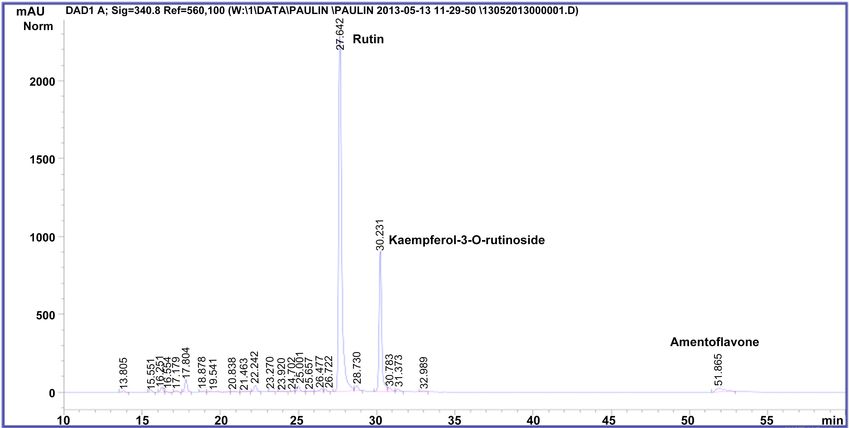
P. M. Kapepula et al.

                              Figure 1. Leaves of Manihot esculenta (Mwambu: A; TEM 419: B) and Manihot glaziovii
                              (C) from Kahemba.

                                 The leaves of M. glaziovii were only consumed in the west of the Democratic
                              Republic of the Congo to the best of our knowledge [6]. Elsewhere M. glaziovii is
                              particularly used as biomass or raw material for bioethanol and natural rubber
                              production [8] [9]. The vegetable sauces constitute the main protein sources for the
                              population of Kahemba, which does not consume meat and fish daily. Based on
                              the very high consumption of Manihot species leaves as a vegetable in DRC, it is
                              worth determining their potential bioactivities and nutritive values. Few reports
                              had documented the bioactivities of the leaves of M. esculenta and M. glaziovii.
                              Tsumbu et al. (2011, 2012) [3] [10] had evaluated the polyphenol content and
                              modulatory activities of M. esculenta, some green vegetables from Kongo Cen-
                              tral. The present paper reports the microscopic features, chromatographic fin-
                              gerprints and the biological activities of leaves of edible Manihot species from
                              DRC used such as traditional vegetables.

                              2. Materiel and Methods
                              2.1. Plant Material
                              The leaves of Manihot esculenta and Manihot glaziovii have been collected from
                              the areas of Kahemba (DR. Congo) in April 2018. The identity of the plant ma-
                              terial was established by Mr. Kombo (Agronomist at the Ministry of Agricul-
                              ture/Kahemba), and was confirmed by Mr. Anthony Kikufi, biologist from the
                              University of Kinshasa (DR. Congo). The leaves were soaked in hot water
                              (100˚C: 1 to 3 minutes), drained and dried at room temperature. The reduction
                              to powder of leaves was done by using a high-speed mill (Retsch ZM 100 Mod-
                              el).

                              2.2. Chemicals
                              All solvents used were of analytical and HPLC grade and purchased from Merck

DOI: 10.4236/jbm.2021.99006                        61                          Journal of Biosciences and Medicines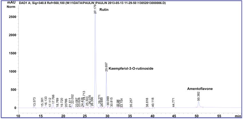
P. M. Kapepula et al.

                              VWR (Leuven, Belgium). 2-Aminoethyldiphenylborat and Phorbol-12-myristate-
                              13-acetate (PMA) were purchased from Sigma (Bornem, Belgium).
                              2’,7’-Dichlorofluorescein-diacetate (DCFH-DA) was purchased from Eastman
                              Kodak (Rochester, NY, USA). L0-12 (8-amino-5-chloro-7-phenylpyrido [3,4-d]
                              pyridazine-1,4(2H, 3H) dione) was purchased from Wako Chemicals Gmbh (Neuss,
                              Germany). Horseradish Peroxidase (HRP) was obtained from Roche (Mannheim,
                              Germany) and human Myeloperoxidase was from Calbiochem, EMD Millipore
                              (Bellirica, MA USA). Gallic acid (purity: 97%) was purchased from Sigma-Aldrich.
                              Rutin (purity ≥ 99%), isoquercitrin (purity ≥ 99%) and, Hyperoside (purity ≥
                              98.5%) were HPLC grade and purchased from Extrasynthese. Water was treated
                              using a Milli-Q water ultra-purification system before use.

                              2.3. Microscopic Analysis
                              Microscopic observations were made using lactic acid reagent [11]. Observations
                              and pictures were done with a Zeiss Primo Star microscope coupled to camera
                              (DP 200).

                              2.4. Preparation of Extracts
                              Methanolic extracts were prepared by percolation with methanol from 10 g of
                              leaf powder to obtain 200 mL of percolate. The evaporation of the solvent was
                              performed under reduced pressure (at 40˚C) followed by 48 - 72 h stay in a va-
                              cuum chamber. The extracts were then weighed and kept in dark hermetic flasks
                              at 4˚C.

                              2.5. Chromatographic Analysis
                              Analytical analysis by Thin Layer Chromatographic of 10 μL of solution for 10
                              mg/mL of methanolic extracts was carried out on normal phase Silica Gel 60 F254
                              plates (Merck, Darmstadt, Germany), using a mixture of solvents as suitable
                              eluents. The plates were visualized at 365 nm with Neu reagent [12].
                                The separation of phenolic compounds of methanolic extract was carried
                              HPLC-DAD out on a Hypersil ODS® RP18 column as described previously [13].

                              2.6. Cellular and Enzymatic Assays
                              2.6.1. Cellular Antioxidant Activity
                                1) Cell culture and treatment
                                Human promyelocytic leukemia cells (HL-60) were obtained from the Amer-
                              ican Type Culture Collection (ATCC, the USA) and cultured in the appropriate
                              medium (IMDM obtained from Biowest, France). Equine Neutrophils were iso-
                              lated as described previously [10].
                                 2) Measurement of Cellular Antioxidant Activity (CAA)
                                a) Measurement of the ROS produced by PMA-Activated HL-60 mono-
                              cytes (fluorescence technique with non-fluorescent DCFH-DA)
                                This technic was based on the method described previously [13] [14].

DOI: 10.4236/jbm.2021.99006                        62                         Journal of Biosciences and Medicines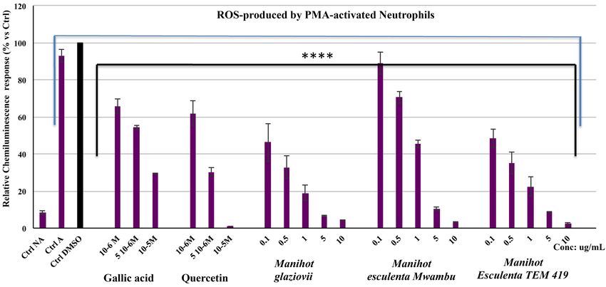
P. M. Kapepula et al.

                                b) Measurement of the total ROS produced by PMA activated neutrophils
                              (chemiluminescence assay)
                                The ROS produced by activated neutrophils were measured by lucigenin-en-
                              hanced chemiluminescence (CL) as reported by Franck et al. (2013) [15].

                              2.6.2. Inhibition of Peroxidase Activity
                                1) Inhibition of myeloperoxidase activity
                                This test was carried out using the SIEFED method that evaluate the capacity
                              of chemical compounds such as phytochemicals extracts from natural products
                              to modulate the activity of MPO [15].
                                2) Inhibition of HRP oxidant activity
                                The used method evaluates the modulatory effect of chemical compounds or
                              extracts from natural products on HRP catalytic activity using L012, a luminol-
                              based chemiluminescent probe as described previously [16].

                              2.7. Cell Viability
                              Cell survivals rate were quantified using a classic colorimetric WST-1 assay to
                              measure mitochondrial activity in viable cells [17] and an exclusion test with
                              Trypan blue [18].

                              2.8. Statistical Analysis
                              The software was performed with GraphPad 7.0 (GraphPad Software, San Diego
                              California, the USA) was used to perform statistical analysis. Two-way analysis
                              (ANOVA), Student’s paired t-test, “Tukey” Multiple Comparisons Test were the
                              test performed. The level of statistical significance was set at p < 0.05.

                              3. Results and Discussion
                              3.1. Botanical Microscopic Characteristics
                              Powders from the leaves of Manihot species showed the following specific bo-
                              tanical microscopic characters. Manihot esculenta (Mwambu) revealed the ab-
                              undance of spherical starch granules ~3 - 18 µm diameter, large underlying pa-
                              lisade cells, calcium oxalate prism ~38 µm long, upper epidermis in surface view,
                              showing thicker-walled cells, abundant lignified fibers, isolated sclereids up to
                              ~53 × 32 µm (L × W), the group of isodiametric sclereids, fibrous sclereids up to
                              ~335 × 20 µm (L × W) usually strung at one end, unicellular non glandular tri-
                              chomes up to ~172 µm long, diacytic stomata, epidermis of polygonal cells, the
                              fragment of bordered pitted vessels (Appendix Figure 1S). M. esculenta cultivar
                              TEM 419 showed large underlying palisade cells and calcium oxalate prism ~27
                              × 15 µm (L × W), few starches granules up to 17 µm diameter, diacytic stomata,
                              unicellular non-glandular trichomes ~146 - 218 µm long, scalariform vessels,
                              lignified fibers, the group of sclereids, isolated elongated sclereids ~35 - 85 µm
                              long, fibrous sclereids ~ up to 290 × 20 µm (L × W), diacytic stomata (Appendix
                              Figure 2S). M. glaziovii revealed abundant lignified fibers, large underlying pa-

DOI: 10.4236/jbm.2021.99006                         63                          Journal of Biosciences and Medicines
P. M. Kapepula et al.
lisade cells ~30 - 11 µm (L × W), parenchyma with clusters crystal of calcium
oxalate ~8 µm diameter, upper epidermis in surface view showing thicker-walled
cells, few starches granules (~6 - 18 µm diameter), numerous smooth unicellular
no glandular trichomes up to 277 µm long, glandular trichomes, cyclocytic sto-
mata, the fragment of helical vessels, elongated sclereids ~92 × 36 µm (L × W),
and fibrous sclereids up to ~248 × 18 µm (L × W) (Appendix Figure 3S). The
stomata, vessels of M. glaziovii are different of those from M. esculenta. The
number and dimension of the starches granules, sclereids, and fibrous sclereids
are not also the same. Sclereids, starches granules from M. esculenta were usual-
ly small than those of M. glaziovii. Microscopy analysis allowing the identifica-
tion of herbal drugs and the detection of individual components of a mixture
[19], the obtained results could contribute to characterize Manihot species.
3.2. Phenolic Compounds
Chromatographic fingerprints of methanolic extracts hinted at the presence of
flavonoids and phenolic acids as major phytochemicals. By comparison with used
standards, it was showed that extracts of M. esculenta contain quercetin-3-rutinoside
(rutin), the most abundant, amentoflavone, isoquercitrin, kaempferol-3-rutinoside
and other non-identified flavonoids (Figure 2 and Figure 3), in accordance with
previous results [3] [20]. M. glaziovii contained equally amentoflavone, querce-
tin-3-rutinoside, kaempferol-3-rutinoside but also quercetin-3-glucoside (Figure
4).
Quercetin has been also identified in the two species. Caffeic acid, gallic acid
and others non-identified acids were also detected in the two species. Ola et al.
(2009) [20] reported that ferulic acid is the main phenolic acid from leaves of M.
esculenta from Nigeria. In our study, there is not ferulic acid in Manihot extracts
Figure 2. HPLC-DAD chromatogram of methanolic extract from Manihot esculenta (cultivar Mwambo).
DOI: 10.4236/jbm.2021.99006 64 Journal of Biosciences and MedicinesP. M. Kapepula et al.
Figure 3. HPLC-DAD chromatogram of methanolic extract from Manihot esculenta (cultivar TEM 419).
Figure 4. HPLC-DAD chromatogram of methanolic extract from Manihot glaziovii.
as shown the TLC and HPLC fingerprints in comparison with standard of ferulic
acid. The chemical composition of plant extracts is related to different parame-
ters such as varieties, genetic, ecology, harvest conditions and the types of ex-
tracts. Chromatographic fingerprints of samples from studied Manihot species
were nearly similar (Figures 2-4). Nevertheless, chromatographic analysis re-
vealed that the two variety of M. esculenta had a chemical fingerprint different
from M. glaziovii.
DOI: 10.4236/jbm.2021.99006 65 Journal of Biosciences and MedicinesP. M. Kapepula et al.
3.3. Cellular Antioxidant Activity
Cell-free antioxidant assays were largely used to evaluate the antioxidant activity
of pure compounds and plant extracts. Cellular models such as those using cells
specialized in the production of reactive species and inflammatory responses al-
low the evaluation of antioxidant and anticatalytic capacities as a complement to
cell-free antioxidant assays. In this study we evaluated the capacities of extracts
to modulate the ROS production resulting mainly from NADPH oxidase activity
by stimulated neutrophil and HL-60 cells [21].
On the one hand, in the range of the concentration of 0.1 to 10 µg·mL −1 for
extracts from Manihot leaves and of 10−6 to 10−4 M for positive controls (gallic
acid and quercetin), we had observed a significant decrease of the neutrophils
ROS production compared to the control test performed with DMSO. Obtained
results showed that the cellular antioxidant activity of extracts is significantly
higher (p < 0.001) in the following order: M. glaziovii > M. esculenta (Mwam-
bu) > M. esculenta (TEM 419) (Figure 5).
The highest inhibitory effect was related to their polyphenolic content and the
obtained IC50 were 0.11 ± 0.05 µg·mL−1, 0.14 ± 0.03 µg·mL−1 and 0.69 ± 0.15
µg·mL−1 for M. glaziovii, M. esculenta (Mwambu) and M. esculenta (TEM 419)
respectively. The total phenol contents of M. glaziovii, M. esculenta (cultivar
Mwambu) were two to three higher than M. esculenta (cultivar TEM 419), which
correlated to their antiradical activity as recently reported [6].
On the other hand, at the concentration of 10, 50 and 100 µg·mL−1 the leaf
Figure 5. Effects of gallic acid, quercetin and methanolic extracts of Manihot species on the CL response produced by PMA acti-
vated equine neutrophils (Means ± SD, n = 6). The CL intensity results from the reaction between lucigenin and the ROS pro-
duced by the non-activated (NA) and activated equine neutrophils (A). The CL response of stimulated neutrophils in the presence
of DMSO used to solubilize the extracts was defined as 100%. P-values (****p < 0.0001) calculated by two-way ANOVA followed
by Sidak Multiple Comparisons Test indicated a significant effect of the extracts vs. DMSO control; ns = not significant vs. DMSO
control.
DOI: 10.4236/jbm.2021.99006 66 Journal of Biosciences and MedicinesP. M. Kapepula et al.
Manihot extracts and of 10−6, 10−5 10−4 M the quercetin have produced a low
dose-dependent decrease of HL-60 ROS production compared to the control test
performed with DMSO. Obtained results with this cellular model showed that
the effect of methanolic extracts is significantly higher (p < 0.05) only at the 100
µg·mL−1 in the following order: M. glaziovii > M. esculenta (Mwambu) > M. es-
culenta (TEM 419) compared to the previous model using lucigenin on neutro-
phils. At 100 µg·mL−1, the percentage of ROS inhibition was of 38.36%, 36.87%
and 30.09% for M. glaziovii, M. esculenta (Mwambu), M. esculenta (TEM 419)
respectively.
Indeed, Manihot extracts were active in the two cell-models assays, and
showed a more pronounced inhibitor effects on ROS production in the lucigenin
CL assay. Tsumbu et al. (2012) had reported the same observations with aqueous
extracts of Manihot esculenta from another area of DRC, i.e. Kongo Central. Lu-
cigenin is considered to be a more specific probe for the detection of superoxide
anions directly produced by the activity of NADPH oxidase and released in the
extracellular media [22]. DCFH-DA makes it possible to indirectly measure the
effect of intracellular antioxidant activities against intracellular ROS production
in fluorescence assay.
Regarding the results obtained in the fluorescence assay, we presumed that
there would be interferences of ions and molecules of plant extracts on the mod-
ulatory effect on intracellular ROS production. For this, assays with EDTA and
Chelex, both being used to complex metallic ions, were performed in the first
hand for excluding the Fenton-like reaction. The reaction between H2O2 and
Fe2+ (Fenton reaction) leads to the formation of hydroxyl radical (•OH) that can
oxidize DCFH to DCF. The Fenton reaction might lead to DCF-amplified fluores-
cence that could lead to the low inhibitory effect on ROS production [23]. On the
other hand, we tested the possible interferences between the probe (DCFH-DA)
and compounds in the extracts. Assays were performed by comparing the fluo-
rescence intensity of the cells (HL60 monocytes), incubated with the probe, and
extracts compared to that obtained when HL60 were incubated with the extracts
without the probe.
The obtained results suggested that there were not any ionic and molecular
interference: This indicated that the components of the tested extracts were not
very good intracellular ROS scavengers. Tested extracts contained glycosylated
flavonoids as major phenolic compounds. Previous studies reported that me-
thanolic extracts of edible Hibiscus and herbal teas from DRC exhibited high ef-
fect on intracellular ROS related to their abundance of phenolic acids [13] [24].
In DCFH-DA fluorescence assay, flavonoids seem to be less active than phenolic
acids. Assays with molecules of flavonoids (quercetin and its glycosides) and
phenolic acids standards in the range concentrations of 10−6 - 10−4 M, showed
that phenolic acids were more active to inhibit intracellular ROS than flavono-
ids. For flavonoids, genins were more active than glycosides and the glycosides
with one sugar are more active than those with several sugars.
DOI: 10.4236/jbm.2021.99006 67 Journal of Biosciences and MedicinesP. M. Kapepula et al.
Nevertheless, Takamatsu et al. (2003) [25] showed that the antioxidant effica-
cy in DCFH-DA fluorescence model depends on the nature of substituents of the
rings of flavonoids and to a great extent on the ability of molecules to penetrate
the cell membranes.
3.4. Inhibition of Peroxidase Activity
The evaluation of the inhibition of peroxidase activity carried out with tests us-
ing as enzymes the Myeloperoxidase (MPO) and the Horseradish Peroxidase
(HRP).
MPO, a pro-oxidant enzyme involved in secondary cell damage and consi-
dered as a marker of inflammation [15]. In SIEFED (Specific Immunological
Extraction Followed by Enzymatic Detection) technique, at the concentrations
of 1, 5 and 10 µg·mL−1, all Manihot extracts showed a significant inhibitory effect
(p < 0.001) on MPO activity in the following order: M. esculenta (TEM 419) >
M. esculenta (Mwambu) > M. glaziovii. The observed effect showed that mole-
cules of Manihot extracts interact better with the active site of MPO. The SIEFED
technic is an immunological test which allows detecting compounds that have
direct interaction with the MPO [15].
Altogether the results of our study showed that the extracts tested have the
highest antioxidant activities and the highest inhibition on the activity of MPO.
As reported by previous studies, molecules or the plant extracts which having a
good antiradical or antioxidant activities are not necessarily good inhibitors of
MPO activity. Gallic acid is less efficient MPO inhibitor compared to quercetin
that is less antiradical [13] [25]. M. glaziovii has showed good antiradical and
antioxidant activities than M. esculenta (TEM 419), but it had a low inhibitory
effect on MPO activity. Phenolic acids and glycosylated flavonoids such as rutin
were found to be the major phenolic compounds of Manihot extracts. Flavono-
ids were reported to be excellent inhibitors of MPO [26] [27]. Previous studies
reported for benzoic acid derivatives, a pyrogallol and the elongation of the car-
boxylic group seem to be essential for the inhibition of MPO activity. These con-
figurations would facilitate interactions of molecules with the MPO active site
[28]. Gallic acid induced a more dose dependent anticatalytic activity on MPO
than caffeic acid and its derivatives [13].
The inhibition of HRP oxidant activity was studied by chemiluminescence
method using L-012 as probes. L0-12 is a chemical analog that has been reported
to gives rise to significantly higher luminescence yield and increased sensitivity
compared to other CL probes, such lucigenin [29]. HRP was used for the inves-
tigation of inhibitor activity of anti-thyroid and anti-inflammatory drugs [30].
At the concentration of 0.1; 1 and 10 µg·mL−1, Manihot extracts showed an ef-
fective inhibition of HRP oxidant activity (Appendix Figure 4S).
At the concentration of 10 μg·mL−1, the percentage of the inhibition effect was
more than 50% for M. glaziovii and M. esculenta (Mwambu); and of 32.53% for
M. esculenta (TEM 419). Regarding to our results with this assay, Manihot ex-
DOI: 10.4236/jbm.2021.99006 68 Journal of Biosciences and MedicinesP. M. Kapepula et al.
tracts exhibited a great capacity to inhibit HRP catalytic activity related to their
phytochemicals. Flavonoids such as quercetin derivatives contained in Manihot
extracts, could be responsible of the inhibition effect on HRP catalytic activity.
Mahfoudi et al. (2017) [30] reported that flavonoids could be promising HRP
inhibitors and can help in developing new molecules to control thyroid diseases.
The uncontrolled stimulation of neutrophils leading to neutrophil degranula-
tion associated with some acute and chronic diseases, could contribute to ampli-
fy or maintain the inflammatory response with the release of peroxidases such as
MPO [31]. The activity of MPO produces highly diffusible reactive oxidants,
which provoke oxidative damage in the host tissues at inflammatory sites. MPO
and its metabolites are as promising biomarkers not only for infectious diseases,
but also for a wide array of non-infectious and neurodegenerative disorders [32].
Our results demonstrated that all tested extracts exerted a noticeable inhibitory
effect on the MPO and on HRP catalytic activity. Polyphenols have by their an-
tioxidant, anti-inflammatory capacities, may to confer health benefit in diverse
neurodegenerative disorders associated with oxidative damage [33]. The inhibi-
tors of HRP and MPO activity are promising therapeutic agents such as an-
ti-inflammatory drugs.
Manihot species contain cyanogenic glycosides (α-hydroxynitrile glucosides)
and leaves have high levels than roots. Cyanogenic glycosides (linnamarin, lo-
taustralin) break down to release toxic cyanide (HCN) when plant tissue is crushed
or chewed, disrupting the cells [1]. The consumption of leaves such as vegetable
needed a better culinary preparation. The processing preparation of Saka-saka,
the sauce from cassava leaves has several stapes: blanching in warm water for a
few minutes, grinding before pounding, boiling 30 minutes before the mixing
with ingredients. The heat treatment and the consistency of pounding play a role
in the reduction of cyanogens. Destruction of the cells leads to contact between
the cyanogenic glucosides and the endogenous linamarase with the subsequent
release of HCN. Ngudi et al. (2003) [34] reported that 96% - 99% of the total
cyanogens were removed after cooking of the cassava leaves. Tested samples of
Manihot species were submitted to heat pretreatment. We estimated that this
treatment allowed the removing of the maximum of cyanogenic compounds be-
fore analysis and it does not affected the bioactivities of Manihot extracts on cel-
lular and enzymatic models, which are essentially related to phenolic compounds
and not to the toxic effect of cyanogens. For this, we performed the cell viability
tests on HL-60 monocytes and neutrophils. The cell viability of HL-60 cells and
equine neutrophils treated with Manihot extracts was significantly superior to the
control group except at the highest concentration tested (10 µg·mL−1). The extracts
of Manihot species caused no cell toxicity and there was no significant difference
of viability between cells incubated with plant extracts and those without extract
solutions (control cells). These results suggest that Manihot extracts do not have
a toxic effect at the high concentration (10 µg·mL−1) and even have a slight protec-
tive effect against cell death at low concentrations (0.5 µg·mL−1). The Manihot ex-
DOI: 10.4236/jbm.2021.99006 69 Journal of Biosciences and MedicinesP. M. Kapepula et al.
tracts did not induce cytotoxicity at doses showing antioxidant and peroxidase
inhibition activities.
Cassava production is growing in the peri-urban areas of Kinshasa for the ex-
ploitation of the roots as raw material for many processing products like liquid
starch, cosettes, chikwange ... by local entrepreneurs. Cassava leaves are for the
Congolese population, great nutritional and economic values as a source of pro-
teins, minerals, and as a source of income for households. The nutritional value
of this vegetable makes its consumption and marketing become more and more
important in the DRC than abroad.
The cellular antioxidant and the inhibition of peroxidases activities of Mani-
hot leaves were positively correlated with their phytochemicals. These bioactivi-
ties justify the benefit effect of Manihot leaves such as traditional vegetable and
potential nutraceutical resources and medicines with beneficial health for Con-
golese people.
4. Conclusion
Microscopic features, chromatographic fingerprints and biological activities of
two Congolese Manihot species were determined. The metabolic profile of M.
glaziovii appears quite similar profiles to those of M. esculenta. Methanolic ex-
tracts tested have the best antioxidant activities. They appeared less efficient on
the inhibition of the production intracellular ROS of HL60 cells, and more effi-
cient as radical scavengers, on the inhibition of the production of extracellular
ROS of neutrophils and the inhibition of MPO and HRP oxidant activities. The
antioxidant and the inhibition of MPO and HRP activities of the leaves of the
studied Manihot species would potential therapeutic interest and could justify
their traditional use as vegetables, potential functional foods or nutraceutical re-
sources and medicines. However, we estimate that further studies are needed,
especially in vivo studies, to demonstrate the benefit of Manihot leaves extracts
in health.
Acknowledgements
The authors thank Mr. Landu and Mr. A. Kikufi of the University of Kinshasa
for identification of the plants studied. We also thank Jean Noel Wauters, Del-
phine Etienne, Jennifer Romainville and A. Niesten for their technical advice
and the community of Kahemba for his collaboration.
Conflicts of Interest
The authors declare no conflict of interest.
References
[1] Burns, A.E., Gleadow, R.M., Zacarias, M., Miller, R.E. and Cavagnaro, T.R. (2012)
Variations in the Chemical Composition of Cassava (Manihot esculenta Crantz)
Leaves and Roots as Affected by Genotypic and Environmental Variation. Journal
DOI: 10.4236/jbm.2021.99006 70 Journal of Biosciences and MedicinesP. M. Kapepula et al.
of Agricultural and Food Chemistry, 52, 1075-1085.
[2] Obadina, A.O., Oyewole, O.B. and Williams, O.E. (2013) Improvement in the Tra-
ditional Processing Method and Nutritional Quality of Traditional Extruded Cassa-
va-Based Snack (Modified Ajogun). Food Science and Nutrition, 1, 350-356.
https://doi.org/10.1002/fsn3.43
[3] Tsumbu, C.N., et al. (2011) Antioxidant and Antiradical Activities of Manihot es-
culenta Crantz (Euphorbiaceae) Leaves and Other Selected Tropical Green Vegeta-
bles Investigated on Lipoperoxidation and Phorbol-12-Myristate-13-Acetate (PMA)
Activated Monocytes. Nutrients, 3, 818-838. https://doi.org/10.3390/nu3090818
[4] Bell, A., Muck, O. and Schuler, B. (2000) Les richesses du sol Les plantes à racines et
tubercules en Afrique: Une contribution au développement des technologies de
récolte et d’après-récolte. INPHO.
[5] Kashala-Abotnes, E., et al. (2018) Dietary Cyanogen Exposure and Early Child Neu-
rodevelopment: An Observational Study from the Democratic Republic of Congo.
PLoS ONE, 13, e0193261. https://doi.org/10.1371/journal.pone.0193261
[6] Kapepula P.M., Tshala-Katumbay, D., Mumba, D., Frédérich, M., Mbemba, T. and
Ngombe, N.K. (2018) Traditional Foods as Putative Sources of Antioxidants with
Health Beneits in Konzo. Antioxidants in Foods and Its Applications, 117-135.
https://doi.org/10.5772/intechopen.74523
[7] Diasolua Ngudi, D., Banea-Mayambu, J.P., Lambein, F. and Kolsteren, P. (2011)
Konzo and Dietary Pattern in Cassava-Consuming Populations of Popokabaka,
Democratic Republic of Congo. Food and Chemical Toxicology, 49, 613-619.
https://doi.org/10.1016/j.fct.2010.06.053
[8] Rodrigues, J.F., Rodrigues, Â.S. and Cardoso, A.L.H. (1991) Characterisation of
Natural Rubber from Manicoba (Manihot glaziovii): Microstructure and Average
Molecular Weight. Journal of Rubber Research, 6, 134-136.
[9] Moshi, A.P., et al. (2014) Characterisation and Evaluation of a Novel Feedstock,
Manihot glaziovii, Muell. Arg, for Production of Bioenergy Carriers: Bioethanol and
biogas. Bioresource Technology, 172, 58-67.
https://doi.org/10.1016/j.biortech.2014.08.084
[10] Tsumbu, C.N., Ginette, D.-D., Monique, T., Luc, A., Thierry, F., Didier, S. and Frank,
T. (2012) Polyphenol Content and Modulatory Activities of some Tropical Dietary
Plant Extracts on the Oxidant Activities of Neutrophils and Myeloperoxidase. In-
ternational Journal of Molecular Sciences, 13, 628-650.
https://doi.org/10.3390/ijms13010628
[11] Bahati, L.M., et al. (2017) Microscopic Features, Chromatographic Fingerprints and
Antioxidant Property of Some Unconventional Green Leafy Vegetables Consumed
in Bandundu, DR Congo. Pharmacognosy Communications, 7, 158-163.
https://doi.org/10.5530/pc.2017.4.23
[12] Wagner, H., Bauer, R., Melchart, D., Xioa, P.-G. and Staudinger, A. (2013) Chro-
matographic Fingerprint Analysis of Herbal Medicinal: Thin-Layer High Perfor-
mance Liquid Chromatography of Chinese Drugs. Springer, Berlin.
[13] Kapepula, P.M., et al. (2017) Comparison of Metabolic Profiles and Bioactivities of
the Leaves of Three Edible Congolese Hibiscus Species. Natural Product Research,
6419, 1-8.
[14] Tsumbu, C.N., Deby-Dupont, G., Tits, M., Angenot, L., Franck, T. and Serteyn, D.
(2011) Antioxidant and Antiradical Activities of Manihot esculenta Crantz (Eu-
phorbiaceae) Leaves and Other Selected Tropical Green Vegetables Investigated on
Lipoperoxidation and Phorbol-12-Myristate-13-Acetate (PMA) Activated Mono-
DOI: 10.4236/jbm.2021.99006 71 Journal of Biosciences and MedicinesP. M. Kapepula et al.
cytes. Nutrients, 3, 818-838. https://doi.org/10.3390/nu3090818
[15] Franck, T., et al. (2013) Differentiation between Stoichiometric and Anticatalytic
Antioxidant Properties of Benzoic Acid Analogues: A Structure/Redox Potential
Relationship Study. Chemico-Biological Interactions, 206, 194-203.
https://doi.org/10.1016/j.cbi.2013.09.009
[16] Ngombe, N., et al. (2019) Peroxidase Inhibition and Antioxidant Activity of Bulk-Mar-
keted Black Tea (Camellia sinensis L.) from the Democratic Republic of the Congo.
Journal of Biological Sciences and Medicine, 7, 66-80.
https://doi.org/10.4236/jbm.2019.79007
[17] Ishiyama, M., Miyazono, Y., Sasamoto, K., Ohkura, Y. and Ueno, K. (1997) A Highly
Water-Soluble Disulfonated Tetrazolium Salt as a Chromogenic Indicator for NADH
as Well as Cell Viability. Talanta, 44, 1299-1305.
https://doi.org/10.1016/S0039-9140(97)00017-9
[18] Tennant, J.R. (1964) Evaluation of the Trypan Blue Technique for Determination of
Cell Viability. Transplantation, 2, 685-694.
https://doi.org/10.1097/00007890-196411000-00001
[19] Jackson Derek, B.P. (1990) Atlas of Microscopy of Medicinal Plants Culinary Herbs
And Spices. CBS HB, Snowdon.
[20] Ola, S.S., Catia, G., Marzia, I., Francesco, V.F., Afolabi, A.A. and Nadia, M. (2009)
“HPLC/DAD/MS Characterisation and Analysis of Flavonoids and Cynnamoil De-
rivatives in Four Nigerian Green-Leafy Vegetables. Food Chemistry, 115, 1568-1574.
https://doi.org/10.1016/j.foodchem.2009.02.013
[21] Derochette, S., Franck, T., Mouithys-Mickalad, A. and Deby-Dupont, G. (2013) In-
tra- and Extracellular Antioxidant Capacities of the New Water-Soluble Form of
Curcumin (NDS27) on Stimulated Neutrophils and HL-60 Cells. Chemico-Biological
Interactions, 201, 49-57. https://doi.org/10.1016/j.cbi.2012.12.010
[22] Li, Y., Zhu, H., Kuppusamy, P., Roubaud, V., Zweier, J.L. and Trush, M.A. (1998)
Validation of Lucigenin (Bis-N-Methylacridinium) as a Chemilumigenic Probe for
Detecting Superoxide Anion Radical Production by Enzymatic and Cellular Sys-
tems. Journal of Biological Chemistry, 273, 2015-2023.
https://doi.org/10.1074/jbc.273.4.2015
[23] Oddvar, M., Jannike, M.A., Aarnes, H. and Fonnum, F. (2003) Evaluation of the
Probes 2’,7’-Dichlorofluorescin Diacetate, Luminol, and Lucigenin as Indicators of
Reactive Species Formation. Biochemical Pharmacology, 65, 1575-1582.
https://doi.org/10.1016/S0006-2952(03)00083-2
[24] Kapepula, P.M., et al. (2017) Antioxidant Potentiality of Three Herbal Teas Con-
sumed in Bandundu Rural Areas of Congo. Natural Product Research, 31, 1940-1943.
https://doi.org/10.1080/14786419.2016.1263844
[25] Takamatsu, S., et al. (2003) Antioxidant Effect of Flavonoids on DCF Production in
HL-60 Cells. Phytotherapy Research, 17, 963-966. https://doi.org/10.1002/ptr.1289
[26] Nyssen, P., et al. (2018) Morphine, a Potential Inhibitor of Myeloperoxidase Activi-
ty. Biochim. Biophys. Acta: General Subjects, 1862, 2236-2244.
https://doi.org/10.1016/j.bbagen.2018.07.007
[27] Shiba, Y., et al. (2008) Flavonoids as Substrates and Inhibitors of Myeloperoxidase:
Molecular Actions of Aglycone and Metabolites. Chemical Research in Toxicology,
21, 1600-1609. https://doi.org/10.1021/tx8000835
[28] Gau, J., et al. (2016) Flavonoids as Promoters of the (Pseudo-)Halogenating Activity
of Lactoperoxidase and Myeloperoxidase. Free Radical Biology and Medicine, 97,
307-319. https://doi.org/10.1016/j.freeradbiomed.2016.06.026
DOI: 10.4236/jbm.2021.99006 72 Journal of Biosciences and MedicinesP. M. Kapepula et al.
[29] Zielonka, J., Lambeth, J.D. and Kalyanaraman, B. (2013) On the Use of L-012, a Lu-
minol-Based Chemiluminescent Probe, for Detecting Superoxide and Identifying
Inhibitors of NADPH Oxidase: A Reevaluation. Free Radical Biology and Medicine,
65, 1310-1314. https://doi.org/10.1016/j.freeradbiomed.2013.09.017
[30] Mahfoudi, R., Djeridane, A., Benarous, K., Gaydou, E.M. and Yousfi, M. (2017) Struc-
ture-Activity Relationships and Molecular Docking of Thirteen Synthesized Flavo-
noids as Horseradish Peroxidase Inhibitors. Bioorganic Chemistry, 74, 201-211.
https://doi.org/10.1016/j.bioorg.2017.08.001
[31] Serteyn, D., Grulke, S., Franck, T., Mouithys-Mickalad, A. and Deby-Dupont, G. (2003)
La myéloperoxydase des neutrophiles, une enzyme de défense aux capacités oxy-
dantes. Annales de Médecine Vétérinaire, 147, 79-93.
[32] Ray, R.S. and Katyal, A. (2016) Myeloperoxidase: Bridging the Gap in Neurodege-
neration. Neuroscience & Biobehavioral Reviews, 68, 611-620.
https://doi.org/10.1016/j.neubiorev.2016.06.031
[33] Vauzour, D., Kerr, J. and Czank, C. (2013) Plant Polyphenols as Dietary Modulators
of Brain Functions. Polyphenols in Human Health and Disease, 1, 357-370.
https://doi.org/10.1016/B978-0-12-398456-2.00027-X
[34] Ngudi, D.D., Kuo, Y.H. and Lambein, F. (2003) Cassava Cyanogens and Free Ami-
no Acids in Raw and Cooked Leaves. Food and Chemical Toxicology, 41, 1193-1197.
https://doi.org/10.1016/S0278-6915(03)00111-X
Appendix
https://drive.google.com/file/d/1K8GUu2Z2S1Rbit5tpos7OD0Ie-veJtmD/view?ts
=6122d15f
DOI: 10.4236/jbm.2021.99006 73 Journal of Biosciences and MedicinesYou can also read