Pyridoxal 5'-phosphate to mitigate immune dysregulation and coagulopathy in COVID-19 - Preprints.org
←
→
Page content transcription
If your browser does not render page correctly, please read the page content below

Preprints (www.preprints.org) | NOT PEER-REVIEWED | Posted: 8 May 2020                    doi:10.20944/preprints202005.0144.v1

             Pyridoxal 5'-phosphate to mitigate immune dysregulation
             and coagulopathy in COVID-19
             Julie Desbarats1

             Abstract

             Although most cases of COVID-19 are paucisymptomatic, severe disease is characterized
             by immune dysregulation, with a decreased type I interferon response,1 increased
             inflammatory indicators, surging IL-6, IL-10 and TNFα suggestive of cytokine storm,2–4
             progressive lymphopenia, and abnormal blood clotting.5,6 Factors determining
             susceptibility to severe disease are poorly understood, although mortality correlates
             with increasing age and co-morbidities including diabetes and cardiovascular disease
             (CVD).7,8 Pyridoxal 5'-phosphate (PLP) tends to be insufficient in populations particularly
             vulnerable to COVID-19, including the elderly,9,10 the institutionalized,11,12 and people
             with diabetes13,14 and CVD,15,16 and PLP becomes further depleted during infection and
             inflammation.17,18 In turn, low PLP results in immune imbalance, as PLP is an essential
             cofactor in pathways regulating cytokine production, in particular type I interferons and
             IL-6, and in lymphocyte trafficking and endothelial integrity.19 Furthermore, normalizing
             PLP levels attenuates abnormalities in platelet aggregation and clot formation.20–22
             Finally, PLP insufficiency induces excess secretion of renin and angiotensin,23 and
             hypertension.24 In inflammatory disease, pharmacological doses of PLP decrease
             circulating TNFα, IL-625 and D-dimer,26 and animal studies demonstrate that
             supplemental PLP shortens the duration and severity of viral pneumonia.27 Severe
             COVID-19 manifests as an imbalance in the immune response1 and the clotting system.5
             Pharmacological PLP supplementation may therefore mitigate COVID-19 symptoms by
             alleviating both the immune suppression underlying viral spread and the pathological
             hypersecretion of inflammatory cytokines, as well as directly bolstering endothelial
             integrity and preventing hypercoagulability.

             Keywords: COVID-19, SARS-CoV-2, pyridoxal 5'-phosphate, pyridoxine, vitamin B6, immune
             response, IL-6, TNF, type I interferon, lymphopenia, blood clotting, coagulopathy, cytokine
             storm, sphingosine-1-phosphate, kynurenine, inflammasome, serine hydroxymethyltransferase
             2 (SHMT2), hypertension, angiotensin

             1
                 ImmunoL0G1C R&D Inc., Montreal, Canada. julie.desbarats@mail.mcgill.ca

           © 2020 by the author(s). Distributed under a Creative Commons CC BY license.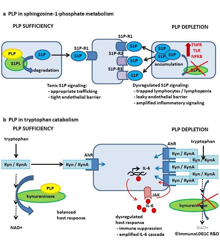
Preprints (www.preprints.org) | NOT PEER-REVIEWED | Posted: 8 May 2020     doi:10.20944/preprints202005.0144.v1

             COVID-19: a disease of systems imbalance

             From the first case series reported in China6 to recent overviews of the clinical
             picture in COVID-19,2,4,5 the heterogeneity of the host response has been striking.
             While most infections are asymptomatic or mild, critically ill patients show
             dysregulation of the immune response, the blood clotting cascade, and the
             vascular system.2,5,6 Plasma IL-6 and TNFα tend to increase with increasing disease
             severity, and in patients requiring ICU admission.2,3 Despite the seeming
             hyperactivity of the immune system in terms of inflammatory cytokine
             production, the majority of hospitalized patients show progressive lymphopenia,
             which may correlate with poor prognosis,6,28 and more recently, low levels of type
             I interferon, a key mediator in the anti-viral response, has been described.1 Thus,
             viral clearance is impaired by the insufficient early interferon response and the
             progressive lymphopenia, while overexpression of inflammatory cytokines leads
             to tissue and organ damage, leaky vasculature, plummeting blood pressure and
             shock. This inappropriate immune response leads to a dilemma for clinicians: to
             treat with immunosupressive drugs and risk aggravating viral pathology, or to let
             immune-mediated damage progress unchecked and exacerbate the patients’
             pathology.4

             In parallel, the blood clotting cascade is also dysregulated, with early reports of
             elevated D-dimer6 confirmed by recent accounts of clotting abnormalities and
             endothelial damage.5 Furthermore, regulation of vascular tone may be
             compromised, exacerbating organ damage particularly in the lung and kidney. 5

             Epidemiology of PLP insufficiency parallels COVID-19 susceptibility

             PLP, the active form of vitamin B6 (pyridoxine), is an essential co-factor in many
             inflammatory pathways and becomes depleted during inflammation.17,29 PLP
             deficiency leads to aberrant function of these inflammatory pathways, and their
             progressive dysregulation; PLP levels are inversely correlated with plasma IL-6 and
             TNFα in chronic inflammatory conditions.25,30 Population studies suggest that PLP
             levels decrease in older people (by 0.9 ng / ml / decade in unsupplemented

             Page 2                                 Desbarats 24/04/2020                   PLP for COVID-19
Preprints (www.preprints.org) | NOT PEER-REVIEWED | Posted: 8 May 2020 doi:10.20944/preprints202005.0144.v1
individuals),10 and up to 24% of Americans without supplemental vitamin intake
may have insufficient levels.31 Institutionalized people, particularly hard hit by
COVID-19 in many outbreaks, are more likely to suffer PLP deficiencies, with up to
70% of the institutionalized elderly in Canada and the US being PLP deficient.11,12
Interestingly, low PLP concentrations are more common in people with
diabetes13,14 and confer an increased and independent risk for CVD.15,29,32
Regardless of pre-existing PLP status, inflammation increases its utilization and
leads to its depletion,19,29,32 suggesting that COVID-19 patients experiencing
inflammation would become acutely depleted of PLP. Furthermore, depletion
tends to occur rapidly at sites of inflammation, leaving those areas particularly
vulnerable to the consequences of PLP deficiency. PLP is an essential cofactor in
several pathways involved in immunity and inflammation, notably sphingosine
metabolism and tryptophan catabolism,19 as well as in the regulation of
endothelial integrity, platelet aggregation, the clotting cascade20–22,26,33, and blood
pressure regulation via renin-angiotensin secretion.23
Sphingosine-1-phosphate (S1P) metabolism
S1P is a potent regulator of inflammation, acting specifically on lymphocyte
trafficking, permeability of the endothelial barrier, and cytokine and chemokine
production.34 PLP directly regulates the abundance of S1P, both as a gatekeeper
for its production and as a bottleneck for its degradation by S1P-lyase.35 PLP is
essential for S1P-lyase function. Inhibition of S1P-lyase results in trapping of
lymphocytes in secondary lymphoid organs and sites of inflammation, with a
consequent loss of lymphocytes in the circulation, resulting in lymphopenia and
systemic immune suppression,36 while the activated lymphocytes trapped in
inflamed tissue may secrete pro-inflammatory cytokines and exacerbate local
tissue damage. Furthermore, a key function of PLP involves tuning the leakiness of
endothelial barriers through regulation of tight junctions. When PLP is limiting,
dysregulated S1P turnover may allow signaling via S1P receptors 2 and 3 (S1PR2 /
S1PR3), resulting in leaky endothelial barriers, hypovolemic shock, and increased
fluid retention in inflamed tissues34 such as the lung in COVID-19. Finally, S1P also
Page 3 Desbarats 24/04/2020 PLP for COVID-19Preprints (www.preprints.org) | NOT PEER-REVIEWED | Posted: 8 May 2020 doi:10.20944/preprints202005.0144.v1
acts as an intracellular rheostat for inflammatory signaling. Changes in PLP
abundance modulate intracellular cascades involved in TNF receptor, nuclear
factor KB (NFKB), and some Toll-like Receptors (TLR) signaling, contributing to
excessive cytokine production and inflammatory tissue damage34 (Figure 1a).
Pharmacological S1P modulators are used to treat autoimmune disease,37 and
have shown promise in animal models of influenza- and SARS-induced cytokine
storm.38 However, pharmacological modulation of the S1P system typically acts by
inducing lymphocyte sequestration and immune suppression, which in the
context of COVID-19 could trigger viral resurgence. In contrast, restoring the
homeostatic function of the S1P system through PLP repletion may allow immune
function to move toward normal balance, enabling the control of viral replication
without uncontrolled cytokine expression. Similarly, loss of PLP during
inflammation, especially in patients with a pre-existing deficiency, may
exacerbate the loss of immune equilibrium characteristic of COVID-19 infection.
Tryptophan catabolism and the kynurenine system
PLP is a cofactor in the tryptophan catabolism pathway (Figure 1b). When PLP is
limiting, tryptophan catabolism to nicotinamide adenine dinucleotide (NAD+) is
impaired and kynurenines accumulate.39,40 Kynurenines are a family of tryptophan
catabolites which signal through the Aryl Hydrocarbon Receptor (AhR) and play
numerous roles in inflammation.40,41 When PLP is limiting, NAD+ production
though this pathway is compromised, limiting energy supply and stress
resilience.39,40 Concomitantly, kynurenine metabolites, including kynurenine,
kynurenic acid, 3-hydroxykynurenine, and quinolinic acid are overproduced,
resulting in pleotropic effects on the immune system which depend on the
relative abundance of the specific metabolites.40,41 Kynurenine tends to produce
immune suppression by decreasing NK activity and T cell proliferation and
increasing T cell apoptosis, while kynurenic acid may increase cytokine
production, in particular IL-6, TNFα, IL-10 and IL-1, and amplify signaling through
the IL-6 / JAK / STAT axis.40,41 Recently kynurenines have been reported to
stimulate an IL-6 positive feedback cycle which could amplify harmful
inflammation.42
Page 4 Desbarats 24/04/2020 PLP for COVID-19Preprints (www.preprints.org) | NOT PEER-REVIEWED | Posted: 8 May 2020 doi:10.20944/preprints202005.0144.v1
Figure 1. PLP regulates inflammation via S1P and tryptophan pathways.
a. PLP is a cofactor for S1P-lyase (S1PL) and regulates S1P abundance and tissue
distribution. S1P gradients control lymphocyte trafficking, including retention at sites of
inflammation and within lymphoid tissues, which can lead to lymphopenia in the
circulation. S1P receptors (S1P-R1,2,3) differentially modulate tight junctions in the
endothelium, and reduced S1PL function results in loss of barrier integrity. S1P may
Page 5 Desbarats 24/04/2020 PLP for COVID-19Preprints (www.preprints.org) | NOT PEER-REVIEWED | Posted: 8 May 2020 doi:10.20944/preprints202005.0144.v1
also amplify TNFα, TLR, and NFΚB intracellular signaling cascades, further
exacerbating leaky vasculature / shock and boosting inflammation, in turn further
depleting PLP.
b. PLP is a cofactor for kynureninase in the production of NAD+ from tryptophan. When
PLP is limiting, NAD+ production decreases, limiting energy supply. Concomitantly,
kynurenine metabolites (including kynurenine, Kyn, and Kynurenic Acid, KynA)
accumulate. Kyn and KynA signal through the Aryl Hydrocarbon Receptor (AhR),
resulting in immune suppression and paradoxically increased cytokine production (IL-6,
TNFα and IL-10) and amplified signaling through the IL-6 / JAK / STAT axis, potentially
resulting in an IL-6 positive feedback loop. In addition, other neurotoxic kynurenine
metabolites are produced, including neuroinflammatory 3-hydroxykynurenine and
excitotoxic quinolinic acid (not shown).40,43
This illustration leaves out the complex cross-talk and cross-amplification between the
S1P and AhR downstream effectors. In addition, PLP deficiency also upregulates IL-1β
and ROS production via the NLRP3 inflammasome and suppresses type I interferons
via serine hydroxymethyltransferase 2 (not shown).
Other inflammatory pathways
PLP suppresses IL-1β production and the production of reactive oxygen species
(ROS) by inhibiting the NLRP3 inflammasome, and thus PLP deficiency may
predispose to excessive IL-1β secretion and ROS-mediated tissue damage.44
PLP regulates serine hydroxymethyltransferase 2 (SHMT2), an enzyme best known
for its role in folate metabolism.19,45 In addition, SHMT2 also regulates type I
interferon production.45 Type I interferons are critical in the early host response
to viral infections and may underlie successful host control of viral replication.46
However, unregulated expression of type I interferons may result in significant
pathology.45,46 PLP modulates the availability and conformation of SHMT2, and
thus is a critical regulator in the type I interferon pathway.45 Here again,
availability of PLP determines the balance between host control of viral
replication, and cytokine-mediated pathology.
Page 6 Desbarats 24/04/2020 PLP for COVID-19Preprints (www.preprints.org) | NOT PEER-REVIEWED | Posted: 8 May 2020 doi:10.20944/preprints202005.0144.v1
Blood clotting and vascular effects of PLP
In 1963, a report in Nature designated vitamin B6 as a new anticoagulant.47
Treatment with physiological or supraphysiological doses of PLP prolongs
bleeding time, reduces platelet aggregation, and decreases thrombin
generation.20–22 In patients with homocysteinuria and clotting abnormalities, high-
dose PLP treatment restored antithrombin III activity to normal, increased factor
VII, and decreased beta-thromboglobulin,48 while lower doses of PLP were
sufficient to reduce D-dimer levels,26 although these effects were not seen at the
lowest levels of supplementation.49
PLP deficiency may also disrupt blood pressure regulation and compromise
cardiac energy metabolism. An animal model of hypertension may be produced
simply by decreasing PLP in the diet.24 In PLP deficient rats, production of both
renin and angiotensin is significantly increased.23 In COVID-19, angiotensin-
converting enzyme 2 (ACE2) serves as a receptor for the SARS-CoV-2 virus, and
hypertension is a risk factor for severe COVID-19 pathology. Furthermore, even
marginal PLP deficiency significantly attenuated citric acid cycle metabolite levels
in the heart, suggesting that low PLP levels impair cardiac energy metabolism.50
Finally, critically ill patients with inflammation presented about six times more
risk of cardiovascular complications when they were PLP-deficient, compared
with matched non-PLP-deficient patients.29
PLP as a weapon against COVID-19
Good nutrition and adequate vitamin intake have been recommended for COVID-
19 prevention,51 with vitamin D specifically implicated in immune protection.52
However, PLP deserves particular investigation because it controls the pathways
which are specifically dysregulated in COVID-19; because it becomes rapidly and
critically depleted during inflammation; and because in supraphysiological doses,
it has previously been shown to decrease pro-inflammatory cytokine levels and
indicators of coagulopathy.
Page 7 Desbarats 24/04/2020 PLP for COVID-19Preprints (www.preprints.org) | NOT PEER-REVIEWED | Posted: 8 May 2020 doi:10.20944/preprints202005.0144.v1
Figure 2. The systemic interactions of PLP deficiency with COVID-19 pathology
a Adequate PLP levels allow homeostatic function of 1. the innate immune response,
where type I interferons (IFN) restrain viral proliferation and spread (areas of viral
infection shown highlighted in yellow), and opposes excessive proinflammatory cytokine
secretion; 2. the adaptive immune response, where PLP supports lymphocyte
proliferation, appropriate trafficking, and regulated cytokine production; 3. the vascular
system, where PLP is essential for endothelial barrier integrity (shown in close-up view),
which prevents edema and maintains blood pressure (BP); and acts as an anti-
coagulant by regulating platelet aggregation and components of the clotting cascade.
b Insufficient PLP leads to dysregulated physiological systems as biochemical flow
through multiple pathways is disrupted, intermediates back up, and end-products are
not formed. 1. Viral spread (yellow) escapes Type I IFN control due to compromised
regulation of SHMT2. 2. Lymphocytes are trapped in lymph nodes and inflamed tissues
as SP1-lyase becomes hypofunctional, leading to lymphopenia in the circulation, and
Page 8 Desbarats 24/04/2020 PLP for COVID-19Preprints (www.preprints.org) | NOT PEER-REVIEWED | Posted: 8 May 2020 doi:10.20944/preprints202005.0144.v1
exacerbating tissue damage through excessive cytokine secretion and ROS production
via multiple dysregulated pathways, including S1P, kynurenines, inflammasomes, and
lack of downregulatory opposition from Type I IFN. Lymphopenia (from compromised
S1P-lyase) and hypofunctional lymphocytes (from immunosuppressive kynurenine
accumulation) further contribute to viral spread. 3. The vascular system, already
susceptible to direct viral pathology due to expression of ACE2, is further compromised
by PLP deficiency as endothelial barrier function fails under the dual assault of S1P
dysregulation and excessive proinflammatory cytokines, leading to edema and
eventually plummeting BP, shock and organ failure; clots form as the anticoagulant
effects of PLP on platelet aggregation and clotting factor activation are lost, in
combination with endothelial damage from cytokines and viral cytopathology; and PLP
deficiency stimulates hypersecretion of renin and angiotensin, leading to constriction of
susceptible vascular beds despite systemic low blood pressure. Callouts illustrate how
systemic dysregulation come together to exacerbate organ damage when PLP is
deficient. In the brain, seizures refractory to antiepileptic medications may occur in
critically ill patients with low PLP, as PLP is required for the biosynthesis of γ-
aminobutyric acid (GABA),53,54 a major inhibitory neurotransmitter; stokes may occur
due to clot formation and emboli; and delirium is exacerbated in ICU patients by the
accumulation of neurotoxic kynurenine metabolites,55 including neuroinflammatory 3-
hydroxykynurenine and excitotoxic quinolinic acid.40,43 In the lung, viral cytopathology is
exacerbated by cytokine- and ROS-mediated tissue damage, and gas exchange is
compromised by edema and vascular constriction. In the kidneys, excessive
proinflammatory cytokines with or without local viral cytopathology may cause tissue
damage, low systemic BP leads to hypoperfusion, and vascular constriction may further
compromise perfusion.
PLP regulates homeostatic lymphocyte trafficking, numbers and functions;
cytokine secretion and signaling; endothelial barrier integrity, platelet
aggregation, and blood clotting parameters (Figure 2). As PLP becomes depleted
during infection, normal regulation of these systems is lost – lymphocytes are
trapped at inflammatory sites, causing local tissue damage and edema,
exacerbated by uncontrolled cytokine release; lymphocytes in the circulation
disappear and function poorly; endothelial barrier function deteriorates under the
dual assault of S1P dysregulation and IL-6 / TNFα overproduction, causing blood
pressure to plummet; increasing renin and angiotensin worsens constriction in
lung and kidney vascular beds, leading to organ dysfunction. PLP repletion, or
Page 9 Desbarats 24/04/2020 PLP for COVID-19Preprints (www.preprints.org) | NOT PEER-REVIEWED | Posted: 8 May 2020 doi:10.20944/preprints202005.0144.v1
supraphysiological supplementation, may therefore represent a safe, inexpensive
and readily available method of restoring some balance to the dysregulated
systems of COVID-19 patients.
Clinical trials in rheumatoid arthritis patients showed that doses of 100 mg of PLP
daily,25 but not 50 mg,56 were sufficient to decrease plasma TNFα and IL-6 levels.
In studies on patients with elevated homocysteine, commonly used dosages of
PLP (3 to 25 mg daily) had only moderate effects on bleeding time and platelet
aggregation, but doses of 50 mg reduced D-dimer26 and 300 mg normalized
antithrombin III, factor VII and beta-thromboglobulin in patients with elevated
homocysteine and coagulopathy.48 In ICU patients with low PLP levels who
developed seizures refractory to antiepileptic medications, intravenous PLP (100
mg every 12 hours) controlled the seizures, and the patients remained seizure-
free on 100 mg oral PLP daily.53
PLP has an excellent safety profile, although prolonged supraphysiological
supplementation may lead to progressive peripheral neuropathy57,58, and a dose
of 43 mg/kg per day (> 1 gram per day) in a 4 year old patient with hemophilia
was associated with recurrent spontaneous hemarthroses.59 It should also be
noted that very high doses of pyridoxine inhibit the active PLP form in vitro.60
Therefore, it may be prudent to attempt treatment and trials, at least initially,
with the PLP form of the vitamin.
Taken together, the biology and epidemiology of PLP suggest that PLP status may
be an additional factor along with age, comorbidities, genetics, and dose and
route of infection, affecting susceptibility to SARS-CoV-2 and COVID-19 severity.
Only randomized controlled trials at multiple dosages of PLP can establish
definitively whether PLP repletion or high-dose supplementation decreases
COVID-19 severity, length of hospitalization, requirements for oxygen, ventilation,
ICU admission, and mortality. Even a small beneficial effect would reduce the
burden on health care systems, as well as to individual patients. However, it will
also be important to determine whether pre-existing PLP status affects disease
severity and outcome. It would be interesting to correlate PLP levels in banked
Page 10 Desbarats 24/04/2020 PLP for COVID-19Preprints (www.preprints.org) | NOT PEER-REVIEWED | Posted: 8 May 2020 doi:10.20944/preprints202005.0144.v1
patient sera with their length of hospitalization, particularly in vulnerable
subgroups such as the very elderly, institutionalized, or people with hypertension.
Such data might inform a preventative strategy for vulnerable populations. In
vitro experiments could determine whether supplemental PLP amiliorates the
deficient type I interferon response to SARS-CoV-2,1 which would suggest a role
for PLP in improving early immunological control and clearance of the virus and
therefore in shortening the often protracted course of COVID-19. Improved
immunological control could reduce the time during which virus is shed, as well as
the magnitude of viral shedding, in both hospitalized and community-dwelling
patients, with important implications for public health.
References
1. Blanco-Melo, D. et al. Imbalanced host response to SARS-CoV-2 drives development of COVID-19.
Cell (2020) doi:10.1016/j.cell.2020.04.026.
2. Pedersen, S. F. & Ho, Y.-C. SARS-CoV-2: A Storm is Raging. J. Clin. Invest. (2020)
doi:10.1172/JCI137647.
3. WHO R&D Blueprint. Informal consultation on the potential role of IL-6/IL-1 antagonists in the
clinical management of COVID 19 infection. (2020).
4. Cao, X. COVID-19: immunopathology and its implications for therapy. Nat. Rev. Immunol. (2020)
doi:10.1038/s41577-020-0308-3.
5. Wadman, M. How does coronavirus kill? Clinicians trace a ferocious rampage through the body,
from brain to toes. Science (2020) doi:10.1126/science.abc3208.
6. Wang, D. et al. Clinical Characteristics of 138 Hospitalized Patients With 2019 Novel Coronavirus-
Infected Pneumonia in Wuhan, China. JAMA (2020) doi:10.1001/jama.2020.1585.
7. Richardson, S. et al. Presenting Characteristics, Comorbidities, and Outcomes Among 5700 Patients
Hospitalized With COVID-19 in the New York City Area. JAMA (2020) doi:10.1001/jama.2020.6775.
Page 11 Desbarats 24/04/2020 PLP for COVID-19Preprints (www.preprints.org) | NOT PEER-REVIEWED | Posted: 8 May 2020 doi:10.20944/preprints202005.0144.v1
8. Fang, L., Karakiulakis, G. & Roth, M. Are patients with hypertension and diabetes mellitus at
increased risk for COVID-19 infection? Lancet Respir. Med. 8, e21 (2020).
9. Kim, D.-Y., Kim, C.-O. & Lim, H. Quality of diet and level of physical performance related to
inflammatory markers in community-dwelling frail, elderly people. Nutrition 38, 48–53 (2017).
10. van der Widen, R. P. J. et al. Vitamin B-6 Malnutrition Among Elderly Europeans: The SENECA Study.
J. Gerontol. A. Biol. Sci. Med. Sci. 51A, B417–B424 (1996).
11. Chen, L. H. & Fan-Chiang, W. L. Biochemical evaluation of riboflavin and vitamin B6 status of
institutionalized and non-institutionalized elderly in Central Kentucky. Int. J. Vitam. Nutr. Res. Int. Z.
Vitam.- Ernahrungsforschung J. Int. Vitaminol. Nutr. 51, 232–238 (1981).
12. Lengyel, C. O., Whiting, S. J. & Zello, G. A. Nutrient inadequacies among elderly residents of long-
term care facilities. Can. J. Diet. Pract. Res. Publ. Dietit. Can. Rev. Can. Prat. Rech. En Diet. Une Publ.
Diet. Can. 69, 82–88 (2008).
13. Merigliano, C., Mascolo, E., Burla, R., Saggio, I. & Vernì, F. The Relationship Between Vitamin B6,
Diabetes and Cancer. Front. Genet. 9, 388 (2018).
14. Nix, W. A. et al. Vitamin B status in patients with type 2 diabetes mellitus with and without incipient
nephropathy. Diabetes Res. Clin. Pract. 107, 157–165 (2015).
15. Friso, S. et al. Low plasma vitamin B-6 concentrations and modulation of coronary artery disease
risk. Am. J. Clin. Nutr. 79, 992–998 (2004).
16. Ulvik, A., Midttun, Ø ., Ringdal Pedersen, E., Nygård, O. & Ueland, P. M. Association of plasma B-6
vitamers with systemic markers of inflammation before and after pyridoxine treatment in patients
with stable angina pectoris. Am. J. Clin. Nutr. 95, 1072–1078 (2012).
17. Sakakeeny, L. et al. Plasma pyridoxal-5-phosphate is inversely associated with systemic markers of
inflammation in a population of U.S. adults. J. Nutr. 142, 1280–1285 (2012).
Page 12 Desbarats 24/04/2020 PLP for COVID-19Preprints (www.preprints.org) | NOT PEER-REVIEWED | Posted: 8 May 2020 doi:10.20944/preprints202005.0144.v1
18. Morris, M. S., Sakakeeny, L., Jacques, P. F., Picciano, M. F. & Selhub, J. Vitamin B-6 Intake Is Inversely
Related to, and the Requirement Is Affected by, Inflammation Status. J. Nutr. 140, 103–110 (2010).
19. Ueland, P. M., McCann, A., Midttun, Ø . & Ulvik, A. Inflammation, vitamin B6 and related pathways.
Mol. Aspects Med. 53, 10–27 (2017).
20. Sermet, A., Aybak, M., Ulak, G., Güzel, C. & Denli, O. Effect of oral pyridoxine hydrochloride
supplementation on in vitro platelet sensitivity to different agonists. Arzneimittelforschung. 45, 19–
21 (1995).
21. van Wyk, V., Luus, H. G. & Heyns, A. D. The in vivo effect in humans of pyridoxal-5’-phosphate on
platelet function and blood coagulation. Thromb. Res. 66, 657–668 (1992).
22. Undas, A., Domagala, T. B., Jankowski, M. & Szczeklik, A. Treatment of hyperhomocysteinemia with
folic acid and vitamins B12 and B6 attenuates thrombin generation. Thromb. Res. 95, 281–288
(1999).
23. DeLorme, C. B., Lupien, P.-J. & Despointes, R. H. Influence of Vitamin B-6 on the Renin-Angiotensin
System in Rats. J. Nutr. 105, 1192–1198 (1975).
24. Dakshinamurti, K., Lal, K. J. & Ganguly, P. K. Hypertension, calcium channel and pyridoxine (vitamin
B6). Mol. Cell. Biochem. 188, 137–148 (1998).
25. Huang, S.-C., Wei, J. C.-C., Wu, D. J. & Huang, Y.-C. Vitamin B(6) supplementation improves pro-
inflammatory responses in patients with rheumatoid arthritis. Eur. J. Clin. Nutr. 64, 1007–1013
(2010).
26. Klerk, M. et al. Effect of homocysteine reduction by B-vitamin supplementation on markers of
clotting activation. Thromb. Haemost. 88, 230–235 (2002).
27. Mirick, G. S., Leftwich, W. B. & With the Technical Assistance of Miss Elizabeth I. Corddry. The effect
of diet on the susceptibility of the mouse to pneumonia virus of mice; influence of pyridoxine
Page 13 Desbarats 24/04/2020 PLP for COVID-19Preprints (www.preprints.org) | NOT PEER-REVIEWED | Posted: 8 May 2020 doi:10.20944/preprints202005.0144.v1
administered in the period before as well as after the inoculation of virus. J. Exp. Med. 89, 175–184
(1949).
28. Reporter’s Notebook: Life and death in a Wuhan coronavirus ICU, East Asia News & Top Stories -
The Straits Times. https://www.straitstimes.com/asia/east-asia/reporters-notebook-life-and-death-
in-a-wuhan-coronavirus-icu.
29. Molina-López, J. et al. Pyridoxal-5′-phosphate deficiency is associated with hyperhomocysteinemia
regardless of antioxidant, thiamine, riboflavin, cobalamine, and folate status in critically ill patients.
Clin. Nutr. 35, 706–712 (2016).
30. Roubenoff, R. et al. Abnormal vitamin b 6 status in rheumatoid cachexia association with
spontaneous tumor necrosis factor α production and markers of inflammation: VITAMIN B 6 IN
RHEUMATOID CACHEXIA. Arthritis Rheum. 38, 105–109 (1995).
31. Morris, M. S., Picciano, M. F., Jacques, P. F. & Selhub, J. Plasma pyridoxal 5′-phosphate in the US
population: the National Health and Nutrition Examination Survey, 2003–2004. Am. J. Clin. Nutr. 87,
1446–1454 (2008).
32. Friso, S., Jacques, P. F., Wilson, P. W. F., Rosenberg, I. H. & Selhub, J. Low Circulating Vitamin B 6 Is
Associated With Elevation of the Inflammation Marker C-Reactive Protein Independently of Plasma
Homocysteine Levels. Circulation 103, 2788–2791 (2001).
33. Church, F. C. et al. Structural and functional properties of human alpha-thrombin,
phosphopyridoxylated alpha-thrombin, and gamma T-thrombin. Identification of lysyl residues in
alpha-thrombin that are critical for heparin and fibrin(ogen) interactions. J. Biol. Chem. 264, 18419–
18425 (1989).
34. Spiegel, S. & Milstien, S. The outs and the ins of sphingosine-1-phosphate in immunity. Nat. Rev.
Immunol. 11, 403–415 (2011).
Page 14 Desbarats 24/04/2020 PLP for COVID-19Preprints (www.preprints.org) | NOT PEER-REVIEWED | Posted: 8 May 2020 doi:10.20944/preprints202005.0144.v1
35. Bourquin, F., Capitani, G. & Grütter, M. G. PLP-dependent enzymes as entry and exit gates of
sphingolipid metabolism. Protein Sci. 20, 1492–1508 (2011).
36. Schwab, S. R. Lymphocyte Sequestration Through S1P Lyase Inhibition and Disruption of S1P
Gradients. Science 309, 1735–1739 (2005).
37. Tsai, H.-C. & Han, M. H. Sphingosine-1-Phosphate (S1P) and S1P Signaling Pathway: Therapeutic
Targets in Autoimmunity and Inflammation. Drugs 76, 1067–1079 (2016).
38. Oldstone, M. B. A. & Rosen, H. Cytokine Storm Plays a Direct Role in the Morbidity and Mortality
from Influenza Virus Infection and is Chemically Treatable with a Single Sphingosine-1-Phosphate
Agonist Molecule. in Sphingosine-1-Phosphate Signaling in Immunology and Infectious Diseases
(eds. Oldstone, M. B. A. & Rosen, H.) vol. 378 129–147 (Springer International Publishing, 2014).
39. Crepaldi, G., Allegri, G., De Antoni, A., Costa, C. & Muggeo, M. Relationship between tryptophan
metabolism and vitamin B6 and nicotinamide in aged subjects. Acta Vitaminol. Enzymol. 29, 140–
144 (1975).
40. Cervenka, I., Agudelo, L. Z. & Ruas, J. L. Kynurenines: Tryptophan’s metabolites in exercise,
inflammation, and mental health. Science 357, eaaf9794 (2017).
41. Bessede, A. et al. Aryl hydrocarbon receptor control of a disease tolerance defence pathway. Nature
511, 184–190 (2014).
42. Guarnieri, T., Abruzzo, P. M. & Bolotta, A. More than a cell biosensor. Aryl hydrocarbon receptor at
the intersection of physiology and inflammation. Am. J. Physiol.-Cell Physiol. ajpcell.00493.2019
(2020) doi:10.1152/ajpcell.00493.2019.
43. Lovelace, M. D. et al. Recent evidence for an expanded role of the kynurenine pathway of
tryptophan metabolism in neurological diseases. Neuropharmacology 112, 373–388 (2017).
44. Zhang, P. et al. Vitamin B6 Prevents IL-1β Protein Production by Inhibiting NLRP3 Inflammasome
Activation. J. Biol. Chem. 291, 24517–24527 (2016).
Page 15 Desbarats 24/04/2020 PLP for COVID-19Preprints (www.preprints.org) | NOT PEER-REVIEWED | Posted: 8 May 2020 doi:10.20944/preprints202005.0144.v1
45. Walden, M. et al. Metabolic control of BRISC-SHMT2 assembly regulates immune signalling. Nature
570, 194–199 (2019).
46. Snell, L. M. & Brooks, D. G. New insights into type I interferon and the immunopathogenesis of
persistent viral infections. Curr. Opin. Immunol. 34, 91–98 (2015).
47. Mandel, E. H. A New Anticoagulant, Vitamin B6: Results of a Comparative Study with
Hydroxychloroquine Sulphate, in vitro. Nature 200, 590 (1963).
48. Palareti, G. et al. Blood coagulation changes in homocystinuria: effects of pyridoxine and other
specific therapy. J. Pediatr. 109, 1001–1006 (1986).
49. Dusitanond, P. et al. Homocysteine-Lowering Treatment With Folic Acid, Cobalamin, and Pyridoxine
Does Not Reduce Blood Markers of Inflammation, Endothelial Dysfunction, or Hypercoagulability in
Patients With Previous Transient Ischemic Attack or Stroke: A Randomized Substudy of the
VITATOPS Trial. Stroke 36, 144–146 (2005).
50. Kumrungsee, T. et al. Novel metabolic disturbances in marginal vitamin B6-deficient rat heart. J.
Nutr. Biochem. 65, 26–34 (2019).
51. Calder, P. C., Carr, A. C., Gombart, A. F. & Eggersdorfer, M. Optimal Nutritional Status for a Well-
Functioning Immune System Is an Important Factor to Protect against Viral Infections. Nutrients 12,
(2020).
52. Grant, W. B. et al. Evidence that Vitamin D Supplementation Could Reduce Risk of Influenza and
COVID-19 Infections and Deaths. Nutrients 12, (2020).
53. Gerlach, A. T. et al. Vitamin B6 deficiency: a potential cause of refractory seizures in adults. JPEN J.
Parenter. Enteral Nutr. 35, 272–275 (2011).
54. Akiyama, T. et al. Vitamin B6 in acute encephalopathy with biphasic seizures and late reduced
diffusion. Brain Dev. 42, 402–407 (2020).
Page 16 Desbarats 24/04/2020 PLP for COVID-19Preprints (www.preprints.org) | NOT PEER-REVIEWED | Posted: 8 May 2020 doi:10.20944/preprints202005.0144.v1
55. Voils, S. A. et al. Intensive Care Unit Delirium in Surgical Patients Is Associated with Upregulation in
Tryptophan Metabolism. Pharmacotherapy (2020) doi:10.1002/phar.2392.
56. Chiang, E.-P. I., Selhub, J., Bagley, P. J., Dallal, G. & Roubenoff, R. Pyridoxine supplementation
corrects vitamin B6 deficiency but does not improve inflammation in patients with rheumatoid
arthritis. Arthritis Res. Ther. 7, R1404-1411 (2005).
57. Vitamin B6 toxicity: a new megavitamin syndrome. Nutr. Rev. 42, 44–46 (1984).
58. Berger, A. R., Schaumburg, H. H., Schroeder, C., Apfel, S. & Reynolds, R. Dose response, coasting,
and differential fiber vulnerability in human toxic neuropathy: a prospective study of pyridoxine
neurotoxicity. Neurology 42, 1367–1370 (1992).
59. Borst, A. J. & Tchapyjnikov, D. B 6 and Bleeding: A Case Report of a Novel Vitamin Toxicity. Pediatrics
141, S430–S433 (2018).
60. Vrolijk, M. F. et al. The vitamin B6 paradox: Supplementation with high concentrations of pyridoxine
leads to decreased vitamin B6 function. Toxicol. Vitro Int. J. Publ. Assoc. BIBRA 44, 206–212 (2017).
Page 17 Desbarats 24/04/2020 PLP for COVID-19You can also read