ACTIVITY DISCLAIMER - Endocrine Disorders in Pregnancy - Aafp
←
→
Page content transcription
If your browser does not render page correctly, please read the page content below

Endocrine Disorders in Pregnancy

      David Glenn Weismiller, MD, ScM, FAAFP

               ACTIVITY DISCLAIMER
The material presented here is being made available by the American Academy of Family
Physicians for educational purposes only. Please note that medical information is constantly
changing; the information contained in this activity was accurate at the time of publication. This
material is not intended to represent the only, nor necessarily best, methods or procedures
appropriate for the medical situations discussed. Rather, it is intended to present an approach,
view, statement, or opinion of the faculty, which may be helpful to others who face similar
situations.

The AAFP disclaims any and all liability for injury or other damages resulting to any individual
using this material and for all claims that might arise out of the use of the techniques
demonstrated therein by such individuals, whether these claims shall be asserted by a
physician or any other person. Physicians may care to check specific details such as drug
doses and contraindications, etc., in standard sources prior to clinical application. This material
might contain recommendations/guidelines developed by other organizations. Please note that
although these guidelines might be included, this does not necessarily imply the endorsement
by the AAFP.

                                                                                                      1
DISCLOSURE
     It is the policy of the AAFP that all individuals in a position to control content disclose
     any relationships with commercial interests upon nomination/invitation of
     participation. Disclosure documents are reviewed for potential conflict of interest
     (COI), and if identified, conflicts are resolved prior to confirmation of participation.
     Only those participants who had no conflict of interest or who agreed to an identified
     resolution process prior to their participation were involved in this CME activity.

     All individuals in a position to control content for this session have indicated they have
     no relevant financial relationships to disclose.

     The content of my material/presentation in this CME activity will not include
     discussion of unapproved or investigational uses of products or devices.

David Glenn Weismiller, MD, ScM, FAAFP
 Professor, Department of Family and Community Medicine, University of Nevada, Las Vegas School of
 Medicine

 Dr. Weismiller is a graduate of Jefferson Medical College of Thomas Jefferson University in Philadelphia,
 Pennsylvania, and completed his residency at the University of Virginia Health Sciences Center in
 Charlottesville. Subsequently, he completed a fellowship in maternal-child health and earned a graduate
 degree in epidemiology at Brown University School of Medicine, Providence. A professor of family medicine at
 the new medical school of the University of Nevada, Las Vegas, he provides full-scope care that includes
 inpatient and maternity care. A proponent of “reflection in practice” and “learner-centered instruction,” he is
 recognized nationally for his work in continuing medical education and faculty development.
 Having taught board review programs for the AAFP for more than 20 years, Dr. Weismiller is the founding and
 current chair of the AAFP Family Medicine Board Review Express™, as well as the AAFP's annual Family
 Medicine Update live course. He is a frequent presenter at AAFP Family Medicine Experience (FMX) and
 teaches American Board of Family Medicine (ABFM) Knowledge Self-Assessments throughout the country.
 He is the author of numerous publications on issues related to women’s and children’s health, and he is an
 advocate for empowering individuals to make sound health care choices.

                                                                                                                   2
Learning Objectives
1.       Develop screening protocols to identify patients at risk for
         developing pregnancy-related endocrine disorders.

2.       Order appropriate laboratory or radiologic tests to confirm
         diagnosis us suspected endocrine disorders.

3.       Recognized indication for referral and possible admission,
         coordinating care and follow-up as necessary.

4.       Develop collaborative care plans that foster patient adherence
         to prescribed lifestyle modifications and pharmacotherapy.

     Audience Engagement System
     Step 1                  Step 2                    Step 3

                                                                          3
Affects 7% of pregnancies7

    Fetus of a diabetic woman in
        excellent glucose control

Diabetes mellitus
                                              Fetus of a diabetic woman in
                                                      poor glucose control

                                                                             4
Diabetes mellitus
 Pregestational                            Gestational
 • 10% of pregnancies complicated          • 90% of pregnancies complicated
   by DM                                     by DM
    • Major congenital malformations         • > 50% eventually develop type 2
      remain the leading cause of              DM
      mortality and serious morbidity in
      infants of mothers with type 1 and
      type 2 diabetes
 • Because few well-designed studies
   have been performed, many of the
   guidelines are based on expert
   and consensus opinion

AES Question #1

Prepregnancy counseling for women with pregestational
diabetes mellitus has been reported to be beneficial and cost
effective and should be encouraged. (Level B evidence)
Which one of the following targets is recommended as the
optimal HbA1c entering pregnancy?
    A. < 5.7%
    B. < 6%
    C. < 6.5%
    D. < 7%

                                                                                 5
Pregestational Diabetes
                                                                Prepregnancy visit
• Counsel – potential complications in             • Evaluate for baseline complications:
  pregnancy                                          hypertension, nephropathy, retinopathy,
   • Fetal anomalies                                 CVD
      • most common cause of neonatal death in
        children of mothers known to have DM       • Ensure adequate contraception if NOT
        before pregnancy is congenital anomalies
                                                     planning pregnancy immediately (LARC
   • PTD
                                                     preferred)
   • Preeclampsia
      • 15-20% of pregnancies                      • Plan to optimize HbA1c (< 6% - anomaly
   • Fetal macrosomia                                rate 2-3%)
      • Shoulder dystocia - >2x                       • HbA1c near 10% - anomaly rate 20-25%
   • Mode of delivery
   • Hyperglycemia
                                                   • Discuss plan to increase folic acid when
   • Worsening diabetic retinopathy and              attempting to get pregnant (800 ug to 1
     nephropathy                                     mg)
   • Neonatal complications

 Pregestational DM
 Neonatal Consequences
    • Poorly controlled Pregestational DM
       • Profound hypoglycemia
       • Increased rate of RDS
       • Polycythemia
       • Organomegaly
       • Electrolyte disturbances
       • Hyperbilirubinemia
       • Long-term outcomes
           • Obesity
           • CHO intolerance

                                                                                                6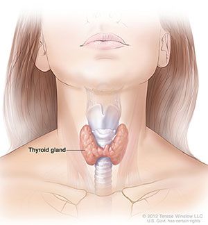
Pregestational Diabetes
                                                                             First trimester
• Prenatal labs/tests include HgbA1c,TSH, 24-hour urine (if no
  baseline), EKG
• Evaluation
  • Ophthalmologist

  • Dietician
     • dietary approach to glycemic control is focused on careful carbohydrate
       counting and allocation of appropriate ratios of carbohydrates to meals and
       snacks (Level B)
  • Possibly endocrinologist, cardiologist, nephrologist

                               Level B—Recommendations are based on limited or inconsistent scientific evidence.

                                      Pregestational Diabetes
                                                                     Second trimester

  • Start low-dose aspirin 12-28 weeks of gestation
    – optimally 16 weeks EGA (Level B)
     • high-risk factor for the development of preeclampsia
  • US including a detailed anatomical survey
  • Consider fetal echocardiography

                               Level B—Recommendations are based on limited or inconsistent scientific evidence.

                                                                                                                   7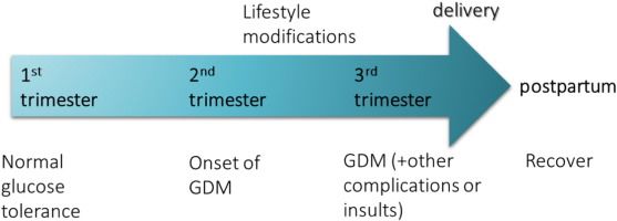
Pregestational Diabetes
                                                                  Third trimester

 • Evaluate fetal growth
 • Start low-dose aspirin by 28 weeks of gestation if
   NOT started in the second trimester
 • Fetal monitoring (nonstress test, MBPP, BPP)
   • usually once or twice per week (Level B)

                      Level B—Recommendations are based on limited or inconsistent scientific evidence.

Pregestational Diabetes

•Treatment (Level B)
   • Use of all oral hypoglycemic agents for control of
     pregestational type 2 diabetes mellitus during pregnancy
     should be limited and individualized until data regarding
     the safety and efficacy of these drugs become available
   • Insulin is the preferred treatment for pregestational
     diabetes in pregnancy not controlled by diet and exercise

                      Level B—Recommendations are based on limited or inconsistent scientific evidence.

                                                                                                          8
Pregestational Diabetes
                                                 Delivery

 • If EFW > 4500 g, consider cesarean delivery (Level C)
 • Without vascular complications and well controlled
   blood glucose levels, deliver at 39 0/7 weeks to 39 6/7
   weeks EGA
 • In women with vascular complications or poorly
   controlled blood glucose, consider delivery at 36 weeks
   0/7 weeks to 38 6/7 weeks EGA, and in rare cases,
   even earlier

AES Question #2

Gestational diabetes has been associated with each of the
following perinatal complications EXCEPT:

  A. Increased frequency of maternal hypertensive
     disorders
  B. Increased risk of operative delivery
  C. Increased frequency of neonatal hyperglycemia
  D. Increased risk of intrauterine fetal death during
     last 4-8 weeks of gestation

                                                             9
Gestational Diabetes
• Condition is increasing as obesity and older age at
pregnancy become more common
• Increased risk:
• Gestational hypertension
• Preeclampsia
• Cesarean delivery
• 7-fold increased risk of developing diabetes later in life
Why All the Fuss …
Adverse Outcomes
Maternal Fetal
• Increased frequency • Excessive fetal growth
o Maternal hypertensive disorders (macrosomia)
o Cesarean delivery o Increased risk for operative delivery
o Shoulder dystocia
• Increased risk of intrauterine fetal
death during last 4-8 weeks of o Birth trauma
gestation • Neonatal morbidity
o Fasting hyperglycemia o Hypoglycemia
(> 105 mg/dL) o Hypocalcemia
o Hyperbilirubinemia
o Polycythemia
10Detection of GDM
Organization Recommendation Comments
ACOG (2018) • Use a 2-step method at 24-28 weeks (Level B). Use a blood glucose level of either 135
• Screen for undiagnosed type 2 diabetes at the or 140 mg/dL with factors such as
first prenatal visit in those with risk factors community prevalence rates of GD
determining the cutoff.
USPSTF • Screen asymptomatic women after 24 weeks Goal was not to look at the performance
(2014) (Grade B). or whether one method was better than
• Current evidence is insufficient to assess the another for screening.
balance of benefits and harms of screening for Found treating can significantly reduce
GD in asymptomatic pregnant women before the risk of preeclampsia, macrosomia,
24 weeks of gestation (Grade I). shoulder dystocia.
ADA (2014) • Screen for undiagnosed type 2 diabetes at the Updated Guidelines: Use either:
first prenatal visit in those with risk factors 1. 1-step method (75g OGTT)
• Screen at 24-28 weeks if not previously known 2. 2-step method
to have diabetes.
RISK FACTORS (Screen early)…ACOG, NIDDK,ADA
• Patient is overweight with BMI of 25 (23 in Asian Americans), and ONE or more of the
following:
• Physical inactivity
• First-degree relative with diabetes
• Known impaired glucose metabolism
• Previous pregnancy history of
• GDM
• Macrosomia (>4000 g)
• Stillbirth
• Hypertension (>140/90 or being treated)
• HDL cholesterol < 35 mg/dL
• Fasting TG > 250
• PCOS, acanthosis nigricans, nonalcoholic steatohepatitis, morbid obesity and OTHER conditions
associated with insulin resistance
• HgbA1C>5.7%, impaired glucose tolerance or impaired fasting glucose on previous testing
• ASCVD
• High risk ethnicity
ACOG Practice Bulletin No. 190: Gestational Diabetes Mellitus. Obstet Gynecol. 2018;131(2):e49-e64.
11Two-Step Approach in USA
ACOG 2018, ADA 2014
• 24-28 weeks (routine)
• Initial screening: 50 g oral glucose load (glucose challenge
test)
• > 135 or > 140 mg/dL* 3-hour OGTT
• Note: > 190, > 90% abnormal 3-hour
• 3-hour OGTT ± 2 or more abnormal values = (+) GDM
• Overnight fast, 100 g glucose polymer
• Abnormal plasma blood glucose: > fasting
95 mg/dL, 1h 180, 2h 155, 3h 140
*Either threshold acceptable, ACOG 2018 (Level C).
± Can also be used as a 1-step method for high-risk women or in areas in which the prevalence of insulin resistance
is 5% or higher (eg, southwestern and southeastern US).
One abnormal value on 3-hour OGTT?
• One abnormal value - significantly increased risk of adverse
perinatal outcomes compared with women without GDM.
• Although a higher level of scrutiny may be focused on this
subset of women, further research is needed to clarify the
risk of adverse outcomes in patients with one abnormal
value on the 100-g, 3-hour OGTT and whether they would
benefit from treatment.
Cheng YW, Block-Kurbisch I, Caughey AB. Carpenter-Coustan criteria compared with the national diabetes data group
thresholds for gestational diabetes mellitus.Obstet Gynecol 2009;114:326–32.
12Gestational Diabetes Mellitus Treatment
ACOG 2018
• Initial management (Level A)
• Nutritional counseling by registered dietician
• Advice on moderate exercise program (if possible); minimum of 150 minutes per week
• No conclusive evidence for the threshold value at which clinicians
should start pharmacologic therapy
• Pharmacologic treatment
• Insulin is considered PREFERRED treatment in pregnancy (Level A)
• Glyburide treatment should NOT be recommended as a first-CHOICE pharmacologic
treatment because, in most studies, it DOES NOT yield equivalent outcomes to insulin
(worse outcome including macrosomia and birth injury (Level B)
• In women who decline insulin therapy or if unable to safely administer, metformin is a
reasonable second-line choice (Level B)
ACOG Practice Bulletin No. 190: Gestational Diabetes Mellitus. Obstet Gynecol. 2018;131(2):e49-e64.
Gestational Diabetes Mellitus
Maternal Surveillance – Glucose Monitoring
• Glucose Target Levels
• Fasting or preprandial < 95 mg/dL
• 2-hour postprandial BG < 120 mg/dL (1 hour < 140mg/dL)
• 1-2 times per week versus daily; review weekly
• Pharmacologic Treatment (if on more than 3 occasions)
• > 95 mg/dL fasting whole blood glucose or
• > 120 mg/dL 2 h postprandial
• Daily glucose monitoring
13Gestational Diabetes Mellitus
Fetal Surveillance/Assessment (Level C)
• Third trimester • Antenatal Testing
• Increased risk for fetal demise • Modified Biophysical Profile (MBPP)
• Preexisting DM • NST and AFI
• Fasting glucose >105 mg/dL • Biophysical Profile (BPP)
• Delay delivery safely in order • Contraction Stress Test (CST)
for the fetus to mature • Ultrasound
• Abnormal results are rare • Amniotic Fluid Index (AFI)
when diabetes well-controlled, • Asymmetric fetal growth
no vascular disease or
• Estimated fetal weight (EFW)
hypertension
Gestational Diabetes Mellitus
Fetal Surveillance/Antepartum
• Well-controlled A1 GDM (Level C)
• no consensus regarding criteria for initiation and frequency (MBPP, BPP)
• beginning 34-40 weeks versus none
• More intensive biophysical testing
• beginning at 32-34 weeks, twice weekly?
• insulin requirement (A2, B)
• hypertension
• previous stillbirth or other adverse obstetrical history
14Gestational Diabetes Mellitus
Fetal Surveillance/Ultrasound
• Assessment for asymmetric fetal growth (early third trimester) may aid in identifying
fetuses that can benefit from maternal insulin therapy
• Aid in the timing and route of delivery????
• Estimate fetal size
• CPD and birth trauma increase after 4000g
• > 4500 g - C-section may be best option (Level C; previously Level B)*
• May reduce likelihood of permanent brachial plexus injury in the infant
• 4000 to 4500 g – consider:
• Past delivery history
• Clinical pelvimetry
• Progress of labor
*ACOG Practice Bulletin No. 190: Gestational Diabetes Mellitus. Obstet Gynecol. 2018;131(2):e49-e64
Gestational Diabetes Mellitus
Timing of Delivery?
• Timing of delivery in women with GDM that is controlled with
only diet and exercise (A1GDM) should NOT be before 39
weeks gestation, unless otherwise indicated. Expectant
management up to 40 6/7 weeks of gestation in the setting of
indicated antepartum testing is generally appropriate (Level
C)
• GDM well controlled on medications (A2GDM) or Type 2,
delivery is recommended at 39 0/7 to 39 6/7 weeks of
gestation (Level C)
ACOG – 2018
15Gestational Diabetes Mellitus
Timing of Delivery?
• Poorly controlled – Expert guidance supports earlier delivery
but data lacking regarding precise timing
• Delivery between 37 weeks 0 days and 38 weeks 6 days
may be justified
• Delivery between 34 weeks 0 days and 36 weeks 6 days
reserved for (1) failure of in-hospital glycemic control or (2)
abnormal fetal testing
• Council regarding risks/benefits of scheduled cesarean
delivery when EFW > 4500 g (Level C) ACOG – 2018
Long-Term Considerations
• Increased risk for recurrence of GD
• 33%-50% likelihood
• Increased risk for development of diabetes after
pregnancy
• Up to 1/3 will have diabetes or impaired glucose metabolism at
postpartum screening
• 35% of women 5-10 years after parturition
• Offspring – increased risk
• Obesity
• Glucose intolerance
• Diabetes in late adolescence and young adulthood
16Postpartum
• Reclassification of maternal glycemic status at least 4
weeks after delivery (preferred 4-12 weeks, ACOG
2018) [Level C]
• FPG or 2-hr OGTT
• Reassessment of glycemia every one (USPSTF) to
three years (ADA) [SOR:C], if above normal; yearly
assessment (ADA) if impaired fasting glucose or
impaired glucose tolerance at 6-12 weeks
ACOG – 2018
Management of Postpartum Screening Results
Gestational diabetes
FPG or 75-g, 2-hr OGTT at 4-12 weeks postpartum
FPG > 125 mg/dL or FPG 100-125 mg/dL or FPG < 100 mg/dL or
2-hr glucose > 199 mg/dL 2-hr glucose 140-199 mg/dL 2-hr glucoseBest Practice Recommendations
• Nutritional Counseling • Programs of moderate
• Insulin is the preferred physical activity have been
pharmacologic therapy shown to lower maternal
• Human preferable glucose concentrations
• Insulin analogs have not been • Impact on neonatal
adequately tested complications awaits rigorous
clinical trials
• Oral agents have not been
generally recommended • Delivery during the 38th
week is recommended
• Glucose monitoring
18Thyroid Disorders
Thyroid Disorders
• Fetus dependent on maternal thyroxine (T4) in early pregnancy.
• Maternal thyroid gland is required to increase thyroid hormone
synthesis by up to 50% to meet the increased demands of
pregnancy due to placental transfer of T4, increased thyroid
hormone metabolism, increased renal iodine losses and
changes in levels of T4 binding proteins. These changes also
affect laboratory thyroid function test assays.
• Thyroid stimulating hormone (TSH) is the most reliable measure
of thyroid function during pregnancy. Pregnancy- and trimester-
specific laboratory reference ranges should be used if available.
19Stimulation
Regulation of Thyroid Function Inhibition
1. TRH from hypothalamus stimulates anterior Hypothalamus
pituitary to release TSH
2. Release of TSH stimulates all thyroid function
3. T3 and T4 release increases basal metabolic rate TRH
4. Negative feedback loop: T3 and T4 act on 1
hypothalamus and pituitary to suppress further
TRH and TSH release Anterior pituitary
TSH 4
2
Tyrosine
and Thyroid gland
Iodine
3
Biologic effects T3 + T4
Changes in Thyroid Function Test Results in
Normal Pregnancy and in Thyroid Disease
Maternal Status TSH Free T4
Pregnancy Varies by trimester* Raised to no change
Overt Hyperthyroidism Decrease Increase
Subclinical Decrease No change
hyperthyroidism
Overt hypothyroidism Increase Decrease
Subclinical hypothyroidism Increase No change
*The level of TSH decreases in early pregnancy because of weak TSH receptor stimulation
due to substantial quantities of human chorionic gonadotropin during the first 12 weeks of
gestation. After the first trimester, TSH levels return to baseline values.
20Physiological changes in pregnancy and their impact on the thyroid
Increase in circulating hCG Raised free T4, suppression of TSH
stimulating TSH receptors
Increase in hepatic
Raised Total T4
production of TBG
Increased urinary iodine Higher iodine requirement; goiter and
excretion hypothyroidism in iodine deficient areas
Pregnancy
Activation of placental Peripheral degradation of T4 and T3; demand for
deiodinase type 3 enzyme increased thyroid hormone production
Increased Plasma volume Increase in T4 and T3 pool
Decreased thyroid Ab titers; improvement in
Immunological changes
Graves’ disease
Thyroid Gland and Pregnancy
• Glandular hyperplasia and increased vascularity result in
moderate thyroid enlargement but not thyromegaly
• Thyroid function tests are NOT indicated in asymptomatic pregnant
women with slightly enlarged thyroid glands (Level B)
• Maternal thyroid volume is 30% larger in the third trimester than in
the first
• Any goiter or nodule recognized during pregnancy should
be considered pathologic
21Thyroid function and the Fetus
• Maternal T4 is transferred to the fetus throughout the entire
pregnancy and is important for normal fetal brain
development
• The high placental content of D3 inactivates most maternal T3 and T4,
and very little free hormone reaches fetal circulation
• It is especially important before the fetal thyroid gland begins
concentrating iodine and synthesizing pituitary TSH and
thyroid hormone at approximately 12 weeks of gestation
• After 15-18 weeks, the fetus controls most of its own thyroidal
secretion
Whom to screen in early pregnancy for
thyroid dysfunction
• No evidence for universal screening • High Risk
• History of previous thyroid dysfunction
• Women at high risk – TSH BEFORE • Current symptoms suggestive of hyper- or
conception and as soon as pregnancy hypothyroidism
is confirmed. • Known (+) thyroid antibodies
• TSH > 2.5 IU/L – obtain FT4 and Thyroid • Age > 30 years
peroxidase antibody (TPOAb) • Any history of autoimmune disease
• History of previous pregnancy loss, preterm
delivery, or infertility
• Use of lithium, amiodarone or recent
iodinated contrast use
• History of head and neck radiation
• Molar pregnancy
• Goiter
22An approach to the management of abnormal TSH levels in pregnancy6
Pregnant women at high risk for thyroid dysfunction
*Strong recommendation, moderate-to-
high-quality evidence
Measure TSH levels in early pregnancy. If > 2.5 mIU/L measure T4 levels
TSH level TSH level lower than TSH level between TSH level > 10
undetectable reference range but 2.5 and 10 mIU/L mIU/L or low free T4
still detectable level
Check TRAb, Repeat in 4 weeks Check TPOAb status Check TPOAb status
T3 and T4 to confirm the cause
levels in autoimmune
TSH level undetectable TPOAb (+) TPOAb (-) hypothyroidism
If T3 or T4 levels If TSH levels
If TSH levels 2.5 to 4 >4 mIU/L, *Commence
elevated and/or TRAb If TSH levels >4
mIU/L, *consider *consider levothyroxine and
positive, refer patient to mIU/L, *treat with
treatment with treatment with refer patient to an
an endocrinologist levothyroxine
levothyroxine levothyroxine endocrinologist
TRAb = TSH receptor antibody
AES Question #3
In considering a pregnant patient with hyperthyroidism,
which one of the following statements is true?
A. Serum Free T4 will be decreased
B. Inadequately treated, it is associated with a
greater risk of preterm delivery
C. Toxic nodular goiter is the most common cause
D. It often improves in the second and third
trimester due to the immunosuppressive effects
of pregnancy
23Hyperthyroidism
• Etiology
• Graves’ Disease (30-80 per 100,000 person-years) – accounts for 95% of cases
• Toxic nodular goiter (1-2 per 100,000 person-years)
Often improves in the second and third trimester due to the
immunosuppressive effects of pregnancy
• Signs and Symptoms are similar to the nonpregnant state
• Problem is that some symptoms of hyperthyroidism are similar to symptoms of
pregnancy
• Serum TFTs differentiate thyroid disease from nonthyroid disease
• Best treated prior to pregnancy
• Goal of Treatment:
• Control thyrotoxicosis while avoiding fetal or neonatal transient hypothyroidism
Hyperthyroidism
Diagnosis
• Clinical suspicion
• Infertility
• Hyperemesis gravidarum
• Failure of nonobese women to gain weight
• Classic Signs of Graves Disease
• Highly sensitive third generation tests
• TSH (Low)
• Free T4 or FTI (Increased)
• Monitored to manage thyroid disease in pregnancy
24Thyroid Disease
Effects of Pregnancy and Hyperthyroidism on Tests commonly used to evaluate Thyroid Function
Test Normal Pregnancy Hyperthyroidism
TSH No change Decreased
TBG Increased No change
Total T4 Increased Increased
Free T4 No change Increased
FTI No change Increased
Total T3 Increased Increased or no change
Free T3 No change Increased
RT3U Decreased Increased
_______________________________________________________________________
• Most of the pregnancy-induced changes in thyroid physiology are stimulated by hyperestrogenemia
which in turn causes production of altered TBG.
• Changes in structure and function of the gland during pregnancy can mimic some of the effects of
hyperthyroidism
Transient gestational hyperthyroidism
• Common cause of mild hyperthyroidism secondary to
thyroid stimulation by beta human chorionic gonadotrophin
• Generally limited to the first half of pregnancy
• Seen more often in women with hyperemesis and those
with high beta human chorionic gonadotrophin levels due
to molar pregnancy or multiple gestation
• Antithyroid medications are NOT indicated for women with
gestational hyperthyroidism
25Thyrotoxicosis
Management
• Controlled medically, does not pose a serious threat to the mother
• Use the least amount of medication required to achieve clinical
euthyroidism (Level B)
―may take 3-4 weeks to be reflected in labs
• Aim of treatment with antithyroid medications - maintain a free T4 level at the
upper end (or within 10%) of the nonpregnant reference range
• Mechanisms:
―directed at blocking thyroid hormone production
• e.g., thioureas
―directed at peripheral manifestations of disease
• e.g., beta blockers
Maternal thyrotoxicosis
• Inadequately treated – associated with a greater
risk:
• Preterm delivery
• Including medically indicated preterm deliveries
• Severe preeclampsia
• Heart failure
• Miscarriage – (no data to support this claim)
• Fetal
• LBW
26Hyperthyroidism
Treatment
• Propylthiouracil (PTU)
• Start 100-150 mg q 8 hours (300-450 mg/day)
Increase to control thyrotoxicosis
May require doses of 600-900 mg per day
Monitor free T4 and TSH q month
Titrate downward as soon as possible
• The birth defects associated with propylthiouracil are generally milder and more
easily corrected so it is used preferentially before a planned pregnancy and during
the first trimester
• Methimazole – Second and third trimesters (if needed, when risk of
malformation lower, preferred due to lower risk of hepatotoxicity)
• Beta blockers may be used for rapid control of adrenergic symptoms
Hyperthyroidism
Effects on Fetus and Infant
• Thioureas cross the placenta
• can cause fetal hypothyroidism and goiter
• appear to have no subsequent growth and development adverse effects
• Morbidity and mortality - infants born to women:
• who remain thyrotoxic despite therapy
• who do not receive adequate prenatal care/treatment
• Fetal Thyrotoxicosis
• in about 1% of mothers with Graves’ disease
• Consider in all women with a history of Graves’ disease
• If diagnosed – consultation with a clinician with expertise in such conditions
warranted
27History of Graves’ Disease
Treated with surgery or radioactive iodine
• TRAb levels measured in early pregnancy – if (+), repeat at 18-22
weeks’ gestation
• Can cross placenta cause fetal hyperthyroidism and neonatal Graves’
disease
• Women with active Graves’ disease or (+) TRAb at 18-22 weeks –
monitoring for fetal hyperthyroidism (MFM specialist)
• TRAb level is elevated at 18 to 22 weeks’ gestation or in women with
active Graves’ disease on treatment, measurement of TRAb levels at
30 to 34 weeks’ gestation can guide decisions about neonatal and
postnatal monitoring
Hypothyroidism in Pregnancy
• Difficult to diagnose
• Subclinical disease (elevated TSH with normal free T4) more common than overt
disease
• Subfertility
• Poor pregnancy outcomes: increased risk of SpAB, PTD, preeclampsia, GDM,
IUGR, PROM
• Untreated hypothyroidism Increased:
• Low-birth-weight infants
• Medically indicated preterm delivery, preeclampsia, placental abruption
• ? IUGR (not clear if independent of other complications)
• Pregnancy loss
• Impaired fetal neurocognitive development
• Hashimoto's thyroiditis is the most common cause of hypothyroidism in pregnancy
28Hypothyroidism
Diagnosis
• Rise in the level of circulating T4 expected during
pregnancy fails to take place (low free T4) and
level of TSH is elevated OR
• TSH > 10 mIU/L regardless of free T4 level
• NOTE: There is insufficient data to warrant routine
screening of asymptomatic pregnant women for
hypothyroidism (Level C)
Thyroid Disease
Effects of Pregnancy and Hypothyroidism on Tests commonly used to evaluate Thyroid Function
Test Normal Pregnancy Hypothyroidism
TSH No change Increased
TBG Increased No change
Total T4 Increased Decreased
Free T4 No change Decreased
FTI No change Decreased
Total T3 Increased Decreased or no
change
RT3U Decreased Decreased
_____________________________________________________________________________________
• Most of the pregnancy-induced changes in thyroid physiology are stimulated by hyperestrogenemia which in
turn causes production of altered TBG
• Changes in structure and function of the gland during pregnancy can mimic some of the effects of
hyperthyroidism
29Using TPOAb
• Women who are (+) TPOAb – increased rates if
miscarriage and PTD – independent of thyroid
function
• Thus, measurement recommended to assist with
decision making on when to treat subclinical
hypothyroidism
• High-quality randomized clinical trials on
levothyroxine replacement to treat subclinical
hypothyroidism during pregnancy are limited
Levothyroxine in pregnancy – data is mixed
Trial Results Lmitations
Controlled Antenatal Thyroid No significant difference in Late commencement of
Screening (CATS)-I and CATS- intelligence quotient in children aged levothyroxine at 13 weeks of
II trials 3 and 9.5 years of mothers with gestation
Hales C, Taylor PN, Channon S, et al. Controlled antenatal
thyroid screening II: effect of treating maternal suboptimal subclinical hypothyroidism
thyroid function on child cognition. J Clin Endocrinol Metab
2018; 103: 1583-1591. 8. Lazarus JH, Bestwick JP, Channon randomized to levothyroxine
S, et al. Antenatal thyroid screening and childhood cognitive
function. N Engl J Med 2012; 366: 493-501. treatment or placebo
Treatment of subclinical No improvement in cognitive Late commencement of
hypothyroidism or outcomes in children of mothers levothyroxine at 17 to 18 weeks of
hypothyroxinemia in pregnancy treated for subclinical hypothyroidism gestation
Casey BM, Thom EA, Peaceman AM, et al. Treatment of
subclinical hypothyroidism or hypothyroxinemia in pregnancy. at 5 years of age
N Engl J Med 2017; 376: 815-825.
Effects of levothyroxine Levothyroxine replacement may
treatment on pregnancy reduce the rates of preterm delivery in
outcomes in pregnant women women with subclinical
with autoimmune thyroid hypothyroidism and positive TPOAb
disease
Nazarpour S, Tehrani FR, Simbar M, Tohidi M, Majd HA, Azizi
F. Effects of levothyroxine treatment on pregnancy outcomes
in pregnant women with autoimmune thyroid disease. Eur J
Endocrinol 2016; EJE-16-0548.
30Hypothyroidism detected in early pregnancy
Treatment6
• TSH > 10mIU/L
• Begin full dose* replacement
TSH Level Initial dose
(mIU/L)
Upper limit of 1 to 1.5 mcg/kg daily (range 50 to 75 mcg daily)
normal to 5
5-10 1 to 1.7 mcg/kg daily (range 75 to 100 mcg daily)
>10 1.7 to 2.5 mcg/kg daily (range 100 to 200 mcg daily)
and consider referral to endocrinologist
* Based on lean bodyweight
Hypothyroidism detected in early pregnancy
Treatment
• TSH > 10mIU/L
• Begin full dose replacement
• Goal is to maintain the TSH in the low normal range
• Monitor TSH every 4-6 weeks until stable then every 8 weeks with a final check at
28-32 weeks
• Thyroxine requirement usually increases as the pregnancy advances
• Fetal surveillance????
• Following delivery, dose of levothyroxine can be halved, or ceased if on
50mcg daily or less during pregnancy, and thyroid function checked two
to three months’ postpartum
31Treatment of pre-existing hypothyroidism
• Levothyroxine dose increase
• 20-30% increase when pregnancy is confirmed
• 50% increase if no thyroid tissue (congenital hypothyroidism, post
total thyroidectomy, post radioactive iodine ablation)
• Thyroid function monitored q four to six weeks until the TSH
level is stable, then q 8 weeks with a final check at about 28
to 32 weeks’ gestation
• Postpartum
• Levothyroxine dose returned to the prepregnancy dose
• Thyroid function should be checked two to three months’ postpartum
Hypothyroidism
Effects on Fetus and Infant
•No evidence of thyroid dysfunction
•Mass screening to minimize sequelae of
congenital hypothyroidism (1/4,000 infants)
and prompt aggressive T4 replacement
• Sequelae can be prevented with treatment in the
first few weeks of life
32Nodular Thyroid Disease
• Evaluate by ultrasound and fine-needle aspiration or
tissue biopsy
• Avoid radioiodine scanning
• (+) thyroid cancer
• Differentiated (papillary or follicular) – surgery can be
delayed until postpartum period as such a delay is unlikely
to affect long-term prognosis
• Advanced differentiated, medullary, or poorly differentiated
– surgery in the second trimester may be considered
Postpartum Thyroid Dysfunction
Postpartum Thyroiditis
• Autoimmune inflammation
• Affects 5-10% of women in postpartum period
• Presents as new-onset:
• Painless hypothyroidism (25%) or
• Transient thyrotoxicosis (50%) or
• Thyrotoxicosis followed by hypothyroidism within one year postpartum
• Eventual return to euthyroidism
• Transient and recurrent in subsequent pregnancies
• 70% risk of recurrence
• May occur after pregnancy loss
33Postpartum Thyroid Dysfunction
Postpartum Thyroiditis
• Risk
• (+) TPOAb – 50% risk of developing
• Past history of PP Thyroiditis – 70% risk of developing
• Treatment unclear
• Permanent hypothyroidism is uncommon
• Thyrotoxicosis and hypothyroidism are mild
• Toxic phase – no antithyroid medications
• Check TSH q 2 months AFTER toxic phase
• May try to wean from replacement at 6-12 months after
initiating treatment
Postpartum Thyroiditis
Diagnosis/Screening
• Document new-onset abnormal levels of TSH or FT4
or both
• Screening with TFTs and antimicrosomal antibodies
in asx women NOT warranted
34Could she have Graves’ Disease?
• This is the main differential diagnosis
• (+) TRAb
• Signs – Goiter with a bruit or ophthalmopathy
• Uncertainty about diagnosis – technetium uptake scan
• Breastfeeding – breastmilk expressed and discarded during scan + 48
hours afterwards
• (+) thyrotoxicosis > 6months postpartum – Graves’
is most likely diagnosis
Antithyroid medications are needed?
• Graves’ disease in Breastfeeding mothers
• Lowest effective dose
• Ingest FOLLOWING a breastfeed
• Doses – safe in breastfeeding; less than 1% of
parent drug transferred to breastmilk
• PTU 300 mg
• Methimazole 20 mg
35What can we say -
• TSH goal in pregnancy < 3.0
• Levothyroxine is indicated for subclinical
hypothyroid with (+) thyroid peroxidase antibodies
• Gravid women with subclinical hypothyroidism not
treated
• Check TSH and T4 q 4 weeks until 16-20 weeks and
then AT LEAST once between 26 and 32 weeks
• No radioactive iodine scanning or ablation
Best Practice Recommendations
• Women at high risk of thyroid dysfunction should undergo screening with
measurement of thyroid stimulating hormone (TSH) levels in early
pregnancy.
• If the TSH level is 2.5mIU/L or more on early pregnancy screening, levels
of thyroid peroxidase antibodies should be measured to identify women
who may benefit from treatment for subclinical hypothyroidism.
• Transient gestational hyperthyroidism is a common cause of mild
hyperthyroidism in early pregnancy. Referral of the patient to an
endocrinologist is recommended if TSH levels remain persistently
undetectable and/or T3 or T4 levels are elevated and/or TSH receptor
antibodies (TRAb) are positive.
• Women with active Graves’ disease or a history of Graves’ disease treated
with surgery or radioactive iodine may be at risk of fetal hyperthyroidism. If
TRAb level is elevated at 18 to 22 weeks’ gestation, endocrinology and
maternal-fetal medicine input are required.
36Speaker Contact Information
David Glenn Weismiller, MD, ScM, FAAFP
david.weismiller@unlv.edu
References
1. ACOG Practice Bulletin No. 201. Pregestational Diabetes Mellitus. Obstet Gynecol. 2018; 132(6):e228-e248.
2. ACOG Practice Bulletin No. 190. Gestational Diabetes Mellitus. Obstet Gynecol. 2018; 131(2):e49—e64.
3. ACOG Practice Bulletin No. 148. Thyroid Disease in Pregnancy. Obstet Gynecol. 2015; 125(4):996-1005. (Reaffirmed
2017)
4. Carney LA, Quinlan JD, West JM. Thyroid Disease in Pregnancy. Am Fam Physician. 2014;89(4):273-278.
5. Alexander EK, Pearce EN, Brent GA, et al. 2017 Guidelines of the American Thyroid Association for the diagnosis and
management of thyroid disease during pregnancy and the postpartum. Thyroid. 2017;27:315-389.
6. Thillainadesan S, Gargya A. Thyroid disorders in Pregnancy and postpartum. Endocrinology Today. 2019;8(1);8-12.
7. Correa A, Bardenheier B, Elixhauser A, Geiss LS, Gregg E. Trends in prevalence of diabetes among delivery
hospitalizations, United States, 1993-2009. Matern Child Health J 2015;19:635-42.
8. Brown J, Grzeskowiak L, Williamson K, Downie MR, Crowther CA. Insulin for the treatment of women with gestational
diabetes. Cochrane Database of Systematic Reviews 2017, Issue 11. Art. No.: CD012037.
9. Farrar D, Simmonds M, Bryant M, Sheldon TA, Tuffnell D, Golder S, et al. Treatments for gestational diabetes: a
systematic review and meta-analysis. BMJ Open 2017;7:e015557.
37Thank you
38Questions
39You can also read