The Effect of Molecular Properties on Active Ingredient Release from Electrospun Eudragit Fibers - MDPI
←
→
Page content transcription
If your browser does not render page correctly, please read the page content below
pharmaceutics

Article
The Effect of Molecular Properties on Active
Ingredient Release from Electrospun Eudragit Fibers
Kieran Burgess, Heyu Li, Yasmin Abo-zeid, Fatimah and Gareth R. Williams *              ID

 UCL School of Pharmacy, University College London, 29-39 Brunswick Square, London WC1N 1AX, UK;
 kieran.burgess.16@ucl.ac.uk (K.B.); heyu.li@ucl.ac.uk (H.L.); y.abozeid@ucl.ac.uk (Y.A.-z.);
 fatimah.14@ucl.ac.uk (F.)
 * Correspondence: g.williams@ucl.ac.uk; Tel.: +44-207-753-5868

 Received: 16 March 2018; Accepted: 16 May 2018; Published: 24 July 2018

 Abstract: The formation of nanoscale fibers from pH-sensitive polymers is a route which has been
 widely explored for targeted drug delivery. In particular, the Eudragit L100 and S100 families of
 polymers have received significant attention for this purpose. However, while in some cases it is
 shown that making drug-loaded Eudragit polymers effectively prevents drug release in low-pH
 media where the polymer is insoluble, this is not always the case, and other studies have reported
 significant amounts of drug release at acidic pHs. In this study, we sought to gain insight into the
 factors influencing the release of active ingredients from Eudragit S100 (ES100) fibers. A family of
 materials was prepared loaded with the model active ingredients (AIs) benzoic acid, 1-naphthoic
 acid, 1-naphthylamine, and 9-anthracene carboxylic acid. Analogous systems were prepared with
 an AI-loaded core and an ES100 sheath. The resultant fibers were smooth and cylindrical in the
 majority of cases, and X-ray diffraction and differential scanning calorimetry showed them to
 comprise amorphous solid dispersions. When AI release from the monolithic fibers was probed,
 it was found that there was significant release at pH 1 in all cases except with 9-anthracene carboxylic
 acid. Analysis of the results indicated that both the molecular weight of the AI and its acidity/basicity
 are important in controlling release, with lower molecular weight AIs and basic species released
 more quickly. The same release trends are seen with the core/shell fibers, but AI release at pH 1
 is attenuated. The most significant change between the monolithic and core/shell systems was
 observed in the case of 1-naphthylamine. Mathematical equations were devised to connect molecular
 properties and AI release under acidic conditions.

 Keywords: electrospinning; Eudragit; nanofibers; drug release

1. Introduction
     Electrospinning is a technique which has attracted great attention in the pharmaceutical
technology field [1,2]. It most commonly involves the preparation of a polymer solution in a volatile
solvent. This is loaded into a syringe and is slowly ejected through a narrow bore needle (the spinneret).
A high voltage power supply is used to charge the needle, and the solution expelled towards
a grounded collector plate. The application of the electrical energy causes drawing of the polymer
solution into a fine jet, and ultimately results in the production of fibers with diameters typically on the
nanoscale. The inclusion of a drug molecule in the solution generally yields drug-loaded fibers in the
form of amorphous solid dispersions. In the simplest embodiment of the experiment, a single liquid is
processed, but more advanced derivatives including coaxial electrospinning (which uses two needles
nested concentrically one inside the other to process two liquids) and triaxial spinning (three needles,
three solutions) have also been reported. The use of coaxial spinnerets results in core/shell structures,
and triaxial spinnerets give three-compartment architectures.

Pharmaceutics 2018, 10, 103; doi:10.3390/pharmaceutics10030103             www.mdpi.com/journal/pharmaceutics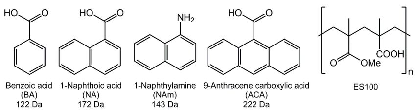
Pharmaceutics 2018, 10, 103                                                                          2 of 14

       Electrospun nanofibers have been explored for a wide range of drug delivery applications,
including preparing fast-dissolving oral drug delivery systems designed for very rapid release in the
mouth [3,4], extended release systems allowing the drug cargo to be freed over a number of hours or
weeks [5–8], and systems able to respond to external stimuli such as temperature [9–12]. Given that
the pH of the human gastrointestinal tract varies from 1–3 in the stomach, 6–8 in the small intestine,
and 4–7 in the large intestine, materials able to respond to changes in pH are particularly useful for oral
delivery systems. A range of polymers exist which are selectively soluble above or below a particular
pH. One clinically used family of such polymers is the Eudragits, methacrylate-based polymers with
tunable pH sensitivity. Eudragit L100-55 is soluble above pH 5.5, L100 above pH 6, and S100 above
pH 7. This allows different sections of the intestinal tract to be targeted depending on the polymer
chosen to fabricate a formulation. A number of authors have explored the electrospinning of Eudragits,
as well as other pH-sensitive polymers such as shellac [13,14].
       The studies in the literature exploring Eudragit have investigated both monolithic fibers from
monoaxial electrospinning and core/shell materials. Shen et al. were the first to electrospin Eudragit,
preparing L100-55 fibers loaded with diclofenac sodium [15]. They found that drug release at pH 1.0
was below 3%, confirming that pH-sensitive fibers can be produced. Other studies have built on this
and reported similar conclusions, for instance with Eudragit fibers containing mebeverine HCl [16],
ketoprofen [17], indomethacin [18] and helicid [19]. However, it is not always the case that simply
making a drug-loaded Eudragit fiber formulation prevents drug release in the acidic pHs typical of
the stomach. For instance, Karthikeyan et al. reported blend fibers of zein and Eudragit S100 (ES100)
loaded with pantoprazole and aceclofenac gave 25% release of the latter after 2 h immersion in 0.1 M
HCl [20]. The same has been shown to be true for spironolactone-loaded Eudragit FS fibers (up to 30%
release at pH 1.2) [21], and 5-fluorouracil (5-FU)-loaded ES100 fibers, which showed some 80% release
at pH 1 [22].
       Analogous findings have been found when preparing core/shell systems. Whilst in some instances
it is reported that drug release at pH 1–3 can be virtually completely obviated in fibers with a Eudragit
shell and drug-loaded core (for instance containing a Gd-based contrast agent or indomethacin [23,24]),
similar fibers containing 5-FU in the core release up to 70% of their drug loading under these
conditions [22]. The reasons behind the different findings reported in the literature are not completely
clear, but it seems that both the molecular weight and acidity or basicity of the drug incorporated are
important. Most recently, Jia et al. prepared fibers with an ES100 shell and a poly(ethylene oxide) core
containing either mebeverine HCl or indomethacin (model basic and acidic drugs with similar molecular
weights (MWs)) [25]. It was observed that release of both the basic mebeverine and acidic indomethacin
at pH 1.2 was restrained by the presence of the ES100 shell, but that there was a noticeably greater extent
of release in the former case (up to 20%) than the latter (ca. 1%).
       In this work, we sought to obtain a fundamental understanding of the factors underlying drug
release from Eudragit fibers at acidic pHs. To do this, we assembled a training set of four model active
ingredients (AIs; Figure 1). While none has any direct applications in drug delivery, the incremental
variation in their structures is ideal for a fundamental study of this type. Benzoic acid (BA), 1-naphthoic
acid (NA) and 9-anthracene carboxylic acid (ACA) all contain one carboxylic acid functional group
and have similar pKa s (BA: 4.20; NA: 3.67; ACA: 3.68), but the number of aromatic rings in the series
increases from one to three. 1-Naphthylamine (NAm) has an identical structure to NA except that the
carboxylic acid is replaced by an amine group; the pKa of NAm conjugate acid is very similar to that
of NA at 3.92, and thus the influence of acidity/basicity can be elucidated through this molecule pair.
       A series of ES100-based fibers was prepared containing the training set AIs, using both monoaxial
electrospinning to generate monolithic composites and also coaxial spinning to produce core/shell
systems with the AI confined to the core. The fibers were subject to a detailed examination of their
physicochemical properties, and AI release explored at pH 1.0 and 6.8 following pharmacopoeia
protocols. Correlations between molecular properties and the release profiles were sought.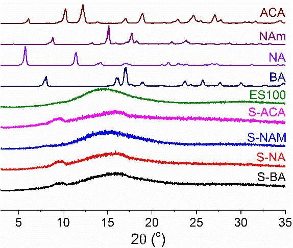
Pharmaceutics 2018, 10, 103                                                                                           3 of 14

      Figure 1. The chemical structures of the active ingredients used in this work and their molecular
      weights, together with the structure of the polymer ES100. BA: Benzoic acid; NA: 1-naphthoic acid;
      NAm: 1-Naphthylamine; ACA: 9-anthracene carboxylic acid; ES100: Eudragit S100.

2. Materials and Methods

2.1. Materials
      Benzoic acid (BA), 1-naphthoic acid (NA), 1-naphthylamine (NAm), 9-anthracene carboxylic
acid (ACA), absolute ethanol and dimethylacetamide (DMAc) were purchased from Sigma-Aldrich
(Gillingham, UK). Eudragit S100 (ES100) was a kind gift from Evonik GmbH (Darmstadt, Germany).
Analytical grade hydrochloric acid and trisodium phosphate dodecahydrate were obtained from Fisher
Scientific (Loughborough, UK). All water was deionised before use.

2.2. Methods

2.2.1. Monoaxial Electrospinning
     Following a series of optimisation experiments, a solvent mixture of ethanol/water/DMAc
(15/1/4 v/v/v) was selected as the most appropriate for electrospinning. A series of solutions was
then prepared for monoaxial electrospinning (see Table 1). 1.2 g of ES100 and 0.1 g of the AI of interest
were dissolved in 10 mL of the ethanol/water/DMAc solvent system. These were magnetically stirred
for a minimum of 24 h to ensure a homogenous solution was formed.

                        Table 1. Details of the electrospun formulations prepared in this work.

                                               Theoretical Fiber AI   Observed Fiber AI    Entrapment            Fiber
     ID            Active Ingredient (AI)
                                                Loading (% w/w) a     Loading (% w/w) b   Efficiency (%) c   Diameter (nm)
  Monoaxial electrospinning
    S-BA               Benzoic acid                    7.69              9.04 ± 0.13          118 ± 2          483 ± 145
    S-NA             1-Naphthoic acid                  7.69              7.05 ± 1.02          92 ± 13          129 ± 102
   S-NAM             1-Naphthylamine                   7.69              6.69 ± 0.27           91 ± 4          214 ± 105
   S-ACA        9-Anthracene carboxylic acid           7.69              6.54 ± 0.38           85 ± 5          585 ± 158
  Coaxial electrospinning
    C-BA               Benzoic acid                    2.70              3.42 ± 0.27         127 ± 10          591 ± 189
    C-NA             1-Naphthoic acid                  2.70              2.38 ± 0.07          88 ± 2           664 ± 171
   C-NAM             1-Naphthylamine                   2.70              2.20 ± 0.37         81 ± 14           547 ± 124
   C-ACA        9-Anthracene carboxylic acid           2.70              2.45 ± 0.16          91 ± 6           621 ± 140
      a Calculated based on the relative masses of the polymer and drug in the system. b Determined experimentally
      through dissolution of the fibers (mean ± S.D.; n = 3). c Calculated as the percentage of the theoretical loading
      observed to be incorporated (mean ± S.D.; n = 3).

      The solutions were loaded into a 5 mL Terumo syringe, with great care taken to avoid the
formation of bubbles. The syringe was then fitted with a blunt-tipped metal needle (internal diameter
0.61 mm; Nordson EFD, Aylesbury, UK), and the positive electrode of a high-voltage power supply
(HCP 35–35000, FuG Elektronik, Schechen, Germany) connected to the tip of the needle via a crocodile
clip. The grounded collector comprised a flat piece of steel coated with aluminium foil. Liquid was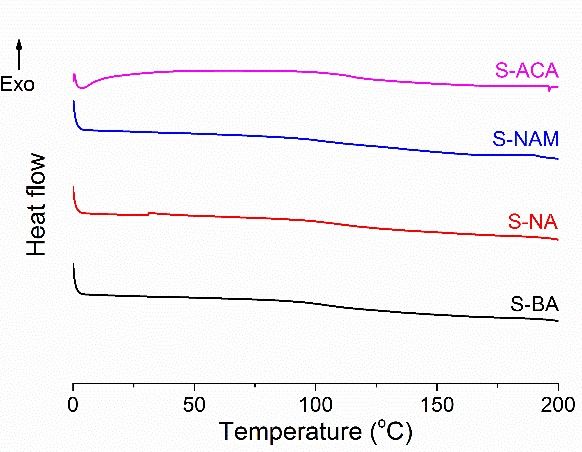
Pharmaceutics 2018, 10, 103                                                                         4 of 14

dispensed using a syringe pump (KDS100, Cole Parmer, London, UK). Electrospinning was performed
at ambient conditions (25 ± 3 ◦ C; relative humidity 38 ± 6%), and processing parameters were as
follows: voltage 16 kV; flow rate 0.5 mL h−1 ; collection distance 18 cm. After fabrication, the fiber
products were stored in a desiccator over silica beads.

2.2.2. Coaxial Electrospinning
      A 12% w/v Eudragit S100 solution in ethanol/water/DMAc (15/1/4 v/v/v) was used as the
shell liquid for coaxial electrospinning. The core solutions were the same as those used for monoaxial
electrospinning (see Table 1). The applied voltage for coaxial spinning was 21 kV, the collection distance
18 cm, and experiments were performed under ambient conditions (25 ± 3 ◦ C; relative humidity
38 ± 6%). The core and sheath solutions were independently dispensed through a coaxial spinneret
(Linari Engineering, Pisa, Italy) using two separate KDS100 syringe pumps. The spinneret had
internal/external diameters for the inner needle of 0.51/0.83 mm and for the outer needle at 1.37/1.83 mm.
The core and shell flow rates were 0.4 and 0.8 mL/h respectively. After production, the fiber products
were stored in a desiccator over silica beads.

2.3. Characterisation

2.3.1. Electron Microscopy
     Small samples (ca. 0.5 × 0.5 cm) were cut from each fiber mat for scanning electron microscopy
(SEM). These were sputter coated with gold and then imaged using a Quanta 200F instrument
(FEI, Hillsboro, OR, USA). The fiber diameters were quantified at 100 points for each sample, using the
ImageJ software (v1.48; National Institutes of Health, Bethesda, MD, USA). The coaxial materials
were also studied with transmission electron microscopy (TEM) on a CM 120 Bio-Twin instrument
(Philips, Amsterdam, The Netherlands). For this, fibers were directly spun onto carbon-coated TEM
grids (TAAB, Aldermaston, UK).

2.3.2. Physical Form Characterisation
     X-ray diffraction (XRD) patterns were collected on a MiniFlex 600 diffractometer (Rigaku,
Tokyo, Japan) supplied with Cu Kα radiation. Data were collected over the 2θ range 3 to 35◦ at a rate of
5◦ min−1 . Differential scanning calorimetry (DSC) was performed on a Q2000 instrument (TA Instruments,
New Castle, DE, USA). Samples weighing between 4–7 mg were placed into T-zero aluminium pans, sealed,
and pinholed. Samples were equilibrated at 0 ◦ C, heated to 100 ◦ C at 10 ◦ C min−1 , and then cooled to 0
◦ C again. A second heating run (to 200 ◦ C in most cases) was finally performed at 10 ◦ C min−1 . All DSC

experiments were undertaken under a nitrogen purge of 50 mL min−1 . IR spectra were obtained with a
Spectrum 100 spectrometer (Perkin Elmer, Waltham, MA, USA) over the wavenumber range 650–4000
cm−1 and with resolution of 1 cm−1 .

2.3.3. Active Ingredient Loading
     After the fibers had been left to dry to remove any residual solvent, samples (ca. 10 mg)
were cut from each sample and dissolved in a mixture of ethanol/water/DMAc (15/1/4 v/v/v).
Calibration curves for each active ingredient (AI) were prepared using a 7315 spectrophotometer
(Jenway, Stone, UK), and the AI loading determined (n = 3).

2.4. Dissolution Studies
     Calibration curves were constructed for each AI at pH 1.0 (0.1 M HCl) and 6.8 (phosphate buffered
saline; PBS) with the aid of a 7315 spectrophotometer (Jenway, Stone, UK). Dissolution studies were
then undertaken following the USPII method on an automated instrument (Caleva, Dorchester, UK).
The dissolution vessels were initially charged with 750 mL of 0.1 M HCl and equilibrated at 37 ± 0.5 ◦ C
under stirring at 50 rpm. Lids on each vessel prevented evaporation. Capsule sinkers were manually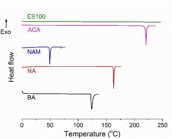
Pharmaceutics 2018, 10, 103                                                                                   5 of 14

filled with ca. 60 mg of a fiber mat and placed into the vessel. Aliquots (5 mL) were periodically
withdrawn from the vessels, and replaced with 5 mL of preheated 0.1 M HCl to maintain a constant
volume. After 2 h of operation, 250 mL of preheated 0.2 M trisodium phosphate dodecahydrate was
added to each vessel, yielding 1 L of a buffer at pH 6.8. Again, 5 mL aliquots were withdrawn at
specific time points, and replenished with 5 mL of preheated PBS at pH 6.8. The AI concentrations
in each aliquot were quantified with a 7315 spectrophotometer (Jenway, Stone, UK), and cumulative
release percentages calculated from these.

3. Results

3.1. Monolithic Fibers
      Scanning electron microscopy (SEM) images of the monolithic fibers are presented in Figure 2.
In all cases, cylindrical fibers have clearly been formed. In the cases of S-BA and S-ACA, the fibers are
homogenous and well formed, with diameters around 500–600 nm (see Table 1). In contrast, the S-NA
and S-NAM materials clearly contain two populations of fibers, with a number of very small branched
fibers visible, likely due to there being some jet instability during spinning. As a result, the average
diameter is rather smaller for these fibers, at 129 ± 102 nm for S-NA and 214 ± 105 nm for S-NAM,
and there is a proportionally greater deviation in the diameters.

      Figure 2. SEM images of the monolithic fibers (top: 10,000× magnification; bottom 40,000× magnification).

     The physical form of the AI in the fibers was explored using XRD and DSC (Figure 3). The XRD
data in Figure 3a demonstrate that the AIs are crystalline materials, with numerous Bragg reflections
visible in their patterns. ES100 is amorphous, and only a broad halo is present in its pattern. No Bragg
reflections can be observed for the fibers, showing them to be amorphous solid dispersions. The DSC
data (Figure 3b,c) concur with these findings. While the AIs exhibit sharp melting endotherms in
their thermograms, these are lacking for ES100 and all the fiber formulations. It should be noted
that the DSC experiments were stopped at 200 ◦ C with the fibers, since in preliminary experiments
degradation was observed to start just above this temperature (data not shown). Thus, the melting of
ACA would not be seen in the data even if crystalline material were present. However, Tg events are
present between 100 and 125 ◦ C for all the fibers, confirming their amorphous nature.
data (Figure 3b–c) concur with these findings. While the AIs exhibit sharp melting endotherms in
their thermograms, these are lacking for ES100 and all the fiber formulations. It should be noted that
the DSC experiments were stopped at 200 °C with the fibers, since in preliminary experiments
degradation was observed to start just above this temperature (data not shown). Thus, the melting of
ACA would
Pharmaceutics not10,be103
2018, seen in the data even if crystalline material were present. However, Tg events
6 ofare
14
present between 100 and 125 °C for all the fibers, confirming their amorphous nature.
(a)
(b) (c)
Figure 3. (a) XRD data and DSC data on (b) the raw materials and (c) formulations from monoaxial
Figure 3. (a) XRD data and DSC data on (b) the raw materials and (c) formulations from monoaxial
electrospinning. The data in (a) are normalized for ease of comparison.
electrospinning. The data in (a) are normalized for ease of comparison.
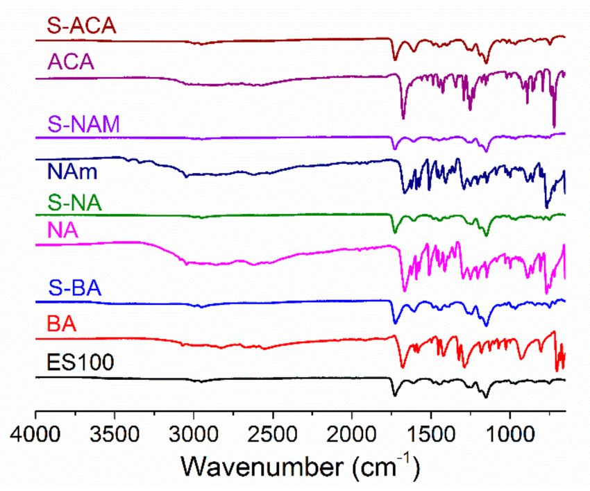
IR spectra are given in Figure 4. ES100 has characteristic peaks at 2950–3000 cm–1 (CH2
IR spectra
stretching), arecm
1727 given in Figure
–1 (C=O 4. ES100
stretching), has1150–1275
and characteristic
cm−1peaks at 2950–3000
(C–O–C cm–1
stretching). The(CH 2 stretching),
spectra of the
1727 cm –1 (C=O stretching), and 1150–1275 cm − 1 (C–O–C stretching). The spectra of the AI-loaded
AI-loaded fibers are very similar to that of ES100, as expected given that the AI loading is relatively
fibers are very similar
low (theoretical loading:to7.69%).that ofHowever,
ES100, as expected
some subtle given
changesthat
canthe
be AI loadingThe
observed. is relatively
C=O stretch lowof
(theoretical loading: 7.69%). However, some subtle changes can be observed.
pure BA is found at 1678 cm−1, and merges with the ES100 C=O stretch in the S-BA fibers to form a The C=O stretch
of purepeak
single BA isatfound
1725 cmat 1678
−1. Thiscm− 1 , and merges with the ES100 C=O stretch in the S-BA fibers to form
shift might be attributed to the formation of intermolecular bonding
ainteractions
single peak(e.g.,
at 1725 − 1
cm . This shift might be attributed to the formation of intermolecular bonding
H-bonding) between BA and ES100. Similarly, NA and ACA display a C=O stretch
interactions (e.g., H-bonding) between BA and ES100. Similarly, NA and ACA
at 1667 and 1674 cm while the S-NA and S-ACA fibers display single C=O bands at 1727 and 1726
−1 display a C=O stretch at
1667 − 1 and 1726 cm 1 −
cm−1 and 1674 cm No
respectively. while the S-NAare
shoulders andvisible
S-ACAonfibers
these display
peaks,single C=O bands
suggesting againat that
1727 intermolecular
respectively. No shoulders
interactions cause the bands aretovisible
merge. onInthese peaks,
the case of suggesting
S-NAM, the again
N–Hthat intermolecular
stretching interactions
vibrations of NAm
cause the bands to merge. In the case of S-NAM, the N–H stretching vibrations
(at 3411 and 3341 cm ) are not visible in the fibers, which might be indicative of intermolecular
−1 of NAm (at 3411 and
3341 cm −1 ) are not visible in the fibers, which might be indicative of intermolecular bonding or simply
bonding or simply the low AI loading in the formulations.
the low AI loading in the formulations.Pharmaceutics 2018, 10, 103 7 of 14
Figure 4. IR spectra of the monolithic fibers.
The AI loading (Table 1) is close to 100% of the theoretical content (>85% in all cases). The differences
observed can be attributed to losses of AI during electrospinning (e.g., through small amounts of
precipitation or adherence to the syringe walls), the presence of some residual solvent in the fibers,
or small inaccuracies in the quantification method.
3.2. Core/Shell Fibers
Core/shell fibers were prepared with an ES100 shell and a core comprising ES100 and the AI.
SEM images of these are given in Figure 5. The fibers are, in general, smooth and homogeneous,
with average diameters of around 550–700 nm as detailed in Table 1. For C-BA and C-ACA there are
a few very fine fibers present as well as the bulk at ca. 600 nm. For C-NA, what appear to be particles
can be seen on the fiber surfaces, suggesting some phase separation may have occurred.
Figure 5. SEM images of the coaxial fibers.Pharmaceutics 2018, 10, 103 8 of 14
TEM images of the fibers from coaxial electrospinning are depicted in Figure 6. The two solutions
used for the core and shell are very similar in their composition, and hence the contrast between the
core and shell is not particularly distinct. However, on close inspection it is clear that the fibers have
separate core and shell compartments.
Figure 6. TEM images of the coaxial fibers.
The physical form of the AI in the fibers was probed using XRD and DSC (Figure 7). Similarly to
the monolithic fibers, there is no evidence for any crystalline material being present in the products of
coaxial electrospinning: the sharp Bragg reflections of the pure AIs are replaced by broad haloes in
the XRD patterns of the fibers, consistent with amorphous AI-in-polymer dispersions. The DSC data
are also typical of amorphous systems, with no melting endotherms visible but instead clear baseline
changes corresponding to Tg s.
Figure 7. (a) XRD and (b) DSC data for the coaxial fibers. The data in (a) are normalized for ease
of comparison.
IR spectra of the core/shell fibers (Figure 8) again are very similar to that of pure ES100, except with
some small shifts in peak position (for instance, the C=O peak shifts from 1727 cm−1 in ES100 to
1725 cm−1 in C-BA and C-NA). Given the low AI loadings of these fibers (theoretical loading: 2.70%) it
is not possible to draw any firm conclusions relating to physical form from the IR spectra alone, but it
appears that intermolecular bonding may be operational here too.Pharmaceutics 2018, 10, 103 9 of 14
Figure 8. IR spectra of the coaxial fibers.
The AI loadings observed (see Table 1) are again close to 100% of the theoretical content (>80%),
with the values close to those found in the monoaxial fibers.
3.3. AI Release
The AI release profiles for the monolithic fibers are shown in Figure 9a. It is clear that the simple
fact of making fibers from ES100 does not prevent AI release at pH 1. This is because the AI is able to
diffuse through the polymer matrix to reach solution. The extent to which this occurs depends on the
nature of the molecule used as the AI: the amount of release in acidic pH decreases in the order S-BA
(86.6 ± 1.9%) > S-NAM (59.4 ± 5.7%) > S-NA (25.5 ± 2.8%) > S-ACA (7.1 ± 4.5%). Both molecular
weight (MW) and the acidity/basicity of the AIs are thus important: the lower MW BA (122 Da)
releases more quickly and to a greater extent than the intermediate NAm (143 Da) and NA (172 Da)
and higher MW ACA (222 Da). S-NAM releases markedly more than S-NA, presumably due to the
basic nature of NAm favoring dissolution in the low pH environment in the former case.
Figure 9. (a) AI release from the monolithic formulations, with data given as mean ± S.D. from three
independent experiments; (b) fitting the Korsmeyer-Peppas model to the experimental data obtained
at pH 1. Percentages are given relative to the theoretical AI loading in the fibers.Pharmaceutics 2018, 10, 103 10 of 14
This hypothesis is supported by a Korsmeyer-Peppas analysis of the release data at pH 1 (Figure 9b
and Table 2). This model takes the form Mt /Minf = ktn , where Mt is the amount of AI released at time
t, Minf is the theoretical AI loading of the fibers, k is a rate constant, and n is an exponent providing
information on the reaction mechanism. For all the monolithic fibers, n isPharmaceutics 2018, 10, 103 11 of 14
before the release percentage is quantified. The maximum extent of release in all cases is reached more
slowly than for the monolithic systems, at around 2 h after the elevation of pH.
Figure 10. (a) AI release from the core/shell formulations, with data given as mean ± S.D. from three
independent experiments; (b) fitting the Korsmeyer-Peppas model to the experimental data obtained
at pH 1. Percentages are given relative to the theoretical AI loading in the fibers.
It proved possible to develop relationships between the molecular weight and the percentage
release attained at pH 1 (Figure 11a). It should be noted that some caution is necessary in extracting
these, since they are based on data from only a small number of formulations, but nevertheless
some trends are clear. We attempted to fit both simple linear and exponential relationships to the
percentage release data. For the monolithic fibers, the best fit to the data is obtained with an exponential
relationship taking the form % release = −6.07 + 92.7e−(MW−122)/51.3 , whereas for the coaxial fibers
a more simple linear equation gives the best fit: % release = 126 − 0.561MW. The influence of MW
on the release percentage after 2 h at pH 1 is thus dulled by the addition of the blank polymer shell.
Considering the Korsmeyer-Peppas rate constants (Figure 11b), an exponential equation of the form
k = 0.146 + 0.324e−(MW−122)/22.3 gives the best fit to the experimental data for the coaxial systems.
In contrast, with the monolithic systems such a relationship can only be constructed for the three
carboxylic acids, for which we find k = 0.00372 + 0.476e−(MW−122)/44.9 . The S-NAM datapoint clearly
does not fit on this trendline, indicating that the basicity of the AI has a major influence on the rate
constant in the case of the monolithic formulations.
Figure 11. The relationships between AI MW and (a) percentage release after 2 h immersion in a pH 1
solution; and (b) the Korsmeyer-Peppas rate constant.Pharmaceutics 2018, 10, 103 12 of 14
4. Discussion
We show in this work that both the molecular weight and the acidity/basicity of an AI are
crucial in determining the extent to which release occurs from Eudragit S100 fibers in acidic media.
The nanoscale nature of the fiber diameters means that they have very large surface area-to-volume
ratios, and thus simply making a fiber from an insoluble polymer does not preclude AI release in
conditions where the polymer is insoluble [22,26]. Rather, it is necessary to consider also the ability
of the AI to diffuse through the matrix, and the thermodynamic solubility driver for this to happen.
It has previously been speculated that lower molecular weight species and those which are more basic
will release to a greater extent at acidic pHs [22,25], and in this work we confirm this to be the case.
For coaxial fibers with AI located in the core only, it is demonstrated that the molecular weight
of the AI is a good predictor for the percentage and rate of release seen at pH 1, and mathematical
relationships can be constructed linking these. For the monolithic fibers, the situation is more complex
and although it is possible to elucidate a mathematical relationship between percentage release and
AI MW, this does not hold true for the rate of release. For the latter, there is a clear linkage with the
MW for the acidic carboxylate AIs considered, but this breaks down for the basic species included
in the study. It thus appears that for the coaxial systems, the rate of diffusion through the polymer
shell, which will be directly correlated with the MW of the AI, is the dominant factor governing release.
For the monolithic analogues, while it is clear that diffusion through the polymer matrix is important,
the solubility of the AI in the release milieu is also vital to consider.
5. Conclusions
We report new insights into the factors governing the release of active ingredients (AIs) from
electrospun Eudragit S100 (ES100) fibers. Monolithic fibers loaded with benzoic acid, 1-naphthoic acid,
1-naphthylamine, and 9-anthracene carboxylic acid were generated, along with analogous systems
with an AI-loaded core and an ES100 shell. The fibers were smooth and cylindrical in the main,
and comprised amorphous solid dispersions. There was significant release at pH 1 with all of the
monolithic fibers except for those containing 9-anthracene carboxylic acid. Both the molecular weight
of the AI and its acidity/basicity are important in controlling release from such materials, with lower
molecular weight AIs and basic species freed from the fibers more quickly. With the coaxial fibers,
since the AI is present only in the core the acidity/basicity appears to be a less important factor, and the
molecular weight, and thus the rate of diffusion through the polymer matrix, is found to be the rate
limiting step in release. Mathematical equations could be constructed to predict the release obtained in
acidic conditions with AIs of differing molecular properties.
Author Contributions: K.B., H.L, F. and G.R.W. conceived and designed the experiments; K.B. and Y.A.-z.
performed the experiments; K.B., H.L., Y.A.-z., F. and G.R.W. analyzed the data; K.B. and G.R.W. wrote
the manuscript.
Acknowledgments: We thank the China Scholarship Council for funding H.L.’s work in UCL, the British Council
and Egyptian Sciences and Technology Development Fund for awarding a Newton-Mosharafa Researcher Links
Travel Grant to Y.A.-z., and the Indonesia Endowment Fund for Education for the award of a PhD studentship
to Fatimah.
Conflicts of Interest: The authors declare no conflict of interest.
References
1. Sebe, I.; Szabo, P.; Kallai-Szabo, B.; Zelko, R. Incorporating small molecules or biologics into nanofibers for
optimized drug release: A review. Int. J. Pharm. 2015, 494, 516–530. [CrossRef] [PubMed]
2. Xie, J.; Jiang, J.; Davoodi, P.; Srinivasan, M.P.; Wang, C.H. Electrohydrodynamic atomization: A two-decade
effort to produce and process micro-/nanoparticulate materials. Chem. Eng. Sci. 2015, 125, 32–57. [CrossRef]
[PubMed]Pharmaceutics 2018, 10, 103 13 of 14
3. Yu, D.G.; Shen, X.X.; Branford-White, C.; White, K.; Zhu, L.M.; Bligh, S.W. Oral fast-dissolving drug delivery
membranes prepared from electrospun polyvinylpyrrolidone ultrafine fibers. Nanotechnology 2009, 20, 055104.
[CrossRef] [PubMed]
4. Illangakoon, U.E.; Gill, H.; Shearman, G.C.; Parhizkar, M.; Mahalingam, S.; Chatterton, N.P.; Williams, G.R.
Fast dissolving paracetamol/caffeine nanofibers prepared by electrospinning. Int. J. Pharm. 2014, 477, 369–379.
[CrossRef] [PubMed]
5. Lu, H.; Wang, Q.; Li, G.; Qiu, Y.; Wei, Q. Electrospun water-stable zein/ethyl cellulose composite nanofiber
and its drug release properties. Mater. Sci. Eng. C 2017, 74, 86–93. [CrossRef] [PubMed]
6. Meng, Z.X.; Zheng, W.; Li, L.; Zheng, Y.F. Fabrication, characterization and in vitro drug release behavior of
electrospun PLGA/chitosan nanofibrous scaffold. Mater. Chem. Phys. 2011, 125, 606–611. [CrossRef]
7. Jia, X.; Zhao, C.; Li, P.; Zhang, H.; Huang, Y.; Li, H.; Fan, J.; Feng, W.; Yuan, X.; Fan, Y. Sustained release of
VEGF by coaxial electrospun dextran/PLGA fibrous membranes in vascular tissue engineering. J. Biomater.
Sci. Polym. Ed. 2011, 22, 1811–1827. [CrossRef] [PubMed]
8. Xie, J.; Wang, C.H. Electrospun micro- and nanofibers for sustained delivery of paclitaxel to treat C6 glioma
in vitro. Pharm. Res. 2006, 23, 1817–1826. [CrossRef] [PubMed]
9. Li, H.; Williams, G.R.; Wu, J.; Lv, Y.; Sun, X.; Wu, H.; Zhu, L.M. Thermosensitive nanofibers loaded with
ciprofloxacin as antibacterial wound dressing materials. Int. J. Pharm. 2017, 517, 135–147. [CrossRef]
[PubMed]
10. Li, H.; Williams, G.R.; Wu, J.; Wang, H.; Sun, X.; Zhu, L.M. Poly(N-isopropylacrylamide)/poly(L-lactic
acid-co-caprolactone) fibers loaded with ciprofloxacin as wound dressing materials. Mater. Sci. Eng. C 2017,
79, 245–254. [CrossRef] [PubMed]
11. Hu, J.; Li, H.-Y.; Williams, G.R.; Yang, H.-H.; Tao, L.; Zhu, L.-M. Electrospun poly(N-isopropylacrylamide)/ethyl
cellulose nanofibers as thermoresponsive drug delivery systems. J. Pharm. Sci. 2016, 105. [CrossRef]
12. Lin, X.; Tang, D.; Cui, W.; Cheng, Y. Controllable drug release of electrospun thermoresponsive
poly(N-isopropylacrylamide)/poly(2-acrylamido-2-methylpropanesulfonic acid) nanofibers. J. Biomed. Mater.
Res. A 2012, 100, 1839–1845. [CrossRef] [PubMed]
13. Wang, X.; Yu, D.G.; Li, X.Y.; Bligh, S.W.; Williams, G.R. Electrospun medicated shellac nanofibers for
colon-targeted drug delivery. Int. J. Pharm. 2015, 490, 384–390. [CrossRef] [PubMed]
14. Salehi, R.; Irani, M.; Eskandani, M.; Nowruzi, K.; Davaran, S.; Haririan, I. Interaction, controlled release,
and antitumor activity of doxorubicin hydrochloride from pH-sensitive p(NIPAAm-MAA-VP) nanofibrous
scaffolds prepared by green electrospinning. Int. J. Polym. Mater. 2014, 63, 609–619. [CrossRef]
15. Shen, X.; Yu, D.; Zhu, L.; Branford-White, C.; White, K.; Chatterton, N.P. Electrospun diclofenac sodium
loaded Eudragit(R) L 100-55 nanofibers for colon-targeted drug delivery. Int. J. Pharm. 2011, 408, 200–207.
[CrossRef] [PubMed]
16. Illangakoon, U.E.; Nazir, T.; Williams, G.R.; Chatterton, N.P. Mebeverine-loaded electrospun nanofibers:
Physicochemical characterization and dissolution studies. J. Pharm. Sci. 2014, 103, 283–292. [CrossRef]
[PubMed]
17. Yu, D.-G.; Williams, G.R.; Wang, X.; Liu, X.-K.; Li, H.-L.; Bligh, S.W.A. Dual drug release nanocomposites
prepared using a combination of electrospraying and electrospinning. RSC Adv. 2013, 3, 4652. [CrossRef]
18. Akhgari, A.; Heshmati, Z.; Afrasiabi Garekani, H.; Sadeghi, F.; Sabbagh, A.; Sharif Makhmalzadeh, B.;
Nokhodchi, A. Indomethacin electrospun nanofibers for colonic drug delivery: In vitro dissolution studies.
Colloids Surf. B 2017, 152, 29–35. [CrossRef] [PubMed]
19. Yu, D.-G.; Liu, F.; Cui, L.; Liu, Z.-P.; Wang, X.; Bligh, S.W.A. Coaxial electrospinning using a concentric Teflon
spinneret to prepare biphasic-release nanofibers of helicid. RSC Adv. 2013, 3, 17775–17783. [CrossRef]
20. Karthikeyan, K.; Guhathakarta, S.; Rajaram, R.; Korrapati, P.S. Electrospun zein/eudragit nanofibers based
dual drug delivery system for the simultaneous delivery of aceclofenac and pantoprazole. Int. J. Pharm.
2012, 438, 117–122. [CrossRef] [PubMed]
21. Balogh, A.; Farkas, B.; Domokos, A.; Farkas, A.; Démuth, B.; Borbás, E.; Nagy, B.; Marosi, G.; Nagy, Z.K.
Controlled-release solid dispersions of Eudragit®FS 100 and poorly soluble spironolactone prepared by
electrospinning and melt extrusion. Eur. Polym. J. 2017, 95, 406–417. [CrossRef]
22. Illangakoon, U.E.; Yu, D.G.; Ahmad, B.S.; Chatterton, N.P.; Williams, G.R. 5-Fluorouracil loaded Eudragit
fibers prepared by electrospinning. Int. J. Pharm. 2015, 495, 895–902. [CrossRef] [PubMed]Pharmaceutics 2018, 10, 103 14 of 14
23. Jin, M.; Yu, D.G.; Wang, X.; Geraldes, C.F.; Williams, G.R.; Bligh, S.W. Electrospun contrast-agent-loaded
fibers for colon-targeted MRI. Adv. Healthcare Mater. 2016, 5, 977–985. [CrossRef] [PubMed]
24. Jin, M.; Yu, D.G.; Geraldes, C.F.; Williams, G.R.; Bligh, S.W. Theranostic fibers for simultaneous imaging and
drug delivery. Mol. Pharm. 2016, 13, 2457–2465. [CrossRef] [PubMed]
25. Jia, D.; Gao, Y.; Williams, G.R. Core/shell poly(ethylene oxide)/Eudragit fibers for site-specific release.
Int. J. Pharm. 2017, 523, 376–385. [CrossRef] [PubMed]
26. Yu, D.G.; Li, X.Y.; Wang, X.; Yang, J.H.; Bligh, S.W.; Williams, G.R. Nanofibers fabricated using triaxial
electrospinning as zero order drug delivery systems. ACS Appl. Mater. Interfaces 2015, 7, 18891–18897.
[CrossRef] [PubMed]
© 2018 by the authors. Licensee MDPI, Basel, Switzerland. This article is an open access
article distributed under the terms and conditions of the Creative Commons Attribution
(CC BY) license (http://creativecommons.org/licenses/by/4.0/).You can also read