Mesoscale nanoparticles encapsulated with emodin for targeting antifibrosis in animal models
←
→
Page content transcription
If your browser does not render page correctly, please read the page content below
Open Chemistry 2020; 18: 1207–1216
Research Article
Lishan Tan, Xiulong Deng, Xuandi Lai, Tao Zeng, Aiqing Li*, Jianqiang Hu*, Zuying Xiong*
Mesoscale nanoparticles encapsulated with
emodin for targeting antifibrosis in animal
models
https://doi.org/10.1515/chem-2020-0163 Em-MNPs possessed specific kidney-targeting ability with
received July 05, 2020; accepted August 17, 2020 relative long retention time in the kidney (∼24 h). In the renal
Abstract: The aim of this study is to explore the kidney- unilateral ureteral obstruction model, Em-MNPs treatment
targeting capability of mesoscale nanoparticles (MNPs)- could significantly alleviate kidney tubule injury and reduce
emodin (Em-MNPs) and its potential antifibrosis in the extracellular matrix deposition compared with free MNPs.
animal model. First, MNPs and Em-MNPs were synthesized Herein, Em-MNPs with specific kidney-targeting and prefer-
via nanoprecipitation method, and their diameters were both able antifibrosis effects in animal model may pave an avenue
∼400 nm with the uniform size. The entrapment efficiency of for treating renal diseases.
MNPs was 45.1% when adding emodin at the concentration Keywords: mesoscale nanoparticles, emodin, kidney-
of 12 mg/mL. Moreover, cytotoxicity assay showed that Em- targeting, anti-fibrosis, UUO model
MNPs presented excellent biocompatibility in rat proximal
tubular cells. Cellular uptake assay demonstrated that Em-
MNPs had high-efficiency uptake, especially in the cyto-
plasm. Ex vivo organ fluorescence imaging revealed that 1 Introduction
Multifunctional nanomaterials have been widely studied in
the biomedical field because of their unique and adjustable
physicochemical properties [1]. Researches focused on their
* Corresponding author: Aiqing Li, Department of Nephrology, application in biosensor, bioimaging and diagnosis, drug
State Key Laboratory of Organ Failure Research, Nanfang Hospital,
delivery, and disease treatment, and tissue regeneration
Southern Medical University, Guangzhou, Guangdong, 510515,
engineering have attracted widespread attention [2–5]. The
China, e-mail: liaiqing@smu.edu.cn
* Corresponding author: Jianqiang Hu, Department of Chemistry, biological functions and applications of nanomaterials are
Nanobiological Medicine Center, Key Lab of Fuel Cell Technology of usually closely related to their structure, size, and surface
Guangdong Province, School of Chemistry and Chemical chemistry (such as surface modification) [6,7]. Thus,
Engineering, South China University of Technology, Guangzhou, appropriate construction and modification of nanomaterials
Guangdong, 510640, China, e-mail: jqhusc@scut.edu.cn
are of significance to broaden their application. It is well
* Corresponding author: Zuying Xiong, Department of Nephrology,
Peking University Shenzhen Hospital, Shenzhen Peking University-
known that shortcomings such as insufficient water solubi-
The Hong Kong University of Science and Technology Medical lity, no renal targeting, and high toxicity of some small
Center, Shenzhen, Guangdong, 518036, China, molecule drugs often restricted the therapy efficiency in a
e-mail: xiongzy2005@163.com variety of diseases. Therefore, it is urgent to develop nontoxic
Lishan Tan: Department of Nephrology, Peking University Shenzhen and specific targeting drug delivery system.
Hospital, Shenzhen Peking University-The Hong Kong University of
Chronic kidney disease (CKD) have attracted increasing
Science and Technology Medical Center, Shenzhen, Guangdong,
518036, China worldwide attention because of its high morbidity (10.8%),
Xiulong Deng: Department of Chemical and Chemical Engineering, and it often progresses to end-stage kidney disease (ESKD),
Key Laboratory of Organo-Pharmaceutical Chemistry, Gannan which required renal replacement therapy [8]. The key
Normal University, Ganzhou, Jiangxi Province, 341000, China procedure of CKD to ESKD is renal fibrosis [9], so finding the
Xuandi Lai: Department of Oncology, Peking University Shenzhen
drugs to reverse or halt the progression of fibrosis is
Hospital, Shenzhen, Guangdong, 518036, China
Tao Zeng: Department of Nephrology, State Key Laboratory of Organ
incredibly essential for CKD patients. Although the therapies
Failure Research, Nanfang Hospital, Southern Medical University, of chronic fibrosis increased, there are several adverse effects
Guangzhou, Guangdong, 510515, China in their clinical practice. Several ingredients of Chinese herbs
Open Access. © 2020 Lishan Tan et al., published by De Gruyter. This work is licensed under the Creative Commons Attribution 4.0
International License.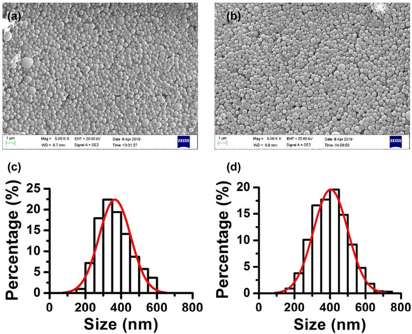
1208 Lishan Tan et al.
(emodin and triptolide) that were verified have been useful was synthesized by the amide reaction between the
in treating chronic kidney fibrosis [10–12]. However, they amino group of mPEG-NH2 terminal and the carboxyl
also have some disadvantages such as low aqueous group of PLGA terminal. First, PLGA (1 g), mPEG-NH2
solubility, low kidney targeting, and short retention that (250 mg), and EDC (75 mg) were dissolved in 5 mL
may limit their application [13]. Recently, several studies anhydrous chloroform and stirred vigorously at room
focus on the medical application of nanomaterials, such as temperature for 12 h. After the reaction, 15 mL ethyl
gold nanoparticles (NPs), polymer NPs, and other NPs with ether/methanol mixture (V/V, 1:1, 0°C) was added to
different sizes [14,15]. Qiao et al. synthesized catechol- precipitate the product. Centrifugation (10,000 rpm,
derived chitosan complex (HCA-Chi) to improve the water 15 min) was performed to collect the bottom residue,
solubility and renal-targeting ability of emodin and to further and the upper liquid was rotated to evaporate and then
alleviate chronic kidney injury, which was an effective way the solvent was removed. Then, all residuals were
to increase the effects of Chinese herbs [16]. However, the dissolved with a small amount of acetonitrile and
retention time of HCA-Chi in the kidneys was relatively short precipitated by ether/methanol (v/v, 1:5, 0°C). The
(∼12 h), and it would be preferable to prolong the kidney products were collected by centrifugation (10,000 rpm,
distribution time with good biocompatibility. Recently, it was 15 min), and this procedure of purification (dissolved
demonstrated that mesoscale nanoparticles (MNPs), with a and precipitated) was repeated for three times. Finally,
diameter of ∼400 nm, showed excellent kidney targeting the product was vacuum dried to constant weight at
ability, high stability, and high efficiency for the treatment of 35°C, and brown solid was obtained, which was stored at
acute kidney injury after loading triptolide [17]. Therefore, −20°C for later use.
MNPs would be a potential nanoplatform for emodin delivery
to the kidney and finally to improve chronic kidney fibrosis.
In this study, we constructed monodispersed MNPs and
loaded with emodin (Em-MNPs, ∼400 nm) for treating 2.3 Preparation of MNPs, Em-MNPs, and
kidney fibrosis. Em-MNPs with good biocompatibility, Cy7-Em-MNPs
kidney-targeting ability, and high uptake efficiency of
kidney cells were synthesized for further pharmac- Under vigorous string, 2 mL PLGA-b-mPEG solution
odynamics study. Finally, we revealed that Em-MNPs had (acetonitrile/tetrahydrofuran (THF) = 1:1, 100 mg/mL)
good therapeutic effects in the renal fibrosis animal model. was added to 2 mL proloxam 188 aqueous solution
(50 mg/mL) at the rate of 0.5 mL/min. After acetonitrile
was completely volatilized, MNPs were collected by
centrifugation (6,600 rpm, 10 min) and washed with
2 Materials and methods water for three times. When Em-MNPs and Cy7-Em-
MNPs were prepared, Em or Cy7 was dissolved with
2.1 Chemicals PLGA-b-mPEG in acetonitrile/tetrahydrofuran (v/v = 1:1,
100 mg/mL), and then, Em or Cy7-Em-MNPs were
prepared according to the preparation and the purifica-
Poly(D,L-lactate-co-glycolic acid) (PLGA; terminal carboxyla- tion method of MNPs. Finally, MNPs, Em-MNPs, or Cy7-
tion, molecular weight 38–54 kDa), EDC, methoxypolyethy- Em-MNPs were dispersed in saline or water and stored at
lene glycol amine (mPEG-NH2, 5 kDa), and FBS were 4°C for later use.
purchased from Sigma (USA). Emodin (Em), poloxam 188,
acetonitrile, methanol, and ethyl acetate were purchased
from Aladdin (Shanghai). Cy7 was purchased from
Lumiprode (USA). Other chemical reagents were purchased 2.4 Determination of encapsulation
from Sinopharm Group Chemical Reagent Co. LTD. Ultrapure efficiency (EE)
water was used throughout the experiments (18.2 MΩ cm).
The content of Em in Em-MNPs was determined by the
UV/visible spectrophotometer. First, lyophilized Em-
2.2 Fabrication of PLGA-b-mPEG MNPs (1 mg) were dissolved in the mixture of acetonitrile
(LC-MS, 50 µL) and methanol (LC-MS, 500 µL). PLGA
The synthesis of PLGA-b-mPEG was based on the precipitation and Em were dissolved in methanol and
method reported in the previous study [18], that is, it centrifuged (13,000 rpm, 10 min). The centrifugation of
MNPs encapsulated with emodin for antifibrosis 1209
residue was repeated (dissolved – precipitated – centri- organic extract was collected, which was repeated for
fuged) for three times. Then, the upper liquid was three times. All the extraction liquid was blow dried with
concentrated in a new centrifuge tube (7 mL) and blow nitrogen, and the residue was added to methanol for
dried with nitrogen. Methanol (LC-MS) was added to dissolution and constant volume to 10 mL. Finally,
dissolve the residue, with a constant volume of 50 mL. 2.0 mL methanol solution was used to remove the large
Finally, 2.0 mL Em methanol solution was removed with particles with a 0.22 µm filter head. The concentration of
a 0.22 nm filter head, and the Em concentration was Em was determined by the UV-vis spectrometer, and the
determined by the UV-vis spectrometer (Thermo Fisher cumulative release rate of Em was calculated.
Multiskan, Finland). Meanwhile, the EE and drug
loading efficiency (DLE) of Em were calculated according
to the following formulas:
Em amount in Em − MNPs 2.6 Cell uptake assay
EE (%) = × 100%. (1)
Em adding amount
MNPs were labeled with Cy7 to track the specificity of
Em amount in Em − MNPs intracellular distribution. Normal rat kidney proximal
DLE (%) = × 100%. (2)
Quantity of MNPs tubular epithelial cells (NRK-52E, American Type Culture
Collection, USA) were planted into coverslip at 37°C
overnight and then incubated with Cy7 and Cy7-Em-
MNPs (equivalent to 50 µg/mL emodin) for 2 h, respec-
tively. Then, the medium was removed, and ice-cold PBS
2.5 In vitro stability and drug release of
was added to wash the Cy7 and Cy7-Em-MNPs for three
Em-MNPs times. Then, the cell nuclear was stained with 4′,6-
diamidino-2-phenylindole for 5 min in the dark.
First, Em-MNPs were dispersed in phosphate buffered
Epifluorescence microscope was used to analyze the
solution (PBS) (10 mM, pH 7.4), 10% FBS, and saline and cellular distribution of Em-MNPs.
placed in a constant temperature metal bath (37°C,
1,000 rpm). Then, 100 µL dispersion was extracted at
different times (1, 3, 6, 12, 24, 48, 72, 120, and 168 h) for
the determination of hydrodynamic diameter (HD) of 2.7 Cytotoxicity assay
Em-MNPs. Before HD of Em-MNPs was determined, the
PBS and saline dispersions of Em-MNPs were diluted NRK-52E cells were cultured with Dulbecco’s Modified
with 2 mL water, and the FBS dispersions of Em-MNPs Eagles’s Medium containing 10% FBS, 100 U/mL peni-
were centrifuged (6,600 rpm, 15 min) to remove FBS, and cillin, and 100 µg/mL streptomycin. Different concentra-
then 2 mL water was added for dispersion. HD was tions of emodin and Em-MNPs solution from 5 to 50 µg/
characterized by quantity value. mL were added to 96-well plates for 12 and 24 h. Finally,
The in vitro release of Em-MNPs was determined by the cell viability was evaluated using a cell counting
dialysis [19]. Fetal bovine serum (FBS; 10%), renal tissue CCK-8 kit and measured by Thermo Fisher Multiskan GO
homogenate (10%), PBS (10 mM, pH 7.4), and saline UV/visible spectrophotometer (Shimadzu, Japan) [20].
were selected as dispersion and dialysate. The prepara-
tion method of renal tissue homogenate was as follows:
twice the volume of saline (g/mL) was added to the
kidney of C57BL/6 mice, and the kidney was homo- 2.8 Kidney targeting evaluation
genized with cell pulverizer. First, the concentration of
Em-MNPs was made to be 2 mg/mL. 2 mL of Em-MNPS The animal protocol was approved by the Animal Ethics
dispersions were placed in a dialysis bag (30 kDa) and Committee of Nanfang Hospital. Balb/c mice were
placed in the corresponding dialytic solution (100 mL), administrated with Cy7 and Cy7-Em-MNPs (equivalent
which was stirred slowly at 37°C. Then, 1 mL dialysate to 1.0 mg/kg emodin) for 12 and 24 h, respectively. All
was taken out at different time points (0.5, 1, 2, 4, 8, 12, mice were sacrificed at corresponding time, and then
and 24 h), 3 mL ethyl acetate (LC-MS) was added for their major organs, namely, heart, liver, spleen, lung,
extraction by rotating for 5 min, the mixture was kidneys, thymus, muscle, small intestine, and brain,
centrifuged at 10,000 rpm for 5 min, and the upper were collected. Before fluorescence imaging, major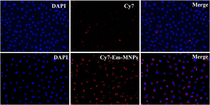
1210 Lishan Tan et al.
Figure 1: Characterization of MNPs and Em-MNPs. (a) 1H NMP spectra of Em, MNPs, and Em-MNPs. (b) FTIR of the Em, MNPs, and Em-MNPs.
(c) UV-vis spectra of MNPs, free Em, and Em-MNPs.
organs were washed with ice-cold PBS for three times to 2.10 Statistical analysis
remove the surface fluorescence. After washing, fresh
organs were placed on the glass plate to image with All experiments were performed for at least three times,
excitation at 710 nm and emission at 790 nm by the and the data were collected. The statistical comparisons
imaging system (Burker Imaging System, USA). between groups were performed by t test using SPSS
software (version 23.0). P < 0.05 was considered as
statistically significant.
2.9 Pharmacodynamics study on renal
unilateral ureteral obstruction (UUO)
model
3 Results and discussion
UUO models were performed based on the procedure
demonstrated in the previous study [21]. 6–8 weeks mice 3.1 Characterization of PLGA-b-mPEG and
were anesthetized using pentobarbital sodium (3%,
Em-MNPs
1.5 mL/kg, i.p.). All mice were randomly divided into
four groups (n = 6 per group): (1) Sham group, (2) UUO +
vehicle (PBS) group, (3) UUO + MNPs group, (4) UUO + The structures of PLGA-b-mPEG and the encapsulation
Em-MNPs (equivalent to 1.0 mg/kg emodin). The mice of Em were confirmed by 1H nuclear magnetic resonance
were injected with corresponding drugs on the first day spectra, Fourier transform infrared (FTIR) and UV-vis
after operation and for 3 days. Then, all mice were spectra. As shown in Figure 1a, the chemical shift peaks
sacrificed, and kidneys were collected for the following at 2.1, 6.5–7.5 and 11.0–12.1 ppm of Em-MNPs were
measurements. consistent with that of Em, and the peaks at 1.5, 3.5 and
After fixing with 10% neutral formaldehyde at 4°C 4.9–5.3 ppm were consistent with that of PLGA-b-mPEG.
overnight, kidneys were dehydrated, hyalinized, and Relative to FTIR spectra of Em-MNPs, there appeared
embed for histology staining. 4 µm kidney slices were cut obvious vibration peaks at 1,750 and 1,627 cm−1, which
and stained with hematoxylin and eosin (H&E) staining were carboxyl characteristic vibration of PLGA-b-mPEG.
to analyze the effect of Em-MNPs in the UUO mice model. Besides, the visible stretching vibration peaks of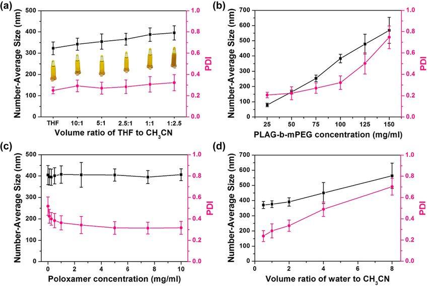
MNPs encapsulated with emodin for antifibrosis 1211 Figure 2: Optimization of the Em-MNPs. Hydrodynamic size of Em-MNPs prepared under different (a) volume ratios of THF to CH3CN, (b) PLAG-b-mPEG concentration, (c) poloxamer concentration, and (d) volume ration of water to CH3CN. Figure 3: Morphology of MNPs and Em-MNPs. SEM images of MNPs (a) and Em-MNPs (b). Diameter distributions of MNPs (c) and Em-MNPs (d) counted in SEM images.
1212 Lishan Tan et al.
index (PDI) of Em-MNPs prepared under different
organic solvent ratio. Along with the decreasing of
THF, the particle size gradually increased, while the PDI
for each sample kept steady. However, with the increase
of PLGA-b-mPEG concentration, the HD and PDI of Em-
MNPs increased significantly, which reached the high
concentration of PLGA-b-mPEG went against to the
monodispersion of NPs (Figure 2b). The effect of
poloxam 188 on particle size and dispersion was
negligible (Figure 2c), while the proportion of organic
solvent showed great influence on the particle size
Figure 4: Encapsulation efficiency (EE) and drug loading efficiency (Figure 2d). Therefore, the optimal condition for the
(DLE). EE and DLE after adding different concentrations of Em preparation of Em-MNPs was as follows: 100 mg/mL
(1–12 mg/mL). PLGA-b-mPEG, 50 mg/mL Proloxam 188, and the volume
ratio of aqueous/solvent phase 1.0.
Then, the morphology of MNPs and Em-MNPs
benzene in Em-MNPs indicated that the Em had been
was observed by scan electronic microscope (SEM). As
encapsulated in NPs successfully (Figure 1b). This was
shown in Figure 3, the as-prepared NPs were uniform
further confirmed by UV-vis spectra (Figure 1c). These
spheres. The diameter observed from SEM images of Em-
three methods were favorable measurements to verify
MNPs (394.0 ± 71.4 nm) was slightly larger than that of
the linkage of Em with MNPs, which was also used in
MNPs (361.3 ± 74.9 nm), and the HD for Em-MNPs was
other studies [22,23].
also greater than MNPs, indicating that the Em encap-
sulation enlarged the size of MNPs and the hydration
layer on the surface of NPs enhanced this trend. It is
3.2 Optimization of the Em-MNPs worth noting that the EE and DLE were closely related to
fabrication the amount of Em added. As shown in Figure 4, the EE
decreased along with the increased Em concentration,
Em-MNPs were prepared by a simple nanometer pre- while the DLE was proportionate to the Em concentra-
cipitation method, in which Poloxam 188 was used as a tion. When the Em concentration was 6 mg/mL, the EE
surfactant to stabilize Em-MNPs. The size and the and DLE reached a balance of 66.9% and 4.0%,
homogeneity of Em-MNPs can be adjusted by changing respectively. The cross point of EE and DLE indicated
PLGA-b-mPEG concentration, poloxam 188 concentra- that the concentration of Em added has reached an
tion, and aqueous/solvent phase volume ratio. Figure 2a optimal value with relatively high drug loading amount
shows the hydrodynamic size (HD) and polydispersity and efficiency.
Figure 5: Stability and in vitro release assays of Em-MNPs in different media. The stability of Em-MNPs (a) measured by HD in PBS (pH 7.4),
10% FBS, and saline (NS) at 37°C. In vitro release assay of Em released from Em-MNPs (b) in 10% FBS and kidney homogenate at 37°C.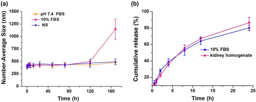
MNPs encapsulated with emodin for antifibrosis 1213
Figure 6: Cytotoxicity assay. Cell viability of rat proximal tubular cells treated with emodin (0–50 µg/mL) and Em-MNPs (equivalent to
0–50 µg/mL emodin) for 12 h (a) and 24 h (b), respectively. *p < 0.05 versus the Em-MNPs group.
Figure 7: Cellular uptake images. NRK-52E cells incubated with Cy7 and Cy7-Em-MNPs for 2 h. Images were captured in 400× magnification.
3.3 Stability and in vitro release assays time, Em exhibited a high release profile from the Em-
MNPs in those two media (Figure 5b). The cumulative
To further investigate the stability of Em-MNPs in release rate reached 80.2% and 86.6% in the FBS and
different media, HDs of Em-MNPs were measured after kidney homogenate for 24 h, respectively. Compared to
incubation with PBS (10 mM, pH 7.4), saline, and 10% covalent modification, Em-MNPs showed higher drug
FBS at different time points. As shown in Figure 5a, the release rate [25], thereby releasing Em from Em-MNPs
HDs of Em-MNPs were stable up to 7 days when Em- and improving drug efficacy.
MNPs were incubated with PBS and saline. After treated
with FBS, a medium enriched with proteins, the HD of
Em-MNPs was nearly same to that of the initial condition 3.4 Cellular uptake study
at 5 days. As reported in the recent study, the HD of Em-
MNPs remained stable for 2.5 times longer than MNPs Cy7 and Cy7-Em-MNPs were used to observe the
[24]. In vitro release assay was conducted in 10% FBS intracellular uptake efficiency. Figure 6 shows the
and kidney homogenate at 37°C. With the increasing fluorescence imaging distribution of NRK-52E cells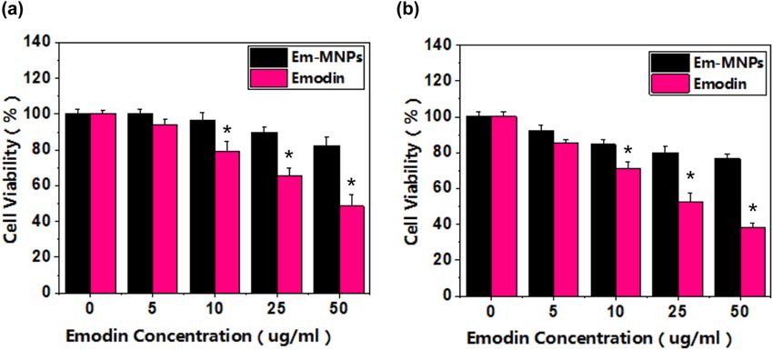
1214 Lishan Tan et al.
incubated with free emodin at the concentration of
10 µg/mL, cell viability presented significant suppres-
sion. With the increasing concentration of emodin, the
toxicity of emodin grow sharply after incubation for 24 h.
However, the viability of cells treated with Em-MNPs
were maintained in relative high levels (>85%). This
result suggested that loading emodin with MNPs could
significantly decrease the cytotoxicity of emodin. More-
over, our previous study found that MNPs reduced the
toxicity of triptolide in other three cell lines [17] and
further demonstrated that MNPs could serve as pro-
mising drug carriers with good biocompatibility.
3.6 Kidney targeting capacity
To further examine the kidney targeting capacity of Cy7-
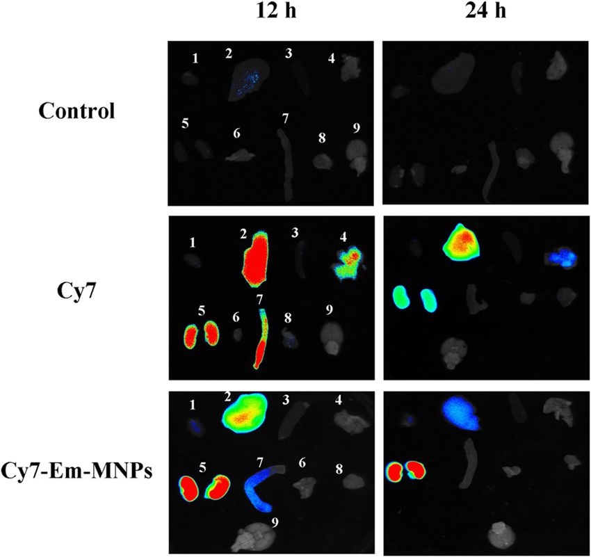
Figure 8: Ex vivo fluorescence imaging. Balb/c mice injected with Em-MNPs, an ex vivo tissue localization was performed
PBS (control), and Cy7 and Cy7-Em-MNPs for 12 h and 24 h, at 12 h and 24 h in Balb/c mice (Figure 8). As a result,
respectively and major organs were collected for imaging. 1, heart;
Cy7-Em-MNPs was mostly accumulated in kidneys rather
2, liver; 3, spleen; 4, lung; 5, kidney; 6, thymus; 7, small intestine;
8, muscle; 9, brain. than other organs (heart, liver, spleen, and lung) after
injection for 12 h. With the increasing time, the fluores-
cence density was increased in kidneys of Cy7-Em-MNPs.
treated with PBS, Cy7, and Cy7-Em-MNPs, respectively. However, free Cy7 treated kidneys had no apparent
After incubation for 2 h, the fluorescence signal of Cy7- fluorescence retention during the period, indicating that
Em-MNPs was significantly stronger than that of free Cy7-Em-MNPs had favorable renal retention ability. The
Cy7. It was suggested that the accumulation of Cy7-Em- slit diaphragm of podocytes (the molecular barrier of the
MNPs was much higher than that of Cy7. The Cy7-Em- nephron, ∼40 nm) are too small for Em-MNPs to pass
MNPs were mainly distributed around nucleus, thereby through [28,29]. So, the kidney-targeting mechanism of
suggesting that Cy7-Em-MNPs were mainly located and MNPs might be attributed to the endothelial cells of the
released drugs in the cytoplasm rather than nucleus. peritubular in the kidneys, which also verified that
Accordingly, these data indicated that Em-MNPs had excellent kidney-targeting capability of free MNPs [24].
higher uptake efficiency in vitro and might be suitable for The pressure in the nephron and the reabsorption ability
kidney diseases. The possible endocytic mechanism was of peritubular capillaries induced endocytosis of Cy7-
the protonation of the surface of Em-MNPs, coinciding Em-MNPs in kidney tubular cells. Therefore, Cy7-Em-
with triptolide-encapsulated MNPs [17], which resulted MNPs was an excellent option for the following
in endosome rupture and then realized lysosome escape pharmacodynamics analysis in the animal model.
[26]. Besides, Williams et al. also demonstrated that free
MNPs performed good cellular uptake and biocompat-
ibility [24].
3.7 Pharmacodynamics study
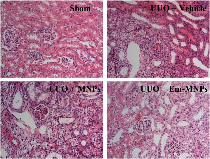
3.5 Cytotoxicity assay UUO mice were a well-recognized and classical animal
model of chronic kidney fibrosis [30]. So, the therapeutic
To compare the cytotoxicity of emodin and Em-MNPs, effect of Em-MNPs was performed in the UUO model.
NRK-52E cells were treated with emodin and Em-MNPs in Figure 9 depicts histology characteristics in different
different concentration gradients (5–50 µg/mL) for 12 and treatment groups for H&E staining. The ligated kidney in
24 h, respectively (Figure 7). The viability of cells was UUO mice showed apparent tubular dilation, lumen
measured by CCK-8, which is a classical method to disappear, fibrosis, and increased inflammatory cells
evaluate cell toxicity of nanomaterials [27]. When cells [31]. After treating with Em-MNPs, the manifestation of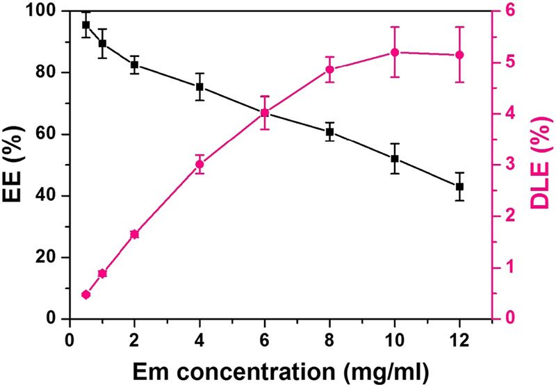
MNPs encapsulated with emodin for antifibrosis 1215
Figure 9: Histology analysis of kidneys. H&E staining images of kidneys from sham-operated, UUO + Vehicle (PBS), UUO + MNPs, and UUO
+ Em-MNPs mice.
ligated kidney was significantly alleviated, which char- Commission (201804020054), Key Project of Guangdong
acterized as decreased inflammation cells and less Natural Science Foundation (2018B030311002), and Key
matrix accumulation in the tubulointerstitium. These Project Science and Technology Planning Project of
results revealed that Em-MNPs could specifically treat Guangdong Province (2017A010103041).
kidney injury, which might be ascribed to the targeting
ability and long retention of Em-MNPs in kidneys. Conflict of interest: The authors declare no conflict of
Furthermore, UUO mice treated with free MNPs could interest.
not perform the therapeutic effects, suggesting that the
effects of Em-MNPs mainly was derived from emodin.
References
4 Conclusion [1] Liu Y, Workalemahu B, Jiang XY. The effects of physicochem-
ical properties of nanomaterials on their cellular uptake in
vitro and in vivo. Small. 2017;13(43):1701815.
In summary, we successfully designed monodispersed
[2] Chen A, Chatterjee S. Nanomaterials based electrochemical
MNPs and loaded with emodin (Em-MNPs). Em-MNPs sensors for biomedical applications. Chem Soc Rev.
possessed low toxicity, high cell uptake efficiency, 2013;42(12):5425–38.
kidney-targeting ability, and long retention time in the [3] Smith BR, Gambhir SS. Nanomaterials for in vivo imaging.
kidneys. In the UUO model, Em-MNPs presented Chem Rev. 2017;117(3):901–86.
[4] Nicolas J, Mura S, Brambilla D, Mackiewicz N, Couvreur P.
excellent antifibrosis effects and may represent a
Design, functionalization strategies and biomedical applica-
potential strategy for the treatment of renal diseases.
tions of targeted biodegradable/biocompatible polymer-
based nanocarriers for drug delivery. Chem Soc Rev.
Acknowledgments: This study was supported by the 2013;42(3):1147–235.
China Postdoctoral Science General Foundation [5] Santoro M, Shah SR, Walker JL, Mikos AG. Poly(lactic acid)
(Nos.2018M643138, 2018M643135), “San-ming” Project nanofibrous scaffolds for tissue engineering. Adv Drug Deliv
Rev. 2016;107:206–12.
of Medicine in Shenzhen (No. SZSM201812097), the
[6] Zhao N, Yan L, Zhao X, Chen X, Li A, Zheng D, et al. Versatile
National Natural Science Foundation of China (Grants types of organic/inorganic nanohybrids: from strategic
81770727, 21673081), GDUPS (2017), Key Project of design to biomedical applications. Chem Rev.
Guangzhou Science Technology and Innovation 2019;119(3):1666–762.1216 Lishan Tan et al.
[7] Ling DS, Hackett MJ, Hyeon T. Surface ligands in synthesis, [20] Geng X, Zhang M, Lai X, Tan L, Liu J, Yu M, et al. Small-sized
modification, assembly and biomedical applications of cationic miRi-PCNPs selectively target the kidneys for high-
nanoparticles. Nano Today. 2014;9(4):457–77. efficiency antifibrosis treatment. Adv Healthc Mater.
[8] Zhang L, Wang F, Wang L, Wang W, Liu B, Liu J, et al. 2018;7(21):e1800558.
Prevalence of chronic kidney disease in China: a cross- [21] Tan L, Lai X, Zhang M, Zeng T, Liu Y, Deng X, et al. A stimuli-
sectional survey. Lancet. 2012;379(9818):815–22. responsive drug release nanoplatform for kidney-specific anti-
[9] Tampe D, Zeisberg M. Potential approaches to reverse or fibrosis treatment. Biomater Sci. 2019;7(4):1554–64.
repair renal fibrosis. Nat Rev Nephrol. 2014;10(4):226–37. [22] Ye P, Wei S, Luo C, Wang Q, Li A, Wei F. Long-term effect
[10] Zhong Y, Menon MC, Deng Y, Chen Y, He JC. Recent advances against methicillin-resistant Staphylococcus aureus of emodin
in traditional Chinese medicine for kidney disease. Am J released from coaxial electrospinning nanofiber membranes
Kidney Dis. 2015;66(3):513–22. with a biphasic profile. Biomolecules. 2020;10(3):362.
[11] Yuan XP, He XS, Wang CX, Liu LS, Fu Q. Triptolide attenuates [23] Song Y, Sheng Z, Xu Y, Dong L, Xu W, Li F, et al. Magnetic
renal interstitial fibrosis in rats with unilateral ureteral liposomal emodin composite with enhanced killing efficiency
obstruction. Nephrology. 2011;16(2):200–10. against breast cancer. Biomater Sci. 2019;7(3):867–75.
[12] Li X, Liu W, Wang Q, Liu P, Deng Y, Lan T, et al. Emodin [24] Williams RM, Shah J, Ng BD, Minton DR, Gudas LJ, Park CY,
suppresses cell proliferation and fibronectin expression via et al. Mesoscale nanoparticles selectively target the renal
p38MAPK pathway in rat mesangial cells cultured under high proximal tubule epithelium. Nano Lett. 2015;15(4):2358–64.
glucose. Mol Cell Endocrinol. 2009;307(1–2):157–62. [25] Yuan ZX, Wu XJ, Mo J, Wang YL, Xu CQ, Lim LY. Renal targeted
[13] Wang S, Chen T, Chen R, Hu Y, Chen M, Wang Y. Emodin delivery of triptolide by conjugation to the fragment peptide of
loaded solid lipid nanoparticles: preparation, characterization human serum albumin. Eur J Pharm Biopharm.
and antitumor activity studies. Int J Pharm. 2015;94:363–71.
2012;430(1–2):238–46. [26] Zhu D, Yan H, Zhou Z, Tang J, Liu X, Hartmann R, et al. Detailed
[14] Daraee H, Eatemadi A, Abbasi E, Fekri Aval S, Kouhi M, investigation on how the protein corona modulates the
Akbarzadeh A. Application of gold nanoparticles in biomedical physicochemical properties and gene delivery of polyethylen-
and drug delivery. Artif Cell Nanomed Biotechnol. imine (PEI) polyplexes. Biomater Sci. 2018;6(7):1800–17.
2016;44(1):410–22. [27] Chen X, Feng B, Zhu DQ, Chen YW, Ji W, Ji TJ, et al.
[15] Kroger APP, Paulusse JMJ. Single-chain polymer nanoparticles Characteristics and toxicity assessment of electrospun
in controlled drug delivery and targeted imaging. J Control Rel. gelatin/PCL nanofibrous scaffold loaded with graphene in vitro
2018;286:326–47. and in vivo. Int J Nanomed. 2019;14:3669–78.
[16] Qiao H, Sun M, Su Z, Xie Y, Chen M, Zong L, et al. Kidney- [28] Johnstone DB, Holzman LB. Clinical impact of research on the
specific drug delivery system for renal fibrosis based on podocyte slit diaphragm. Nat Clin Pract Nephrol.
coordination-driven assembly of catechol-derived chitosan. 2006;2(5):271–82.
Biomaterials. 2014;35(25):7157–71. [29] Weavers H, Prieto-Sánchez S, Grawe F, Garcia-López A,
[17] Deng X, Zeng T, Li J, Huang C, Yu M, Wang X, et al. Kidney- Artero R, Wilsch-Bräuninger M, et al. The insect nephrocyte is
targeted triptolide-encapsulated mesoscale nanoparticles for a podocyte-like cell with a filtration slit diaphragm. Nature.
high-efficiency treatment of kidney injury. Biomater Sci. 2009;457(7227):322–6.
2019;7(12):5312–23. [30] Baues M, Klinkhammer BM, Ehling J, Gremse F, van
[18] Cheng J, Teply BA, Sherifi I, Sung J, Luther G, Gu FX, et al. Zandvoort M, Reutelingsperger CPM, et al. A collagen-binding
Formulation of functionalized PLGA-PEG nanoparticles for in protein enables molecular imaging of kidney fibrosis in vivo.
vivo targeted drug delivery. Biomaterials. 2007;28(5):869–76. Kidney Int. 2020;97(3):609–14.
[19] Huang C, Zeng T, Li J, Tan L, Deng X, Pan Y. Folate receptor- [31] Bi J, Watanabe H, Fujimura R, Nishida K, Nakamura R,
mediated renal-targeting nanoplatform for the specific Oshiro S, et al. A downstream molecule of 1,25-dihydroxy-
delivery of triptolide to treat renal ischemia/reperfusion vitamin D3, alpha-1-acid glycoprotein, protects against mouse
injury. ACS Biomater Sci Eng. 2019;5(6):2877–86. model of renal fibrosis. Sci Rep. 2018;8(1):17329.You can also read