Generative Adversarial Training for MRA Image Synthesis Using Multi-Contrast MRI
←
→
Page content transcription
If your browser does not render page correctly, please read the page content below
Generative Adversarial Training for MRA Image
                                                  Synthesis Using Multi-Contrast MRI

                                                               Sahin Olut, Yusuf H. Sahin, Ugur Demir, Gozde Unal
                                                                                   ITU Vision Lab
                                                                          Computer Engineering Department
arXiv:1804.04366v1 [cs.CV] 12 Apr 2018

                                                                            Istanbul Technical University
                                                                 {oluts, sahinyu, ugurdemir, gozde.unal}@itu.edu.tr

                                                                                      Abstract
                                                  Magnetic Resonance Angiography (MRA) has become an essential MR contrast for
                                                  imaging and evaluation of vascular anatomy and related diseases. MRA acquisitions
                                                  are typically ordered for vascular interventions, whereas in typical scenarios, MRA
                                                  sequences can be absent in the patient scans. This motivates the need for a technique
                                                  that generates inexistent MRA from existing MR multi-contrast, which could be
                                                  a valuable tool in retrospective subject evaluations and imaging studies. In this
                                                  paper, we present a generative adversarial network (GAN) based technique to
                                                  generate MRA from T1-weighted and T2-weighted MRI images, for the first time
                                                  to our knowledge. To better model the representation of vessels which the MRA
                                                  inherently highlights, we design a loss term dedicated to a faithful reproduction of
                                                  vascularities. To that end, we incorporate steerable filter responses of the generated
                                                  and reference images inside a Huber function loss term. Extending the well-
                                                  established generator-discriminator architecture based on the recent PatchGAN
                                                  model with the addition of steerable filter loss, the proposed steerable GAN (sGAN)
                                                  method is evaluated on the large public database IXI. Experimental results show
                                                  that the sGAN outperforms the baseline GAN method in terms of an overlap score
                                                  with similar PSNR values, while it leads to improved visual perceptual quality.

                                         1   Introduction
                                         Due to recent improvements in hardware and software technologies of Magnetic Resonance Imaging
                                         (MRI) as well its non-invasive nature, the use of MRI has become ubiquitous in examination and
                                         evaluation of patients in hospitals. Whereas the most common MRI sequences are T1-weighted and
                                         T2-weighted MRI, which are acquired routinely in imaging protocols for studying in vivo tissue
                                         contrast and anatomical structures of interest, Non-Contrast Enhanced (NCE) time-of-flight (TOF)
                                         MR Angiography (MRA) has become established as a non-invasive modality for evaluating vascular
                                         diseases throughout intracranial, peripheral, abdominal, renal and thoracic imaging procedures
                                         [8, 17, 26]. Early detection of vessel abnormalities has vast importance in treatment of aneurysms,
                                         stenosis or evaluating risk of rupture and hemorrhage, which can be life-threatening or fatal. MRA
                                         technique has shown a high sensitivity of 95.4% in detection of hemorrhage as reported in [28]. High
                                         accuracy values of 85% in detection of vessel abnormalities like aneurysms using MRA are reported
                                         in [32]. In addition to NCE-MRA, Contrast-Enhanced MRA is found to improve assessment of
                                         morphological differences, while no differences were noted between NCE-MRA and CE-MRA in
                                         detection and localization of aneurysms [3]. CE-MRA is less preferred in practice due to concerns
                                         over safety of contrast agents and risks for patients as well as increased acquisition time and costs.
                                         Furthermore, when compared to the invasive modality Digital Subtraction Angiography (DSA), which
                                         is considered to be the gold standard in detection and planning of endovascular treatments, MRA is
                                         reported to present statistically similar accuracy and specificity, with a slightly reduced sensitivity

                                         1st Conference on Medical Imaging with Deep Learning (MIDL 2018), Amsterdam, The Netherlands.
in detection of intracranial aneurysms [34]. As MRA is free of ionization exposure effects, it is a
desired imaging modality for study of vasculature and its related pathologies.
In a majority of the MRI examinations, T1-weighted and T2-weighted MRI contrast sequences are
the main structural imaging sequences. Unless specifically required by endovascular concerns, MRA
images are often absent due to lower cost and shorter scan time considerations. When a need for a
retrospective inspection of vascular structures arises, generation of the missing MRA contrast based
on the available contrast could be a valuable tool in the clinical examinations.
Recent advances in machine learning, particularly emergence of convolutional neural networks
(CNNs), have led to an increased interest in their application to medical image computing problems.
CNNs showed a great potential in medical image analysis tasks like brain tumor segmentation and
lesion detection [7, 14]. In addition to classification and segmentation related tasks, recently, deep
unsupervised methods in machine learning have started to be successfully applied to reconstruction
[35], image generation and synthesis problems [2]. In the training stages of such techniques, the
network learns to represent the probability distributions of the available data in order to generate new
or missing samples from the learned model. The main purpose of this work is to employ those image
generative networks to synthesize a new MRI contrast from the other existing multi-modal MRI
contrast. Our method relies on well-established idea of generative adversarial networks (GANs) [6].
The two main contributions of this paper can be summarized as follows:
• We provide a GAN framework for generation of MRA images from T1 and T2 images, for
the first time to our knowledge.
• We present a dedicated new loss term, which measures fidelity of directional features of
vascular structures, for an increased performance in MRA generation.
2 Related Works
Various methods have been proposed to generate images and/or their associated image maps. Some
examples in medical image synthesis are given by Zaidi et al. [36] who proposed a segmentation
based technique for reconstruction and refinement of MR images. Catana et al. suggested an atlas
based method to estimate CT using MRI attenuation maps [1]. However, as the complexity of the
proposed models was not capable to learn an end-to-end mapping, the performance of those models
were limited [25]. Here we refer to relatively recent techniques based on convolutional deep neural
networks.
Image synthesis, which is also termed as image-to-image translation, relied on auto-encoders or its
variations like denoising auto-encoders [30], variational auto-encoders [16]. Those techniques often
lead to blurry or not adequately sharpened outputs because of their classical loss measure, which is
based on the standard Euclidean (L2) distance or L1 distance between the target and produced output
images [23]. Generative adversarial networks (GANs) [6] address this issue by adding a discriminator
to the network in order to perform adversarial training. The goal is to improve the performance of the
generator in learning a realistic data distribution while trying to counterfeit the discriminator. GANs
learn a function which maps a noise vector z to a target image y. On the other hand, to produce
a mapping from an image to another image, GANs can be conditioned [24]. Conditional GANs
(cGANs) learn a mapping G : x, z → y, by adding the input image vector x to the same framework.
cGANs are more suitable for image translation tasks since the conditioning vector can provide vast
amount of information to the networks.
Numerous works have been published on GANs. DCGAN [27] used convolutional and fractionally
strided convolutional layers (a.k.a. transposed convolution) [37] and batch normalization [10] in
order to improve the performance of adversarial training. Recently, inspired from the Markov
random fields [21], PatchGAN technique, which slides a window over the input image, evaluates and
aggreagates realness of patches, is proposed [11]. Isola et al’s PatchGAN method, also known as
pix2pix, is applied to various problems in image-to-image translation such as sketches to photos,
photos to maps, various maps (e.g. edges) to photos, day to night pictures and so on.
Medical image synthesis is currently an emerging area of interest for application of the latest image
generation techniques mentioned above. Wolterink et al. [33] synthesized Computed Tomography
(CT) images from T1-weighted MR images using the cyclic loss proposed in the CycleGAN technique
[38]. Using 3D fully convolutional networks and available contrasts such as T1, T2, PD, T2SE images,
2Adversarial Loss
Real Image
L1 Loss
Patch Discriminator
ResNet Generator
...
Generated Image
Steerable Filters
...
.
Huber Loss
... .
.
Real Image
Figure 1: The sGAN architecture. ResNet generator takes concatenation of T1- and T2- weighted
MR images and transforms them into an MRA image slice. The quality of the generated image is
measured with three loss functions: (i) Adversarial loss generated by PatchGAN discriminator ; (ii)
Reconstruction loss which evaluates pixel-wise similarity between the original and generated MRA;
(iii) Steerable filter responses through a huber loss function.
Wei et al. [31] synthesized the associated FLAIR image contrast. Nie and Trullo et al. [25] proposed
a context-aware technique for medical image synthesis, where they added a gradient difference as
a loss term to the generator to emphasize edges. Similarly, [4] utilized CycleGAN and pix2pix
technique in generating T1-weighted MR contrast from T2-weighted MR contrast or vice versa.
In this paper, we create a pipeline for generating MR Angiography (MRA) contrast based on multiple
MRI contrast, particularly the joint T1-weighted and T2-weighted MRI using cGAN framework.
As MRA imaging mainly targets visualization of vasculature, we modify the cGAN in order to
adapt it to the MRA generation by elucidating vessel structures through a new loss term in its
objective function. We are inspired from the steerable filters which arose from the idea of orientation
selective excitations in the human visual cortex. Steerable filters involve a set of detectors at different
orientations [18]. Orientation selective convolution kernels are utilized in various image processing
tasks such as enhancement, feature extraction, and vesselness filtering [5, 19]. In our work, the
addition of the steerable filter responses to the cGAN objective tailors the generator features to both
reveal and stay faithful to vessel-like structures, as will be demonstrated.
3 Method
The proposed method for generating a mapping from T1- and T2- weighted MRI to MRA images,
which is named as steerable filter GAN (sGAN), is illustrated in Figure 1. The generator and the
discriminator networks are conditioned on T1- and T2-weighted MRI, which are fed to the network
as two channels of the input. The details of the proposed architecture are described next.
33.1 Generator network
An encoder-decoder type generator network with residual blocks [9] that is similar to the architecture
introduced in [13] is adopted in sGAN. Our network consists of 3 down-sampling layers with strided
convolutions of stride 2, which is followed by 9 residual blocks. In residual blocks, the channel
size of input and output are the same. At the up-sampling part, 3 convolutions with fractional
strides are utilized. All convolutional layers except the last up-sampling layer are followed by batch
normalization [10] and ReLU activation. In the last layer, tanh activation without a normalization
layer is used.
3.2 Discriminator network
In a GAN setting, the adversarial loss obtained from the discriminator network D forces the generator
network G to produce sharper images, while it updates itself to distinguish real images from synthetic
images. As shown in [11], the Markovian discriminator (PatchGAN) architecture leads to more
refined outputs with detailed texture as both the input is divided to patches and the network evaluates
patches instead of the whole image at once. Our discriminator architecture consists of 3 downsampling
layers with strides of 2 which are followed by 2 convolutional layers. In the discriminator network,
convolutional layers are followed by batch normalization and LReLU [22] activation.
3.3 Objective Functions
In sGAN, we employ three different objective functions to optimize parameters of our network.
Adversarial loss, which is based on the original cGAN framework, is defined as follows:
LGAN (G, D) =Ex,y [log D(x, y)] + Ex [log(1 − D(x, G(x))] (1)
where G is generator network and D is discriminator network, x is the two channel input consisting
of T1-weighted and T2-weighted MR images, G(x) is the generated MRA image, and y is the
reference (target) MRA image, respectively. We utilize the PatchGAN approach, where similarly, the
adversarial loss evaluates whether its input patch is real or synthetically generated [11]. The generator
is trained with Ladv which consists of the second term in Equation 1.
Reconstruction loss helps the network to capture global appereance characteristics as well as
relatively coarse features of the target image in the reconstructed image. For that purpose, we utilize
the L1 distance, which is calculated as the absolute differences between the synthesized output and
the target images:
Lrec = ||y − ŷ||1 (2)
where y is the target, ŷ = G(x) is the produced output.
Steerable filter response loss
As MR Angiography specifically targets imaging of the vascular anatomy, faithful reproduction of
vessel structures is of utmost importance. Recently, it is shown that variants of GANs with additional
loss terms geared towards the applied problem can achieve improved performance compared to the
conventional GANs [20]. Hence, we design a loss term that emphasizes vesselness properties in the
output images. We resort to steerable filters that are orientation selective convolutional kernels to
extract directional image features tuned to vasculature. In order to increase the focus of the generator
towards vessels, we propose the following dedicated loss term which further incorporates a Huber
function loss involving a combination of an L1 and L2 distance between steerable filter responses of
the target image and the synthesized output:
K
1 X
Lsteer = ρ(fk ∗ y, fk ∗ ŷ) (3)
K
k=1
where ∗ denotes the convolution operator, K is the number of filters, fk is the k th steerable filter
kernel. The Huber function with its parameter set to unity is defined as:
(x − y)2 ∗ 0.5 if |x − y| ≤ 1
ρ(x, y) =
|x − y| − 0.5 otherwise
4Figure 2: Top two rows: Generated steerable filter kernel weights (5 × 5); Bottom row: two examples
of steerable filter responses (k=7, 18) to the input MRA image on the left.
Figure 2 depicts the K=20 steerable filters of size 5 × 5. We also show sample filter responses to an
MRA image to illustrate different characteristics highlighted by the steerable filters.
In the sGAN setting, the overall objective is defined as follows:
L = λ1 Ladv + λ2 Lrec + λ3 Lsteer (4)
where Ladv , Lrec , Lsteer refer to Equations 1,2,3, respectively, with corresponding weights
λ1 , λ2 , λ3 .
4 Experiments and Results
4.1 Dataset and Experiment Settings
We use IXI dataset1 which includes 578 MR Angiography and T1- and T2-weighted images. As the
images are not registered, we used rigid registration provided by FSL software [29] to register T1-
and T2- contrast to MRA. In training, we utilize 400 MRA volumes of size 512 x 512 x 100, and
randomly selected additional 40 volumes are used for testing.
The MRA scans in the dataset have higher spatial resolution in the axial plan, therefore, the sGAN
architecture is slice-based. The image slices in a volume are normalized according to the mean and
standard deviation of the whole brain. The sGAN model is trained for 50 epochs and learning rate is
linearly decreased after 30 epochs. The parameters used in the model are: learning rate 0.0002, loss
term constants λ1 = 0.005, λ2 = 0.8 and λ3 = 0.145 in Equation 4. Adam [15] optimizer is used
with β1 = 0.5 and β2 = 0.999. PyTorch2 framework is used in all of our experiments which are run
on NVIDIATM Tesla K80 GPUs. We trained both models for a week. A feedforward pass for each
brain generation takes about 10 seconds.
4.2 Evaluation metrics
We utilize two different measures for performance evaluation. First one is the peak signal-to-noise
ratio (PSNR) which is defined by
(max y)2
P SN R = 10 log10 1
Pn 2
,
n i (yi − ŷi )
where n is the number of pixels in an image. The PSNR is calculated between the original MRA and
the generated MRA images.
1
http://brain-development.org/ixi-dataset
2
http://pytorch.org
5In the MRA modality generation, it is important to synthesize vessel structures correctly. We utilize
Dice score as the second measure in order to highlight the fidelity of the captured vascular anatomy
in the synthesized MRA images. The Dice score is defined by
2|y ∩ ŷ|
Dice(y, ŷ) = .
|y| + |ŷ|
In order to calculate the Dice score, the segmentation maps are produced by an automatic vessel
segmentation algorithm presented in [12] over both the original MRA images and the generated
MRA images using the same set of parameters in the segmentation method. As reference vascular
segmentation maps are not available in the IXI dataset, we calculated the Dice score as the overlap
between the vasculature segmented on the original images and the vasculature segmented on the
generated images.
4.3 Quantitative Results
To our knowledge, no previous works attempted synthetic MRA generation. To evaluate our results,
we compare the generated MRA images corresponding to the baseline, which is the PatchGAN with
ResNet architecture against the sGAN, which is the baseline with added steerable loss term. The
PSNR and Dice scores are tabulated in Table 1.
Method PSNR (dB) Dice Score (%)
Baseline: Ladv + Lrec 29.40 74.8
sGAN: Ladv + Lrec + Lsteer 29.51 76.8
Table 1: Performance measures (mean PSNR and mean Dice scores) on the test set: first row
corresponds to the baseline PatchGAN; second row shows the sGAN results.
4.4 Qualitative Results
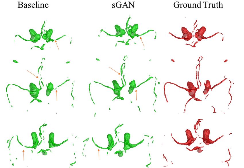
We show sample visual results of representative slices in Figure 3. Sample 3D visual results are given
as surface renderings of segmentation maps in Figure 4.
5 Discussion and Conclusion
MRA is based on different relaxation properties of moving spins in flowing blood inside vessels,
compared to those of static spins found in other tissue. The presented sGAN method is a data-driven
approach to generation of MRA contrast, from the multi-contrast T1- and T2-weighted MRI, which
are based on spin-lattice and spin-spin relaxation effects. It is possible to include other available MR
contrast in patient scans such as Proton Density, FLAIR, and so on, as additional input channels to
the sGAN network.
The sGAN relies on the recent popular PatchGAN framework as the baseline. In the adaptation
of the baseline method to MRA generation, the steerable-filter response based loss term included
in the sGAN method highlights the directional features of vessel structures. This leads to an
enhanced smoothing along vessels while improving their continuity. This is demonstrated qualitatively
through visual inspection. In quantitative evaluations, the sGAN performs similarly with a slight
increase (statistically insignificant) in PSNR values compared to those of the baseline. However, it
is well-known that PSNR measure does not necessarily correspond to perceptual quality in image
evaluations [20, 23]. In terms of the vascular segmentation maps extracted from the generated MRAs
and the original MRA, the sGAN improves the overlap scores by 2% against the baseline. This is a
desirable output, as the MRA targets imaging of vascular anatomy.
The presented sGAN method involves 2D slice generation. This choice is based on the native axial
acquisition plan of the MRA sequences, hence the generated MRA has expectedly higher resolution
in the axial plane. Our future work includes extension of sGAN to a fully 3D architecture. Making
use of 3D neighborhood information, both in generator networks and 3D steerable filter responses is
expected to increase the continuity of vessels in 3D.
6Baseline GAN sGAN Ground Truth
Figure 3: Visual comparison of generated 2D MRA axial slices to the original MRA slices by both
the baseline and the sGAN methods. The last row shows a sagittal slice.
7Figure 4: Visual comparison of segmentation maps over generated MRA to those over the original
MRA in surface rendering format using both the baseline and the sGAN methods.
The proposed sGAN has the potential to be useful in retrospective studies of existing MR image
databases that lack MRA contrast. Furthermore, after extensive validation, it could lead to cost and
time effectiveness where it is needed, by construction of the MRA based on relatively more common
sequences such as T1- and T2-weighted MR contrast.
References
[1] Ciprian Catana, Andre van der Kouwe, Thomas Benner, Christian J Michel, Michael Hamm,
Matthias Fenchel, Bruce Fischl, Bruce Rosen, Matthias Schmand, and A Gregory Sorensen.
Toward implementing an mri-based pet attenuation-correction method for neurologic studies on
the mr-pet brain prototype. Journal of Nuclear Medicine, 51(9):1431–1438, 2010.
[2] Agisilaos Chartsias, Thomas Joyce, Mario Valerio Giuffrida, and Sotirios A Tsaftaris. Multi-
modal mr synthesis via modality-invariant latent representation. IEEE transactions on medical
imaging, 37(3):803–814, 2018.
[3] Mario Cirillo, Francesco Scomazzoni, Luigi Cirillo, Marcello Cadioli, Franco Simionato,
Antonella Iadanza, Miles Kirchin, Claudio Righi, and Nicoletta Anzalone. Comparison of
3d tof-mra and 3d ce-mra at 3 t for imaging of intracranial aneurysms. European journal of
radiology, 82(12):e853–e859, 2013.
[4] Salman Ul Hassan Dar, Mahmut Yurt, Levent Karacan, Aykut Erdem, Erkut Erdem, and Tolga
Çukur. Image synthesis in multi-contrast mri with conditional generative adversarial networks.
arXiv preprint arXiv:1802.01221, 2018.
[5] William T Freeman, Edward H Adelson, et al. The design and use of steerable filters. IEEE
Transactions on Pattern analysis and machine intelligence, 13(9):891–906, 1991.
[6] Ian Goodfellow, Jean Pouget-Abadie, Mehdi Mirza, Bing Xu, David Warde-Farley, Sherjil
Ozair, Aaron Courville, and Yoshua Bengio. Generative adversarial nets. In Advances in neural
information processing systems, pages 2672–2680, 2014.
8[7] Hayit Greenspan, Bram van Ginneken, and Ronald M Summers. Guest editorial deep learning in
medical imaging: Overview and future promise of an exciting new technique. IEEE Transactions
on Medical Imaging, 35(5):1153–1159, 2016.
[8] Michael P Hartung, Thomas M Grist, and Christopher J François. Magnetic resonance angiog-
raphy: current status and future directions. Journal of Cardiovascular Magnetic Resonance,
13(1):19, 2011.
[9] Kaiming He, Xiangyu Zhang, Shaoqing Ren, and Jian Sun. Deep residual learning for image
recognition. In Proceedings of the IEEE conference on computer vision and pattern recognition,
pages 770–778, 2016.
[10] Sergey Ioffe and Christian Szegedy. Batch normalization: Accelerating deep network training
by reducing internal covariate shift. arXiv preprint arXiv:1502.03167, 2015.
[11] Phillip Isola, Jun-Yan Zhu, Tinghui Zhou, and Alexei A Efros. Image-to-image translation with
conditional adversarial networks. arXiv preprint, 2017.
[12] Tim Jerman, Franjo Pernuš, Boštjan Likar, and Žiga Špiclin. Enhancement of vascular structures
in 3d and 2d angiographic images. IEEE transactions on medical imaging, 35(9):2107–2118,
2016.
[13] Justin Johnson, Alexandre Alahi, and Li Fei-Fei. Perceptual losses for real-time style transfer
and super-resolution. In European Conference on Computer Vision, 2016.
[14] Konstantinos Kamnitsas, Christian Ledig, Virginia FJ Newcombe, Joanna P Simpson, Andrew D
Kane, David K Menon, Daniel Rueckert, and Ben Glocker. Efficient multi-scale 3d cnn with
fully connected crf for accurate brain lesion segmentation. Medical image analysis, 36:61–78,
2017.
[15] Diederik P Kingma and Jimmy Ba. Adam: A method for stochastic optimization. arXiv preprint
arXiv:1412.6980, 2014.
[16] Diederik P Kingma and Max Welling. Auto-encoding variational bayes. arXiv preprint
arXiv:1312.6114, 2013.
[17] Andrew JM Kiruluta and R Gilberto González. Magnetic resonance angiography: physical
principles and applications. In Handbook of clinical neurology, volume 135, pages 137–149.
Elsevier, 2016.
[18] Hans Knutsson, Roland Wilson, and Gösta Granlund. Anisotropic nonstationary image estima-
tion and its applications: Part i–restoration of noisy images. IEEE Transactions on Communica-
tions, 31(3):388–397, 1983.
[19] Thomas Markus Koller, Guido Gerig, Gabor Szekely, and Daniel Dettwiler. Multiscale detection
of curvilinear structures in 2-d and 3-d image data. In Computer Vision, 1995. Proceedings.,
Fifth International Conference on, pages 864–869. IEEE, 1995.
[20] Christian Ledig, Lucas Theis, Ferenc Huszár, Jose Caballero, Andrew Cunningham, Alejandro
Acosta, Andrew Aitken, Alykhan Tejani, Johannes Totz, Zehan Wang, et al. Photo-realistic
single image super-resolution using a generative adversarial network. arXiv preprint, 2016.
[21] Chuan Li and Michael Wand. Precomputed real-time texture synthesis with markovian gen-
erative adversarial networks. In European Conference on Computer Vision, pages 702–716.
Springer, 2016.
[22] Andrew L Maas, Awni Y Hannun, and Andrew Y Ng. Rectifier nonlinearities improve neural
network acoustic models. In Proc. icml, volume 30, page 3, 2013.
[23] Michael Mathieu, Camille Couprie, and Yann LeCun. Deep multi-scale video prediction beyond
mean square error. arXiv preprint arXiv:1511.05440, 2015.
[24] Mehdi Mirza and Simon Osindero. Conditional generative adversarial nets. arXiv preprint
arXiv:1411.1784, 2014.
[25] Dong Nie, Roger Trullo, Jun Lian, Caroline Petitjean, Su Ruan, Qian Wang, and Dinggang Shen.
Medical image synthesis with context-aware generative adversarial networks. In International
Conference on Medical Image Computing and Computer-Assisted Intervention, pages 417–425.
Springer, 2017.
[26] Dwight G Nishimura. Time-of-flight mr angiography. Magnetic resonance in medicine,
14(2):194–201, 1990.
9[27] Alec Radford, Luke Metz, and Soumith Chintala. Unsupervised representation learning with
deep convolutional generative adversarial networks. arXiv preprint arXiv:1511.06434, 2015.
[28] Anna MH Sailer, Janneke P Grutters, Joachim E Wildberger, Paul A Hofman, Jan T Wilmink,
and Willem H van Zwam. Cost-effectiveness of cta, mra and dsa in patients with non-traumatic
subarachnoid haemorrhage. Insights into imaging, 4(4):499–507, 2013.
[29] Stephen M Smith, Mark Jenkinson, Mark W Woolrich, Christian F Beckmann, Timothy EJ
Behrens, Heidi Johansen-Berg, Peter R Bannister, Marilena De Luca, Ivana Drobnjak, David E
Flitney, et al. Advances in functional and structural mr image analysis and implementation as
fsl. Neuroimage, 23:S208–S219, 2004.
[30] Pascal Vincent, Hugo Larochelle, Yoshua Bengio, and Pierre-Antoine Manzagol. Extracting and
composing robust features with denoising autoencoders. In Proceedings of the 25th international
conference on Machine learning, pages 1096–1103. ACM, 2008.
[31] Wen Wei, Emilie Poirion, Benedetta Bodini, Stanley Durrleman, Olivier Colliot, Bruno Stankoff,
and Nicholas Ayache. FLAIR MR Image Synthesis By Using 3D Fully Convolutional Networks
for Multiple Sclerosis. In ISMRM-ESMRMB 2018 - Joint Annual Meeting , pages 1–6, Paris,
France, June 2018.
[32] Philip M White, Evelyn M Teasdale, Joanna M Wardlaw, and Valerie Easton. Intracranial
aneurysms: Ct angiography and mr angiography for detection—prospective blinded comparison
in a large patient cohort. Radiology, 219(3):739–749, 2001.
[33] Jelmer M Wolterink, Anna M Dinkla, Mark HF Savenije, Peter R Seevinck, Cornelis AT van den
Berg, and Ivana Išgum. Deep mr to ct synthesis using unpaired data. In International Workshop
on Simulation and Synthesis in Medical Imaging, pages 14–23. Springer, 2017.
[34] Ruifang Yan, Bo Zhang, Long Wang, Qiang Li, Fengmei Zhou, Jipeng Ren, Zhansheng Zhai,
Zheng Li, and Hongkai Cui. A comparison of contrast-free mra at 3.0 t in cases of intracranial
aneurysms with or without subarachnoid hemorrhage. Clinical imaging, 49:131–135, 2018.
[35] Guang Yang, Simiao Yu, Hao Dong, Greg Slabaugh, Pier Luigi Dragotti, Xujiong Ye, Fangde
Liu, Simon Arridge, Jennifer Keegan, Yike Guo, et al. Dagan: Deep de-aliasing generative
adversarial networks for fast compressed sensing mri reconstruction. IEEE Transactions on
Medical Imaging, 2017.
[36] Habib Zaidi, Marie-Louise Montandon, and Daniel O Slosman. Magnetic resonance imaging-
guided attenuation and scatter corrections in three-dimensional brain positron emission tomog-
raphy. Medical physics, 30(5):937–948, 2003.
[37] Matthew D Zeiler, Dilip Krishnan, Graham W Taylor, and Rob Fergus. Deconvolutional
networks. In Computer Vision and Pattern Recognition (CVPR), 2010 IEEE Conference on,
pages 2528–2535. IEEE, 2010.
[38] Jun-Yan Zhu, Taesung Park, Phillip Isola, and Alexei A Efros. Unpaired image-to-image
translation using cycle-consistent adversarial networks. arXiv preprint arXiv:1703.10593, 2017.
10You can also read