Diagnostic Accuracy and Failure Mode Analysis of a Deep Learning Algorithm for the Detection of Cervical Spine Fractures - American Journal of ...
←
→
Page content transcription
If your browser does not render page correctly, please read the page content below
Published June 11, 2021 as 10.3174/ajnr.A7179

                                                                                                                                        ORIGINAL RESEARCH
                                                                                                                                                              SPINE

            Diagnostic Accuracy and Failure Mode Analysis of a Deep
              Learning Algorithm for the Detection of Cervical Spine
                                                          Fractures
                                                                                               A.F. Voter,      M.E. Larson,       J.W. Garrett, and         J.-P.J. Yu

      ABSTRACT
      BACKGROUND AND PURPOSE: Artificial intelligence decision support systems are a rapidly growing class of tools to help manage
      ever-increasing imaging volumes. The aim of this study was to evaluate the performance of an artificial intelligence decision sup-
      port system, Aidoc, for the detection of cervical spinal fractures on noncontrast cervical spine CT scans and to conduct a failure
      mode analysis to identify areas of poor performance.

      MATERIALS AND METHODS: This retrospective study included 1904 emergent noncontrast cervical spine CT scans of adult patients
      (60 [SD, 22] years, 50.3% men). The presence of cervical spinal fracture was determined by Aidoc and an attending neuroradiologist;
      discrepancies were independently adjudicated. Algorithm performance was assessed by calculation of the diagnostic accuracy, and
      a failure mode analysis was performed.

      RESULTS: Aidoc and the neuroradiologist’s interpretation were concordant in 91.5% of cases. Aidoc correctly identified 67 of 122
      fractures (54.9%) with 106 false-positive flagged studies. Diagnostic performance was calculated as the following: sensitivity, 54.9%
      (95% CI, 45.7%–63.9%); specificity, 94.1% (95% CI, 92.9%–95.1%); positive predictive value, 38.7% (95% CI, 33.1%–44.7%); and negative
      predictive value, 96.8% (95% CI, 96.2%–97.4%). Worsened performance was observed in the detection of chronic fractures; differen-
      ces in diagnostic performance were not altered by study indication or patient characteristics.

      CONCLUSIONS: We observed poor diagnostic accuracy of an artificial intelligence decision support system for the detection of
      cervical spine fractures. Many similar algorithms have also received little or no external validation, and this study raises concerns
      about their generalizability, utility, and rapid pace of deployment. Further rigorous evaluations are needed to understand the weak-
      nesses of these tools before widespread implementation.

      ABBREVIATIONS: AI ¼ artificial intelligence; ASiR ¼ adaptive statistical iterative reconstruction; DSS ¼ decision support system; CSFx ¼ cervical spinal fractures

C     ervical spinal fractures (CSFx) are devastating injuries that can
      cause severe morbidity and mortality from damage to the
                                                                                        enclosed spinal cord, the craniocervical junction, and cervical vas-
                                                                                        culature.1 Failure of the osseous spinal column can lead to instabil-
                                                                                        ity and impingement of the underlying spinal cord;2 therefore,
                                                                                        timely identification and stabilization of CSFx are crucial to prevent
Received December 9, 2020; accepted after revision March 14, 2021.
                                                                                        further disability.1,3 In the acute clinical setting, NCCT of the cervi-
From the School of Medicine and Public Health (A.F.V.) and Departments of
Radiology (M.E.L., J.W.G., J.-P.J.Y.), Biomedical Engineering (J.-P.J.Y.), College of   cal spine is the recommended method for detecting CSFx;4 how-
Engineering, and Psychiatry (J.-P.J.Y.), University of Wisconsin School of Medicine     ever, with diagnostic imaging volumes dramatically increasing,5,6
and Public Health, Madison, Wisconsin.
                                                                                        these increased imaging volumes place a burden on radiologists
A.F.V. was supported by National Institutes of Health F30 CA210465 and T32
GM008692; J.W.G. was supported by National Institutes of Health R01 LM013151;           who must maintain diagnostic accuracy and efficiency.7 While there
and J.-P.J.Y. was supported by the Clinical and Translational Science Award
program, through the National Institutes of Health National Center for Advancing
                                                                                        has been great effort to reduce the number of unnecessary scans or-
Translational Sciences, grant UL1TR002373.                                              dered, including the use and implementation of the National
The content is solely the responsibility of the authors and does not necessarily        Emergency X-Radiography Utilization Study Group8 criteria and
represent the official views of the National Institutes of Health.
                                                                                        the Canadian C-Spine Rule9 to reduce the number of unnecessary
Please address correspondence to John-Paul J. Yu, MD, PhD, Department of
Radiology, University of Wisconsin-Madison, 600 Highland Ave, D4-352, Madison,          cervical spinal NCCTs, their effectiveness appears to be modest,10,11
Wisconsin; e-mail: jpyu@uwhealth.org                                                    and diagnostic imaging volumes continue to increase.
     Indicates open access to non-subscribers at www.ajnr.org                               To assist radiologists in managing these rising case volumes,
     Indicates article with online supplemental data.                                   artificial intelligence (AI) decision support systems (DSSs) have
http://dx.doi.org/10.3174/ajnr.A7179                                                    been developed to help prioritize imaging studies with critical
                                                                                                      AJNR Am J Neuroradiol :          2021    www.ajnr.org        1

                                        Copyright 2021 by American Society of Neuroradiology.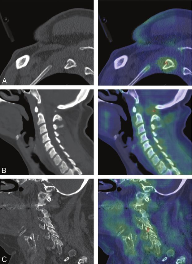
findings.12,13 These DSSs identify and subsequently flag studies
with actionable results, allowing radiologists to prioritize them
over scans with likely negative findings to speed the reporting of
critical findings. However, DSSs that incorrectly flag an excessive
number of studies with negative findings or conversely miss criti-
cal findings might slow the radiologist’s performance. Rigorous
analysis is, therefore, crucial. AI algorithms are known to have
numerous limitations, including the need for large, diverse, and
unbiased datasets,14 which can be difficult to acquire or curate15
and operate in a manner that precludes direct interrogation of
the decision process itself. These issues can lead to poor perform-
ance, which is difficult or impossible to troubleshoot, especially
when the algorithms are implemented in settings beyond their
initial training environment.16–18 While the rapid development
and clinical implementation of DSSs are exciting, this prolifera-
tion risks outstripping our ability to rigorously assess and validate
their performance. This validation and assessment have not been
extensively performed or reported in the literature. Furthermore,
site-specific performance differences without obvious etiologies
have been observed for AI DSSs.16-18 Thus, rigorous studies to
guide AI DSS installations in varied clinical settings and a greater
understanding of the generalizability (or lack thereof) of AI DSSs
are needed to safely translate this important tool into widespread
clinical practice.
    Our institution recently implemented Aidoc (Aidoc Medical),
an FDA-cleared, commercially available AI DSS for the detection
of CSFx.19 While several spine fracture DSSs have been devel-
oped,19-23 their diagnostic accuracy and overall performance
remain unknown. To gain insight into the performance of this            FIG 1. Standards for reporting diagnostic accuracy studies (STARD)
system specifically and AI DSSs more generally, we conducted a          patient flow diagram.
retrospective review of Aidoc as clinically implemented in our
institution. The aim of this study was to characterize the perform-     rotation speed ¼ 5.6 mm/rotation; rotation time ¼ 0.5 seconds;
ance of Aidoc for the detection of CSFx and conduct a failure           automatic exposure control ¼ smart mA (230–750 mA); section
mode analysis to identify areas of poor diagnostic performance.         thickness ¼ 1.25 mm; interval ¼ 0.625 mm. Standard soft-tissue
                                                                        and bone window (Bone Plus algorithm [GE Healthcare]) recon-
                                                                        structions were contemporaneously generated for review by radiol-
MATERIALS AND METHODS
                                                                        ogists (1.25-mm section thickness, sagittal and coronal; 0.625-mm
This Health Insurance Portability and Accountability Act–compliant
                                                                        interval; no adaptive statistical iterative reconstruction [ASiR]).
retrospective study was approved by the institutional review board.
                                                                        Immediately following study acquisition, axial thin bone (Bone
The requirement for informed consent was waived. The data were
                                                                        Plus reconstruction; 0.625-mm section thickness; 0.312-mm inter-
analyzed and controlled by the authors exclusively, none of whom
                                                                        val; no ASiR) and sagittal bone (Bone Plus reconstruction; 1.5-mm
are employees of or consultants to Aidoc Medical or its competitors.
                                                                        section thickness; 0.98-mm interval; no ASiR) series were gener-
Study Population, Data Collection, Imaging Parameters,                  ated and analyzed by the Aidoc algorithm, which then classifies
and AI System                                                           each scan as positive or negative for CSFx. Aidoc-specific image se-
Adult (older than 18 years of age) CT cervical spine studies with-      ries were not available to the interpreting radiologist for review.
out contrast from January 20, 2020, to October 8, 2020, in our radi-    However, because the algorithm was evaluated as clinically imple-
ology information system were identified and contemporaneously          mented, the final Aidoc classification and key image indicating the
processed by Aidoc. Pediatric (younger than 18 years of age) stud-      flagged pathology were available to the radiologist at the time of
ies and examinations with intrathecal contrast were excluded from       initial study interpretation. For the purposes of this study, the final
this study. Scans were performed at an academic level I trauma          neuroradiologist interpretation serves as ground truth data and is
center and associated outreach imaging centers with a fleet of 9        in keeping with prior approaches evaluating the diagnostic per-
models of scanners (GE Healthcare) (summarized in Online                formance of AI-related systems.24,25
Supplemental Data). A total of 1904 adult, noncontrast cervical
spine CT scans were identified in 1923 emergent neck CT scans           Data Processing and Analysis
(mean age, 60 [SD, 22 ] years; 50.3% men). Acquisition parameters       The presence of a cervical spine fracture, type of fracture, vertebra
for noncontrast CT examinations of the cervical spine are as fol-       fractured, estimate of fracture age, and study indication were
lows: 120 kV(peak); axial helical acquisition; pitch ¼ 0.625 mm;        manually extracted from the attending neuroradiologist imaging
2    Voter    2021   www.ajnr.org
Table 1: The impact of patient characteristics on Aidoc performance assumed to be correct; studies with discordant inter-
Aidoc pretations were reviewed by a third independent
Incorrect P reviewer not involved in the initial interpretation (ra-
Factor (No.) % Value
diology resident and attending neuroradiologist with
Total (No.) (%) 1904, 100 161 100 6 years of experience) to make a final ground truth
Indication (No.) (%) .97
Trauma 1796, 94 155 96 determination. Study indication was inferred from
Critical 511, 27 45 28 the report body and imaging order. Critical traumas
Minor 888, 47 74 46 included motor vehicle collisions, falls from heights
Not specified 397, 21 36 22 or stairs, sporting accidents, assaults, and hangings.
Neck pain 27, 1 1 1
Minor traumas largely involved falls from standing
Neurologic deficit 33, 2 2 1
Postoperative 10, 1 1 1 height or lower. Last, traumas were categorized as
Other 38, 2 2 1 “not specified” if there was insufficient information
Sex (No.) (%) .08 regarding the mechanism of trauma.
Male 958, 50 92 57
Female 946, 50 69 43 Statistical Analysis
Imaging location (No.) (%) .86
x 2 tests and 2-sided paired t tests were used for sta-
Academic center 1659. 87 141 88
Outreach center 245, 13 20 12 tistical testing for categoric and quantitative com-
History of cervical spine surgery .57 parisons, respectively, with a significance threshold
(No.) (%) of .05. Diagnostic accuracy (sensitivity, specificity,
Prior surgery 67, 4 7 4 positive predictive value, negative predictive value,
No prior surgery 1837, 96 154 96
and tests for statistical significance were all per-
Age (mean) (yr) .03
Overall 60 (SD, 22) formed in Excel 365 [Microsoft]).
Aidoc incorrect 64 (SD, 21)
Aidoc correct 60 (SD, 22)
RESULTS
To gauge the diagnostic accuracy of Aidoc as clini-
Table 2: Etiology of the false-positive flagged studies cally implemented in our institution, we identified
Percentage of All 1904 noncontrast cervical spine CTs for inclusion during our
Flagged Studies study. A total of 173 (9.1%) of the total studies were flagged by
False-Positive Etiology Count (n = 173) Aidoc as positive for CSFx, and CSFx were identified on 38.7%
Degeneration 55 31.8 (67/173) of the flagged studies. Of the studies not flagged by
Degenerative ossicle 18 10.4 Aidoc, 3.2% (55/1731) contained fractures (Fig 1). Diagnostic
Facet degeneration 14 8.1
performance characteristics with 95% confidence intervals were
Calcified ligament 6 3.5
Cortical irregularity 7 4.0 determined as follows: sensitivity, 54.9% (95% CI, 45.7%–63.9%);
Osteopenia 4 2.3 specificity, 94.1% (95% CI, 92.9%–95.1%); positive predictive
Cystic degeneration 4 2.3 value, 38.7% (95% CI, 33.1%–44.7%); and negative predictive
Atlantodental joint 1 0.6 value, 96.8% (95% CI, 96.2%–97.4%).
Osteophyte 1 0.6
First, we sought to understand how patient factors might
Noncervical pathology 15 8.7
Rib fracture 8 4.6 impact the diagnostic accuracy of Aidoc (Table 1). Because the
Degeneration, thoracic 4 2.3 mechanism of injury can determine the type and severity of
Skull fracture 2 1.2 injury, we calculated the Aidoc false-negative rate based on the
Carotid calcification 1 0.6 indication for the CT examination of the cervical spine (eg,
Anatomic variant 10 5.8
trauma, neck pain, neurologic deficit). No significant differences
Nonunion vertebrae 4 2.3
Transitional anatomy 2 1.2 in Aidoc performance were noted for any of the study indica-
Limbus 2 1.2 tions, study location (ie, academic center or outreach imaging
Bifid spinous process 1 0.6 center), or model of CT scanner (Online Supplemental Data,
Secondary transverse foramen 1 0.6 P ¼ .82). Similarly, the diagnostic error rate of Aidoc was not
Nutrient foramen 9 5.2
Artifact 7 4.0
impacted by either patient sex or history of cervical spine surgery.
Unknown 8 4.6 We did observe, however, that patients incorrectly classified by
Other 2 1.7 Aidoc were older than those correctly classified (mean 64
DISH 1 0.6 [SD, 21] years versus 60 [SD, 22] years, respectively; P ¼ .03).
Occipital suture 1 0.6 Next, we examined whether characteristics of the individual
Total 106 61.3
fractures impacted algorithm performance (Online Supplemental
Note:—DISH indicates diffuse idiopathic skeletal hyperostosis.
Data). Aidoc performance was found to be independent of the
number of vertebrae fractured (single versus multiple) and the
report of each study. To establish the ground truth of the pres- identity of the fractured vertebrae. However, while they were not
ence or absence of an CSFx, we compared the interpretations of significant as a category, we observed a lower rate of incorrect
the neuroradiologist and Aidoc. Concordant interpretations were Aidoc calls with injuries of C2 and a higher rate at C5. We also
AJNR Am J Neuroradiol : 2021 www.ajnr.org 3Data). However, the algorithm missed
50% (5 of 10) of acute fractures involv-
ing the transverse foramen.
Because the number of false-positive
flagged studies exceeded the number of
true-positives (106 versus 67), we next
sought to understand the poor positive
predictive value of Aidoc by exploring
possible failure modes of the false-
positive studies. Each study flagged by
Aidoc is accompanied by a probability
heat map highlighting the suspected frac-
ture identified by Aidoc, thus allowing us
to identify the etiology of each false-
positive finding (Table 2). The most
common etiology was the presence of de-
generative structures such as a degenera-
tive ossicle (Fig 2A), facet degeneration
(Fig 2B), ossification of the ligamentum
flavum (Fig 2C), or other degenerative
cortical irregularities. The next most
common sources of false-positive find-
ings were pathologies outside the cervical
spine and scope of the algorithm, such as
rib or skull fractures (Fig 3A), and nonpa-
thologic anatomic variants (Fig 2B, -C).
False-positives were also found to have
been triggered by motion artifacts or nor-
mal anatomy, and in a small number of
cases, we were unable to identify any
abnormality.
DISCUSSION
A wide range of AI DSSs have been devel-
oped to reduce the risk of missing or
delaying the reporting of time-sensitive
findings.12,13 However, AI algorithms are
known to have limitations and can be dif-
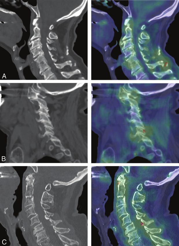
FIG 2. Examples of degenerative findings falsely flagged by Aidoc. Each panel shows the sagittal
noncontrast cervical spine CT (left) and the Aidoc key image indicating the flagged pathology in ficult to generalize to clinical sites with
red (right). A, A chronic ossicle falsely flagged by Aidoc. B, False-positive findings triggered by disease prevalence and imaging protocols
facet degeneration. C, Ossification of the ligamentum flavum incorrectly identified as a fracture that differ from training datasets. Because
by Aidoc. poorly performing DSSs can hinder radi-
ologists, it is crucial that these tools
undergo rigorous evaluation before wide-
spread implementation. While the imple-
observed that the algorithm was significantly more successful at mentation of Aidoc for CSFx has excellent reported diagnostic
identifying acute fractures than nonacute fractures (ie, chronic or characteristics (sensitivity of 91.7% and specificity of 88.6%, as
age-indeterminate). Furthermore, location of the fracture within reported in the initial FDA disclosure),19 to our knowledge, no in-
each vertebra was a significant contribution to algorithm per- dependent evaluations of its performance have been published or,
formance, with fractures of osteophytes or the vertebral body more generally, any data evaluating the diagnostic accuracy of AI
overrepresented in the false-negative studies. DSSs in detecting cervical spine fractures. To this end, we con-
The timely identification of new fractures is of particular clini- ducted a retrospective study to evaluate the diagnostic accuracy of
cal importance, so we explored the performance of Aidoc in the Aidoc, an FDA-cleared AI DSS for the evaluation of CSFx as clini-
detection of acute fractures. We did not find any significant dif- cally implemented at our institution.
ferences between the acute fractures correctly flagged by Aidoc At our institution, Aidoc fared poorly, with a notably lower
and those it missed, though our analysis was limited by the rela- sensitivity and positive predictive value than initially
tively small number of acute fractures (Online Supplemental reported to the FDA.19 To understand this unexpected
4 Voter 2021 www.ajnr.orgBecause the value of this and similar
algorithms stems from the faster detec-
tion of findings that can alter clinical
management, it is especially important
to consider the performance in the
detection of acute fractures. We did not
find any differences between the acute
fractures correctly identified or missed
by Aidoc, though our statistical analysis
was limited by the relatively small num-
ber of acute fractures missed by the
algorithm. However, it is notable that
the 50% of the acute fractures involving
the transverse foramen were missed by
Aidoc. These fractures can indicate
compromise of the underlying vertebral
artery, so rapid detection by the algo-
rithm is especially valuable and more
examples should be included in the
algorithm training set.
In cases with multiple fractures, the
algorithm needs to correctly identify only
a single fracture to score as correct.
Therefore, we hypothesized that these
studies would have a lower false-negative
rate. However, we observed that the miss
rate did not depend on the total number
of fractures present in an imaging exami-
nation, suggesting that fracture identifi-
cation may have been precluded by other
features of the study rather than fracture
characteristics themselves.
We noted a significant and unex-
pected number of false-positive studies
in our dataset, outnumbering the
flagged true CSFx. Spine degeneration
was the most common etiology of false-
positives observed. This is perhaps not
FIG 3. Examples of nondegenerative findings falsely flagged by Aidoc. Sagittal noncontrast cervi-
cal spine CT (left) and the Aidoc key image indicating the flagged pathology in red (right). A, Rib surprising because degeneration occurs
fracture outside of the cervical spine incorrectly flagged by Aidoc. B, Congenital hypoplasia of with aging and generates abnormalities
the posterior arch of the atlas flagged as a fracture. C, A nonpathologic nutrient foramen with such ossicles or irregularities in the
degenerative changes identified as a fracture by Aidoc. bony surface that could be mistaken for
fractures. Accordingly, the age of
patients misclassified by Aidoc was
performance gap, we conducted a failure mode analysis to higher than that in the correctly classified group, and we
identify possible sources of this impaired performance. hypothesize that the increased burden of degeneration may have
Neither imaging location, scanner model, nor study indica- led to impaired performance. Our dataset lacked an accessible
tions were found to be significantly associated with the diag- way to assess the extent of degeneration directly, but this could be
nostic performance of Aidoc. However, the sensitivity was explored in future studies. We speculate that greater representa-
affected by patient age and characteristics of the underlying tion of nonfractured examples of both degeneration and
fracture, specifically the fracture acuity and location of the anatomic variants in the training set would likely reduce the
fracture, with chronic fractures and fractures of osteophytes false-positive burden, given their overrepresentation here in our
and the vertebral body overrepresented among the missed analysis as false-positives. In addition, differences in diagnostic
fractures. Osteophyte formation and compression fractures accuracy may also be attributed to institution-specific differ-
are degenerative in nature, so underperformance in their ences and would be difficult to disentangle. However, in the
detection may contribute to the worsened algorithm per- FDA 510(k) application, the number of cases positive and
formance in older patients. negative for CSFx were adjusted to be roughly equal. Because
AJNR Am J Neuroradiol : 2021 www.ajnr.org 5diagnostic performance is strongly influenced by disease 3. Fischer PE, Perina DG, Delbridge TR, et al. Spinal motion restric-
prevalence, this also likely contributes to the observed differ- tion in the trauma patient: a joint position statement. Prehosp
ences in the reported diagnostic accuracy of Aidoc and our Emerg Care 2018;22:659–61 CrossRef Medline
4. Beckmann NM, West OC, Nunez D Jr, et al. Expert Panel
clinical observations.19 Our observed rate of positive findings on Neurological Imaging and Musculoskeletal Imaging. ACR
is 6.4%, which reflects the true rate of CSFx at our institution. Appropriateness Criteria® Suspected Spine Trauma. J Am Coll
Because positive and negative predictive values depend on the Radiol 2019;16:S264–85 CrossRef Medline
underlying prevalence of the disease, we believe our measurements 5. Hess EP, Haas LR, Shah ND, et al. Trends in computed tomography
will more closely reflect the experience of other users. This discrep- utilization rates: a longitudinal practice-based study. J Patient Saf
2014;10:52–58 CrossRef Medline
ancy highlights an emerging need to standardize study design to
6. Kocher KE, Meurer WJ, Fazel R, et al. National trends in use of com-
allow rigorous and unbiased comparisons across different sites and puted tomography in the emergency department. Ann Emerg Med
for accurate reporting and evaluation of AI DSS algorithms in the 2011;58:452–62 CrossRef Medline
imaging literature. 7. McDonald RJ, Schwartz KM, Eckel LJ, et al. The effects of changes
Our study has limitations that must be considered. First, in utilization and technological advancements of cross-sectional
imaging on radiologist workload. Acad Radiol 2015;22:1191–98
because Aidoc has already been clinically implemented at our insti-
CrossRef Medline
tution, the interpretation by Aidoc of each study was available to the 8. Hoffman JR, Mower WR, Wolfson AB, et al. Validity of a set of clin-
neuroradiologist during the initial read. While this may have ical criteria to rule out injury to the cervical spine in patients
inflated the accuracy of the neuroradiologist’s read, the diagnostic with blunt trauma: National Emergency X-Radiography
accuracy of Aidoc is unaffected. Additionally, while the Aidoc algo- Utilization Study Group. N Engl J Med 2000;343:94–99 CrossRef
Medline
rithm is available to all radiologists at our institution, there is
9. Mower WR, Wolfson AB, Hoffman JR, et al. The Canadian C-spine
marked variation in how it has been incorporated into their individ- rule. N Engl J Med 2004;350:1467–69 CrossRef Medline
ual workflow. We were, therefore, unable to assess whether the algo- 10. Sharp AL, Huang BZ, Tang T, et al. Implementation of the
rithm reduced time to image analysis in cases flagged for CSFx. Canadian CT head rule and its association with use of computed
Nevertheless, given the poor positive predictive value, we suspect tomography among patients with head injury. Ann Emerg Med
2018;71:54–63 CrossRef Medline
that any time savings would be diluted by the number of false-
11. Mower WR, Gupta M, Rodriguez R, et al. Validation of the sensi-
positives. Last, this single-institution study was performed at an aca- tivity of the National Emergency X-Radiography Utilization
demic center equipped with GE Healthcare scanners, potentially Study (NEXUS) head computed tomographic (CT) decision
limiting the generalizability of our findings to institutions in other instrument for selective imaging of blunt head injury patients:
practice settings or those with a different fleet of scanners from an observational study. PLoS Med 2017;14:e1002313 CrossRef
Medline
other vendors.
12. DSI Home. FDA Cleared AI Algorithms. American College of
Radiology. 2020. https://models.acrdsi.org/. Accessed September 11,
CONCLUSIONS 2020
We examined the diagnostic performance of Aidoc for the detec- 13. Benjamens S, Dhunnoo P, Mesko B. The state of artificial intelli-
gence-based FDA-approved medical devices and algorithms: an
tion of CSFx as implemented at our institution and observed
online database. NPJ Digit Med 2020;3:118 CrossRef Medline
meaningful worse diagnostic accuracy than previously reported. 14. Alwosheel A, van Cranenburgh S, Chorus CG. Is your dataset big
Although the nature of neural network algorithms obscures a full enough? Sample size requirements when using artificial neural
understanding of this impairment, our failure mode analysis has networks for discrete choice analysis. J Choice Model 2018;28:167–
identified several potential areas for improvement. Nevertheless, 82 CrossRef
15. Park SH, Han K. Methodologic guide for evaluating clinical per-
the overall performance of this AI DSS at our institution is differ-
formance and effect of artificial intelligence technology for medi-
ent enough and raises potential concerns about the generalizabil- cal diagnosis and prediction. Radiology 2018;286:800–09 CrossRef
ity of AI DSSs across heterogeneous clinical environments and Medline
motivates the creation of data-reporting standards and standar- 16. Kim DW, Jang HY, Kim KW, et al. Design characteristics of stud-
dized study design, the lack of which precludes unbiased compar- ies reporting the performance of artificial intelligence algo-
rithms for diagnostic analysis of medical images: results from
isons of AI DSS performance across both institutions and
recently published papers. Korean J Radiol 2019;20:405–10
algorithms. Adoption of a standardized design for all AI DSS CrossRef Medline
algorithms will help speed the development and safe implementa- 17. Liu X, Faes L, Kale AU, et al. A comparison of deep learning per-
tion of this promising technology as we aim to integrate this im- formance against health-care professionals in detecting diseases
portant tool into clinical workflow. from medical imaging: a systematic review and meta-analysis.
Lancet Digit Health 2019;1:e271–97 CrossRef Medline
18. Zech JR, Badgeley MA, Liu M, et al. Variable generalization per-
Disclosures: Andrew F. Voter—RELATED: Grant: National Institutes of Health.* John
formance of a deep learning model to detect pneumonia in chest
W. Garrett—RELATED: Grant: National Institutes of Health.* John-Paul J. Yu—
RELATED: Grant: National Institutes of Health.* *Money paid to the institution.
radiographs: a cross-sectional study. PLoS Med 2018;15:e1002683
CrossRef Medline
19. U.S. Food and Drug Administration. K190896. 2019 https://www.
REFERENCES accessdata.fda.gov/cdrh_docs/pdf19/K190896.pdf. Accessed February
1. Copley D, Tilliridou D, Jamjoom M. Traumatic cervical spine fractures 19, 2021
in the adult. Br J Hosp Med (Lond) 2016;77:530–35 CrossRef Medline 20. Burns JE, Yao J, Munoz H, et al. Automated detection, localiza-
2. Denis F. The three-column spine and its significance in the classifi- tion, and classification of traumatic vertebral body fractures
cation of acute thoracolumbar spinal injuries. Spine (Phila Pa 1976) in the thoracic and lumbar spine at CT. Radiology 2016;278:64–
1983;8:817–31 CrossRef Medline 73 CrossRef Medline
6 Voter 2021 www.ajnr.org21. Burns JE, Yao J, Summers RM. Vertebral body compression frac- Recognition January 29, 2016. https://arxiv.org/abs/1602.00020.
tures and bone density: automated detection and classification on Accessed September 2, 2020
CT images. Radiology 2017;284:788–97 CrossRef Medline 24. Ginat DT. Analysis of head CT scans flagged by deep learning
22. Tomita N, Cheung YY, Hassanpour S. Deep neural networks for software for acute intracranial hemorrhage. Neuroradiology
automatic detection of osteoporotic vertebral fractures on CT 2020;62:335–40 CrossRef Medline
scans. Comput Biol Med and Med 2018;98:8–15 CrossRef 25. Ojeda PZ, Zawaideh M, Mossa-Basha M, et al. The utility of deep
Medline learning: evaluation of a convolutional neural network for detec-
23. Roth HR, Wang Y, Yao J, et al. Deep convolutional networks tion of intracranial bleeds on non-contrast head computed tomog-
for automated detection of posterior-element fractures raphy studies. In: Proeedings of SPIE Medical Imaging, San Diego
on spine CT. Computer Science . Computer Vision and Pattern California. February 16–21, 2019
AJNR Am J Neuroradiol : 2021 www.ajnr.org 7You can also read