CT image segmentation for inflamed and fibrotic lungs using a multi resolution convolutional neural network - Nature
←
→
Page content transcription
If your browser does not render page correctly, please read the page content below
www.nature.com/scientificreports
OPEN CT image segmentation
for inflamed and fibrotic
lungs using a multi‑resolution
convolutional neural network
Sarah E. Gerard1*, Jacob Herrmann2, Yi Xin3, Kevin T. Martin4, Emanuele Rezoagli5,7,
Davide Ippolito6, Giacomo Bellani5,7, Maurizio Cereda4, Junfeng Guo1,8, Eric A. Hoffman1,8,
David W. Kaczka1,8,9 & Joseph M. Reinhardt1,8
The purpose of this study was to develop a fully-automated segmentation algorithm, robust to
various density enhancing lung abnormalities, to facilitate rapid quantitative analysis of computed
tomography images. A polymorphic training approach is proposed, in which both specifically
labeled left and right lungs of humans with COPD, and nonspecifically labeled lungs of animals with
acute lung injury, were incorporated into training a single neural network. The resulting network is
intended for predicting left and right lung regions in humans with or without diffuse opacification
and consolidation. Performance of the proposed lung segmentation algorithm was extensively
evaluated on CT scans of subjects with COPD, confirmed COVID-19, lung cancer, and IPF, despite
no labeled training data of the latter three diseases. Lobar segmentations were obtained using
the left and right lung segmentation as input to the LobeNet algorithm. Regional lobar analysis
was performed using hierarchical clustering to identify radiographic subtypes of COVID-19. The
proposed lung segmentation algorithm was quantitatively evaluated using semi-automated and
manually-corrected segmentations in 87 COVID-19 CT images, achieving an average symmetric
surface distance of 0.495 ± 0.309 mm and Dice coefficient of 0.985 ± 0.011. Hierarchical clustering
identified four radiographical phenotypes of COVID-19 based on lobar fractions of consolidated and
poorly aerated tissue. Lower left and lower right lobes were consistently more afflicted with poor
aeration and consolidation. However, the most severe cases demonstrated involvement of all lobes.
The polymorphic training approach was able to accurately segment COVID-19 cases with diffuse
consolidation without requiring COVID-19 cases for training.
Computed tomographic (CT) imaging has played an important role in assessing parenchymal abnormalities in
lung diseases such as chronic obstructive pulmonary disease (COPD), and more recently, the novel coronavirus
disease (COVID-19). CT imaging is important for diagnostics as well as quantifying disease involvement and
progression over time. CT-based disease quantification can be used for patient stratification, management, and
prognostication1,2. Automated analysis of images is critical for objective quantification and characterization of
large numbers of CT datasets. In particular, reliable lung and lobe segmentation is an important precursor to
quantifying total lung and regional involvement of the disease.
Conventional lung and lobar segmentation approaches programmatically achieve segmentation using prior
information about voxel intensity and second-order structure in small neighborhoods3–9. More advanced
methods have used shape priors in the form of atlases or statistical shape models10–15. Recently, deep learning
approaches have surpassed the performance of rule-based segmentation by learning important features for
1
Department of Radiology, University of Iowa, Iowa City, IA, USA. 2Department of Biomedical Engineering,
Boston University, Boston, MA, USA. 3Department of Radiology, University of Pennsylvania, Philadelphia,
PA, USA. 4Department of Anesthesiology and Critical Care, University of Pennsylvania, Philadelphia, PA,
USA. 5Department of Medicine and Surgery, University of Milano-Bicocca, Monza, Italy. 6Department of Diagnostic
and Interventional Radiology, San Gerardo Hospital, Monza, Italy. 7Department of Emergency and Intensive Care,
San Gerardo Hospital, Monza, Italy. 8Roy J. Carver Department of Biomedical Engineering, University of Iowa,
Iowa City, IA, USA. 9Department of Anesthesia, University of Iowa, Iowa City, IA, USA. *email: sarah‑gerard@
uiowa.edu
Scientific Reports | (2021) 11:1455 | https://doi.org/10.1038/s41598-020-80936-4 1
Vol.:(0123456789)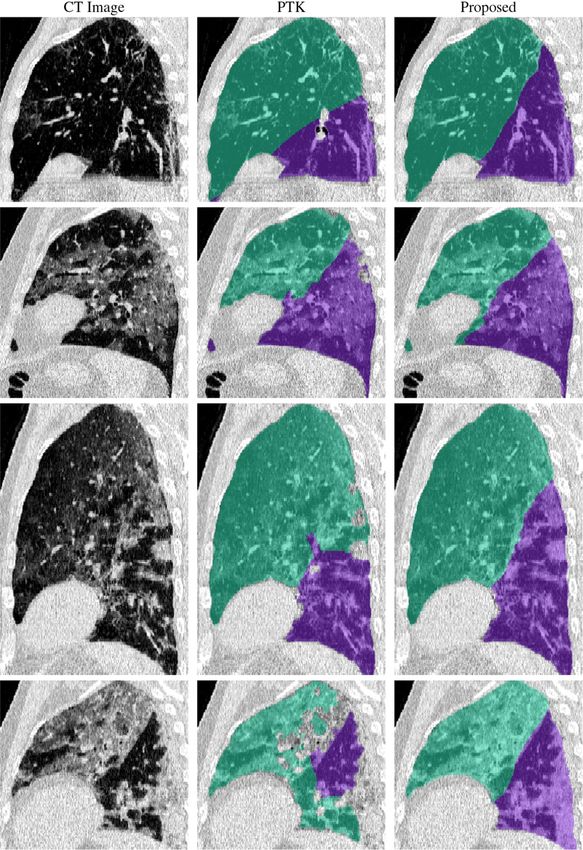
www.nature.com/scientificreports/
Figure 1. (A) Motivation for polymorphic training. In this work, the desired segmentation target is
consolidated cases with specific labels of left lung (LL), right lung (RL), and background (B) (upper right).
However, only normal cases with specific labels (upper left) and consolidated cases with non-specific labels
of lung (L) and background (B) (lower right) are available for training. The proposed polymorphic training
approach allows us to utilize the available training data and generalize to the target domain of consolidated
specifically labeled cases (upper right). (B) Standard training (top) using only specifically labeled COPD images
lacks the consolidation phenotype necessary to successfully segment injured regions in COVID-19 images.
Polymorphic training (bottom) utilizes specifically labeled COPD images with nonspecifically labeled animal
models of acute lung injury to achieve specific lung labels including injured regions in COVID-19 images. The
specific lung labels are depicted in green and blue for left and right lung, respectively. The nonspecific lung label
is depicted in orange.
segmentation from labeled training data. A multi-scale CNN approach for segmentation of acutely injured lungs
in animal models demonstrated that incorporation of global features improved lung segmentation in cases with
diffuse consolidation16. FissureNet is a deep learning based fissure segmentation method which identifies the
boundaries between l obes17, a critical step for lobar segmentation. Preliminary work on extending FissureNet to
segment lobes was proposed, although this method was only evaluated on chronic obstructive pulmonary disease
(COPD) cases without density enhancing pathologies18. Other methods have directly learned lobe segmentation
without first explicitly identifying lungs and fissures19,20.
Automated lung segmentation in patients with COVID-19 is a challenging task, given the multitude of
nonspecific features that appear on CT (i.e., bilateral and peripheral ground-glass opacities and consolidation).
Intensity-based segmentation methods may fail to include infected regions, which is critical for any image
quantitative analysis. Furthermore, lung opacities can obscure the fissure appearance, making it challenging to
identify lobes. CNNs have great potential for automated segmentation due to their ability to identify low-level
and abstract features. However, a challenge with deployment of deep learning methods in medical imaging is the
accessibility to labeled training data representative of all disease phenotypes—for example, a lobar segmentation
network trained only on data from COPD patients is unlikely to perform well in COVID-19 patients with diffuse
lung and focal lung consolidation.
Additional labeled training data may be available, although the labels may not have the desired level of speci-
ficity. For example, a voxel corresponding to parenchymal tissue may simply be labeled as lung (as opposed to
non-lung) or it could be more specifically labeled as left or right lung (see Fig. 1). Although nonspecific labels
may not be directly useful for training networks to predict specific labels, the nonspecific dataset may still contain
important disease phenotypes absent from the dataset with specific labels. We thus hypothesize that data with
generic labels can still be valuable when training a network to predict specific labels. Ideally, training would
accommodate labels with different degrees of specificity (i.e., a hierarchical categorization). In this study, we
propose a solution to accommodate partially labeled training data, wherein “partial” refers to different degrees
of specificity in a hierarchical categorization of labels. We refer to this solution as “polymorphic” training. Poly-
morphism in biology and computer science refers to the ability of organisms and data types to exist as one of
multiple subtypes (e.g., schnauzer is a subtype of dog, dog is a subtype of mammal). We propose a polymorphic
training strategy that injects supervision at different network layers predicting different subtypes of voxel clas-
sification, specifically for data with hierarchical labels.
The specific aim of this work was to develop an algorithm for fully-automated and robust lung segmentation
in CT scans of patients with pulmonary manifestations of COVID-19, to facilitate regional quantitative analysis.
In related work, F issureNet17 and LobeNet18 were proposed for robust segmentation of pulmonary fissures and
lobes. However, FissureNet and LobeNet cannot be applied directly to CT images, but require an initial lung
segmentation which distinguishes left versus right lung. Automated lung segmentation for COVID-19 images
is challenging due to diffuse consolidation obscuring lung boundaries. In this work, we propose a segmentation
method which identifies left and right lungs in COVID-19 images. Given the scarcity of labeled COVID-19 CT
images available for training, two existing datasets with complementary features were used: (1) a dataset from
patients with COPD, with specifically labeled left and right lungs; and (2) a dataset from experimental animal
models of acute lung injury, with only a single nonspecific lung label. The first dataset provides human training
examples with specific left and right lung labels, while the second dataset contains important disease phenotypes
Scientific Reports | (2021) 11:1455 | https://doi.org/10.1038/s41598-020-80936-4 2
Vol:.(1234567890)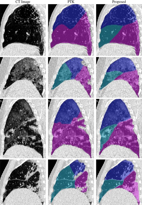
www.nature.com/scientificreports/
Training Evaluation
COPDGene 1000 5986
Animal ARDS 453 –
Cancer – 1620
IPF – 305
COVID-19 – 87
Total 1453 7998
Table 1. Number of 3D CT images used for training and evaluation.
(i.e., ground glass opacification and consolidation) absent from the COPD images (see Fig. 1). The design of
the polymorphic training is motivated by a need to accommodate labeled training data with heterogeneous
degrees of subclassification, since datasets may have a single label for all lung tissue or labels distinguishing left
and right lungs.
Materials and methods
Datasets. The number of images used for training and evaluation are summarized in Table 1. A combina-
tion of human and animal CT datasets with different diseases were utilized for training the lung segmentation
model. Human datasets were acquired from COPDGene21, a multi-center clinical trial with over 10,000 COPD
patients enrolled. Animal datasets of acute lung injury models included canine, porcine, and ovine species (see16
for detailed description of datasets). In total, 1000 human CT images and 452 animal CT images were used
for training the lung segmentation module. Note, only 1000 of the COPD CT images were used for training in
effort to avoid a large imbalance between disease phenotypes in the training data. All training CT images have
a ground truth lung segmentation generated automatically using the Pulmonary Analysis Software Suite (PASS,
University of Iowa Advanced Pulmonary Physiomic Imaging L aboratory22) with manual correction if necessary.
For human datasets, ground truth segmentations distinguished the left and right lungs, whereas the animal
datasets had only a single label for all lung tissue. It is important to note that separation of left and right lungs is
not trivial due to close proximity of the left and right lungs, especially in the three animal species used due to the
accessory lobe adjacent to both the left and right lungs.
A dataset of 133 clinical CT images of COVID-19 patients was acquired from: the Hospital of San Gerardo,
Italy; University of Milan-Bicocca, Italy; Kyungpook National University School of Medicine, South Korea; and
Seoul National University Hospital, South Korea. Patients were included based on confirmed COVID-19 diag-
nosis by nucleic acid amplification tests. Data use was approved by Institutional Review Boards at University of
Milano-Bicocca, the Hospital of San Gerardo, Kyungpook National University School of Medicine, and Seoul
National University Hospital. Given the retrospective nature of the study and in the presence of technical difficult
in obtaining an informed consent of patients in this period of pandemic emergency, informed consent was be
waived and all data was anonymized. All procedures were followed in accordance with the relevant guidelines.
Details from the Korean COVID-19 cases are provided in Nagpal et al.23. Ground truth lung segmentations were
performed for 87 cases using PASS22 or pulmonary toolkit (PTK)24 with manual correction as necessary. Manual
correction required an average of 94 ± 48 min per case.
To evaluate the performance on other pulmonary diseases, three additional evaluation datasets were utilized:
5986 CT images from COPDGene, 1620 CT images from lung cancer patients undergoing radiation therapy,
and 305 CT images from patients with idiopathic pulmonary fibrosis (IPF). Ground truth segmentations were
generated using PASS followed by manual correction.
Multi‑resolution model. The LungNet module used a multi-resolution approach adapted f rom16 to facili-
tate learning both global and local features important for lung segmentation. LungNet consists of a cascade of
two CNN models; the low-resolution model LungNet-LR and the high-resolution model LungNet-HR.
LungNet-LR was trained using low-resolution images. All CT images and target label images are downsam-
pled to 4 mm isotropic voxels using b-spline and nearest-neighbor interpolation for the CT and label images,
respectively. A Gaussian filter was applied to the CT images prior to downsampling to avoid aliasing. LungNet-LR
yields a three-channel image, corresponding to predicted probabilities for left lung, right lung, and background.
LungNet-HR was trained with high-resolution images. The CT image, the output of LungNet-LR, and the
target label image were resampled to have 1 mm isotropic voxels for consistency. The CT image and left/right
probability maps were then combined to produce a three-channel input for training the high-resolution net-
work. Similar to LungNet-LR, the output of LungNet-HR was a three-channel probability image. The final lung
segmentation was obtained by thresholding the left and right probability channels at p = 0.5.
Polymorphic training. We used a novel polymorphic training strategy, illustrated in Fig. 2, which incorpo-
rated all information in partially labeled datasets. The ultimate goal was to train a network that could distinguish
left versus right lung, with or without abnormal pathological features. The three-channel prediction image pro-
duced by the last layer of Seg3DNet, denoted ŶLR , yielded channels corresponding to left lung, right lung, and
background probabilities. To make this output compatible with the animal datasets, which have only a single
lung label, an auxiliary layer with supervision was added to the network after ŶLR . The auxiliary layer performed
Scientific Reports | (2021) 11:1455 | https://doi.org/10.1038/s41598-020-80936-4 3
Vol.:(0123456789)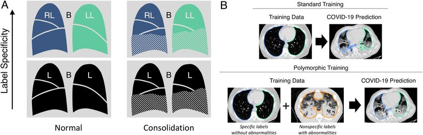
www.nature.com/scientificreports/

                                            Figure 2.  Polymorphic training accommodates labeled data with different degrees of specificity. In this case
                                            some labeled training have specific labels distinguishing left and right lung, while other training data only have a
                                            single label for all lung tissue.

                                            a voxelwise summation of the two channels of ŶLR corresponding to left and right lung prediction. The result-
                                            ing single-channel image was concatenated with the background channel of ŶLR . This produced a two-channel
                                            prediction image, denoted ŶT, with the channels corresponding to lung versus background. During training,
                                            supervision was provided at both ŶLR and ŶT. Equal numbers of human and animal images were sampled for
                                            each batch. Ground truth images were denoted YLR for labeled images that distinguished left versus right lung,
                                            and YT for labeled images that had a single label for total lung. The loss between ŶLR and YLR was computed
                                            using only the human half of the batch, while the loss between ŶT and YT was computed using the entire batch
                                            by converting YLR to YT for human cases. These two losses were equally weighted during each training step.

                                            Lobar analysis. Lobar segmentations were obtained by using the proposed left and right lung segmentation
                                            as input to the FissureNet and LobeNet algorithms, which is currently the leading performer in the LOLA11
                                            grand challenge. No additional training of FissureNet and LobeNet was performed. Regional lobar analysis was
                                            performed using hierarchical clustering to identify subtypes of COVID-19.

                                            Ablation study.      To evaluate the contribution of the polymorphic training approach for lung segmentation,
                                            the proposed approach was compared to a nonpolymorphic model. The nonpolymorphic model only used the
                                            human CT images of COPD for training (i.e., the auxiliary layer and animal training data were not utilized).
                                            Otherwise, there were no differences in the design or training of the polymorphic and nonpolymorphic models.
                                            A two-way analysis of variance was performed with model type as a categorical variable and nonaerated lung
                                            volume fraction as a continuous variable, as well as an interaction term.

                                            Results
                                            Lung segmentation. Lung segmentation results for the polymorphic and nonpolymorphic models are
                                            shown in Fig. 3. Quantitative evaluation of lung segmentations was performed on CT images by comparing the
                                            segmentations to ground truth manual segmentations. The Dice coefficient was used to measure volume over-
                                            lap and the average symmetric surface distance (ASSD) was used to assess boundary accuracy. The ASSD and
                                            Dice coefficient results for each of the four evaluation datasets are shown in Table 2. Overall, on the COVID-19
                                            dataset the polymorphic model achieved an average ASSD of 0.495 ± 0.309 mm and average Dice coefficient
                                            of 0.985 ± 0.011. By comparison, the nonpolymorphic model achieved an average ASSD of 0.550 ± 0.546 mm
                                            and average Dice coefficient of 0.982 ± 0.024. ASSD and Dice coefficient results with respect to nonaerated
                                            lung volume fraction are displayed in Fig. 4. Two-way analysis of variance revealed a significant interaction
                                            between model and nonaerated fraction for each evaluation metric, indicating that the regression coefficients
                                            with respect to nonaerated fraction were significantly different for polymorphic versus nonpolymorphic models.

                                            Lobar segmentation. Lobar segmentation results for the proposed method and PTK are shown in Fig. 5
                                            for right lungs and Fig. 6 for left lungs. For each image in the COVID-19 dataset (133 images in total), the lobar
                                            segmentation result was used to extract the amount of poor aeration (−500 < HU < −100) and consolidation
                                            (HU ≥ −100) in each lobe. Common phenotypes of COVID-19 affected lungs were identified by hierarchical
                                            clustering over the fraction of poorly aerated and consolidated tissue in each lobe. Dendrographic analysis in

          Scientific Reports |   (2021) 11:1455 |                https://doi.org/10.1038/s41598-020-80936-4                                                    4

Vol:.(1234567890)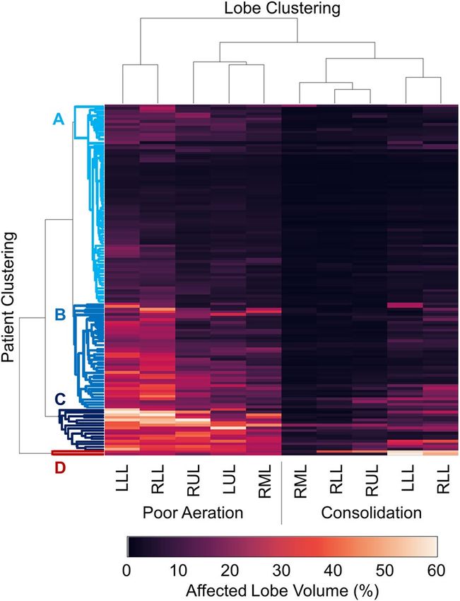
www.nature.com/scientificreports/
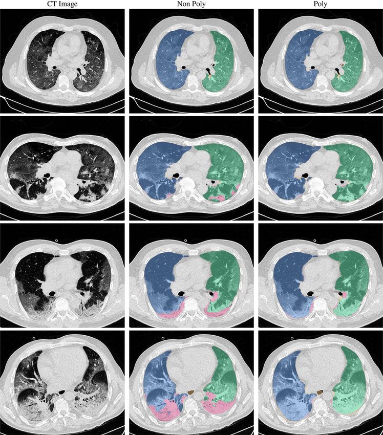
Figure 3. Axial slices of CT images (left column) and lung segmentation results for the nonpolymorphic model
(center column) and the polymorphic model (right column) algorithms for four COVID-19 patients (by row).
Correctly classified voxels are displayed in blue and green for right and left lungs, respectively. False negative and
false positive voxels are illustrated in pink and yellow, respectively.
Fig. 7 reveals four primary clusters of patients that were identified by the hierarchical clustering: (a) mild loss of
aeration primarily in the two lower lobes without consolidation; (b) moderate loss of aeration focused in the two
lower lobes with or without consolidation in lower lobes; (c) severe loss of aeration throughout all lobes with or
without consolidation; and (d) severe loss of aeration and consolidation throughout all lobes.
Scientific Reports | (2021) 11:1455 | https://doi.org/10.1038/s41598-020-80936-4 5
Vol.:(0123456789)www.nature.com/scientificreports/
COPD Cancer IPF COVID-19
N = 5986 N = 1620 N = 305 N = 87
LL RL LL RL LL RL LL RL
ASSD
Non poly 0.339 0.300 0.355 0.485 0.478 0.500 0.514 0.586
Poly 0.378 0.346 0.430 0.513 0.505 0.594 0.480 0.510
Dice
Non Poly 0.990 0.992 0.990 0.987 0.985 0.985 0.982 0.982
Poly 0.989 0.991 0.988 0.986 0.984 0.982 0.984 0.985
Table 2. Lung segmentation results for polymorphic (Poly) and nonpolymorphic (Non-Poly) models. Results
are stratified by lung (LL: left lung, RL: right lung) and the four evaluation datasets.. ASSD results are in mm.
Discussion
In this study, we proposed and implemented a novel polymorphic training algorithm for lung and lobar seg-
mentation in a fully automated pipeline. The pipeline was independently evaluated on CT scans of subjects with
COVID-19, lung cancer, and IPF—however, no COVID-19, lung cancer, or IPF scans were utilized for training
the CNNs. Additionally, the pipeline was extensively evaluated on CT scans of patients with COPD. The COVID-
19 scans are considered very challenging cases for lung and lobe segmentation. Peripheral and diffuse opacities
result in little contrast at the lung boundary. In many cases, the fissure appearance was irregular due to close
proximity of infection. Furthermore, these are clinical scans with some cases having slice thickness greater than
3 mm. Fissure segmentation is especially challenging on such cases. Success of the proposed algorithm on these
cases lends to the generalizability of the proposed approach.
Out lung segmentation algorithm was quantitatively evaluated on 7998 CT images, consisting of four dis-
tinct pulmonary pathologies. To our knowledge, this is the most extensive evaluation of a lung segmentation
algorithm to date. The polymorphic and nonpolymorphic models both achieve sub-voxel lung segmentation
accuracy and demonstrate generalizability across datasets and diseases which were not used for training. The
polymorphic and nonpolymorphic models achieved similar performance on COPD, IPF, and lung cancer cases
and on COVID-19 cases without consolidation. The ablation studied demonstrated that the polymorphic model
was able to accurately segment COVID-19 cases with severe consolidation, whereas the nonpolymorphic model
failed on such cases.
Gerard et al proposed a transfer learning approach for lung segmentation in animal images, using a network
pre-trained on human d atasets16. This resulted in two networks that performed well in their respective domains:
humans with COPD, and animals with diffuse opacities. However, neither network was developed to performed
adequately in the domain of humans with diffuse opacities. In this study, we utilized the human and animal data-
sets for training in a combined domain, which led to accurate performance on human datasets with diffuse opaci-
ties and consolidation (COVID-19). This was achieved using novel polymorphic training to accommodate both
human and animal datasets with different degrees of label specificity. The lung module trained only with COPD
datasets (i.e., nonpolymorphic training) performed poorly on COVID-19 cases with consolidation. By contrast,
the fissure and lobar modules showed high performance despite being trained on COPD datasets exclusively.
Our lung segmentation which identifies left and right lungs can be used as input to the LobeNet algorithm to
achieve lobar segmentation. The lobar segmentations can be used to quantify involvement of disease at the lobar
level, and thus may identify clusters of patients with similar phenotypes indicative of disease stage or prognosis.
Pan et al. reported predominant lower lobe involvement in early disease that progresses to all lobes at the peak
of disease s everity25. Inui et al. reported similar findings in the Diamond Princess cohort and also found that
83% of asymptomatic patients have more ground glass opacities than consolidation compared to only 59% of
symptomatic patients26. The four quantitatively identified clusters in our study match the results of qualitative
scoring performed by radiologists in these studies25,26. Cluster (a) is similar to early disease phenotype with
predominantly ground glass opacities in the lower lobes; cluster (d) is similar to peak disease phenotype with
large amounts of consolidation and ground glass opacities in all lobes; and clusters (b) and (c) may represent
transitional phenotypes. Clinical information could be used to validate this analysis. Huang et al. performed a
similar lobar analysis using a deep learning approach and also reported increasing opacification with disease
progress. However, they did not show lobar segmentation results in a manner that allows us to qualitatively
assess their a ccuracy27.
Our computational pipeline required an average of 2.5 min to run on a GPU. By comparison, manual seg-
mentation of lungs and lobes takes several hours, which is not feasible in clinical settings. Our approach thus
allows regional quantification of disease at the lobar level, which would otherwise not be possible in such a short
time frame. Lobar characterization of disease involvement may also assist in identifying subtypes of COVID-19
for treatment stratification.
A limitation of the current work is lack of comparison to other lung segmentation methods. Given this is the
first attempt to handle training data with different levels of specificity, other comparisons would be limited to
Scientific Reports | (2021) 11:1455 | https://doi.org/10.1038/s41598-020-80936-4 6
Vol:.(1234567890)www.nature.com/scientificreports/
Figure 4. Quantitative evaluation of lung segmentation on the COVID evaluation dataset (N = 87). The
proposed polymorphic model (black) is compared to a nonpolymorphic model (white) using ASSD and the
Dice coefficient. Results are stratified by nonaerated lung volume percent in the right panel. Left and right lung
results are denoted using left- and right-facing triangles, respectively (left: ◭⊳, right: ◮⊲). Linear regression for
polymorphic (solid) and nonpolymorphic (dashed) models revealed significantly different coefficients for ASSD
in mm %−1 (polymorphic: 0.073, nonpolymorphic: 0.138, p < 0.001) and Dice coefficient in %−1 (polymorphic:
− 0.003, nonpolymorphic: − 0.006, p < 0.001).
training on only the COPD dataset. This would not be an appropriate comparison for evaluation on COVID-19
cases, as demonstrated by the ablation study in this work. Another limitation is the number of COVID-19 cases
available, making it difficult to draw conclusions from the regional analysis. We only proposed a type of analysis
that can be performed, and did not make any conclusions regarding disease prognosis and stratification. In this
work, polymorphic training approach was applied to identifying left versus right lung. However, this approach
could be generalized to other problems with hierarchical labels. A natural extension of this work is to apply the
polymorphic training to lobes, which can be explored in the future.
Scientific Reports | (2021) 11:1455 | https://doi.org/10.1038/s41598-020-80936-4 7
Vol.:(0123456789)www.nature.com/scientificreports/
Figure 5. Sagittal slices of CT images (left column) and right lobe segmentation results for the PTK (center
column) and proposed (right column) algorithms for four COVID-19 patients (by row).
Scientific Reports | (2021) 11:1455 | https://doi.org/10.1038/s41598-020-80936-4 8
Vol:.(1234567890)www.nature.com/scientificreports/
Figure 6. Sagittal slices of CT images (left column) and left lobe segmentation results for the PTK (center
column) and proposed (right column) algorithms for four COVID-19 patients (by row).
Scientific Reports | (2021) 11:1455 | https://doi.org/10.1038/s41598-020-80936-4 9
Vol.:(0123456789)www.nature.com/scientificreports/
Figure 7. Hierarchical clustering results showing disease subtypes of COVID-19 patients. Each row
corresponds to one patient. The left five columns show percent of lobe volume with poor aeration
(−500 < HU < −100) and the right five columns show percent of lung lobe volume with consolidation
(HU ≥ −100). Poor aeration is used as an approximation of ground glass opacities. The dendrogram
visualization shows four subtypes of patients: (A) mild loss of aeration primarily in the two lower lobes without
consolidation, (B) moderate loss of aeration focused in the two lower lobes with or without consolidation in
lower lobes, (C) severe loss of aeration throughout all lobes with or without consolidation, and (D) severe loss of
aeration and consolidation throughout all lobes.
Conclusion
In summary, we have demonstrated a robust deep learning pipeline for lung and lobar segmentation of CT images
in patients with COVID-19, without requiring previously segmented COVID-19 datasets for training. A novel
polymorphic algorithm was proposed to accommodate training data with different levels of label specificity.
Our approach accurately segmented lungs and lobes across various pulmonary diseases, including challenging
cases with diffuse consolidation seen in critically-ill COVID-19 patients. Automated and reliable segmentation is
critical for efficient and objective quantification of infection from CT images, and may be valuable for identifying
subtypes and monitoring progression of COVID-19.
Received: 23 September 2020; Accepted: 29 December 2020
References
1. Zhou, F. et al. Clinical course and risk factors for mortality of adult inpatients with COVID-19 in wuhan, china: a retrospective
cohort study. Lancet 395, 1054–1062 (2020).
2. Huang, C. et al. Clinical features of patients infected with 2019 novel coronavirus in Wuhan, China. Lancet 395, 497–506 (2020).
3. Hu, S., Hoffman, E. A. & Reinhardt, J. M. Automatic lung segmentation for accurate quantitation of volumetric X-Ray CT images.
IEEE Trans. Med. Imaging 20, 490–498 (2001).
4. van Rikxoort, E. M. et al. Automatic segmentation of pulmonary lobes robust against incomplete fissures. IEEE Trans. Med. Imaging
29, 1286–1296 (2010).
5. Kuhnigk, J. .-M. et al. New tools for computer assistance in thoracic CT. Part 1. Functional analysis of lungs, lung lobes, and
bronchopulmonary segments. Radiographics 25, 525–536 (2005).
6. Zhou, X. et al. Automatic segmentation and recognition of anatomical lung structures from high-resolution chest CT images.
Comput. Med. Imaging Graph. 30, 299–313 (2006).
7. Ukil, S. & Reinhardt, J. M. Anatomy-guided lung lobar surface detection in X-ray CT images. IEEE Trans. Med. Imaging 28,
202–214. https://doi.org/10.1109/TMI.2008.929101 (2009).
8. Lassen, B. et al. Automatic segmentation of the pulmonary lobes from chest CT scans based on fissures, vessels, and bronchi. IEEE
Trans. Med. Imaging 32, 210–222 (2013).
9. Pu, J. et al. Pulmonary lobe segmentation in CT examinations using implicit surface fitting. IEEE Trans. Med. Imaging 28, 1986–
1996 (2009).
Scientific Reports | (2021) 11:1455 | https://doi.org/10.1038/s41598-020-80936-4 10
Vol:.(1234567890)www.nature.com/scientificreports/
10. Sun, S., Bauer, C. & Beichel, R. Automated 3-d segmentation of lungs with lung cancer in CT data using a novel robust active shape
model approach. IEEE Trans. Med. Imaging 31, 449–460 (2012).
11. Sofka, M. et al. Multi-stage learning for robust lung segmentation in challenging CT volumes. In International Conference on
Medical Image Computing and Computer- Assisted Intervention, 667–674 (Springer, 2011).
12. Sluimer, I., Prokop, M. & Van Ginneken, B. Toward automated segmentation of the pathological lung in CT. IEEE Trans. Med.
Imaging 24, 1025–1038 (2005).
13. Zhang, L., Hoffman, E. A. & Reinhardt, J. M. Atlas-driven lung lobe segmentation in volumetric X-Ray CT images. IEEE Trans.
Med. Imaging 25, 1–16 (2006).
14. van Rikxoort, E. M., de Hoop, B., Viergever, M. A., Prokop, M. & van Ginneken, B. Automatic lung segmentation from thoracic
computed tomography scans using a hybrid approach with error detection. Med. Phys. 36, 2934–2947 (2009).
15. Pinzón, A. M., Orkisz, M., Richard, J.-C. & Hoyos, M. H. Lung segmentation in 3D CT images from induced acute respiratory
distress syndrome. In 11th IEEE International Symposium on Biomedical Imaging (2014).
16. Gerard, S. E. et al. Multi-resolution convolutional neural networks for fully automated segmentation of acutely injured lungs in
multiple species. Med. Image Anal. 60, 101592 (2020).
17. Gerard, S. E., Patton, T. J., Christensen, G. E., Bayouth, J. E. & Reinhardt, J. M. FissureNet: a deep learning approach for pulmonary
fissure detection in CT images. IEEE Trans. Med. Imaging 38, 156–166. https: //doi.org/10.1109/TMI.2018.285820 2 (2019) (PMID:
30106711).
18. Gerard, S. E. & Reinhardt, J. M. Pulmonary lobe segmentation using a sequence of convolutional neural networks for marginal
learning. In 2019 IEEE 16th International Symposium on Biomedical Imaging (ISBI 2019), 1207–1211, https://doi.org/10.1109/
ISBI.2019.8759212 (2019).
19. George, K., Harrison, A. P., Jin, D., Xu, Z. & Mollura, D. J. Pathological pulmonary lobe segmentation from CT images using
progressive holistically nested neural networks and random walker. In Deep Learning in Medical Image Analysis and Multimodal
Learning for Clinical Decision Support, 195–203 (Springer, 2017).
20. Imran, A.-A.-Z. et al. Automatic segmentation of pulmonary lobes using a progressive dense v-network. In Deep Learning in
Medical Image Analysis and Multimodal Learning for Clinical Decision Support, 282–290 (Springer International Publishing, Cham,
2018).
21. Regan, E. . A. et al. Genetic epidemiology of COPD (COPDGene) study design. COPD J. Chronic Obstr. Pulm. Dis. 7, 32–43 (2011).
22. Guo, J., Fuld, M. K., Alford, S. K., Reinhardt, J. M. & Hoffman, E. A. Pulmonary analysis software suite 9.0: integrating quantitative
measures of function with structural analyses. In Brown, M. et al. (eds.) First International Workshop on Pulmonary Image Analysis,
283–292 (2008).
23. Nagpal, P. et al. Imaging of COVID-19 pneumonia: patterns, pathogenesis, and advances. Br. J. Radiol. 93, 20200538 (2020).
24. Doel, T. Pulmonary toolkit. https://github.com/tomdoel/pulmonarytoolkit (2017). Accessed 23 September 2020.
25. Pan, F. et al. Time course of lung changes on chest CT during recovery from 2019 novel coronavirus (COVID-19) pneumonia.
Radiology 295, 200370 (2020).
26. Inui, S. et al. Chest CT findings in cases from the cruise ship “Diamond Princess’’ with coronavirus disease 2019 (COVID-19).
Radiol. Cardiothorac. Imaging 2, e200110 (2020).
27. Huang, L. et al. Serial quantitative chest CT assessment of COVID-19: deep-learning approach. Radiol. Cardiothorac. Imaging 2,
e200075 (2020).
Acknowledgements
We thank Parth Shah, Shiraz Humayun, Paolo Delvecchio, Debanjan Haldar, Gayatri Maria Schur, Noah Mcqueen
who have worked on the manual segmentation. We thank Dr. Chang Hyun Lee of Seoul National University
and Dr. Kyung Min Shin of Kyungpook National University School of Medicine, Daegu, South Korea for con-
tributing CT scans of COVID-19 patients. We thank Guido Musch, Ana Fernandez-Bustamante, and Brett A.
Simon for providing ovine animal datasets. This work was supported in part by NIH Grants R01-HL142625 and
R01-HL137389, and by a grant from the Carver Charitable Trust. This work was supported by the Office of the
Assistant Secretary of Defense for Health Affairs through the Peer-Reviewed Medical Research Program under
Award No. W81XWH-16-1-0434. Opinions, interpretations, conclusions, and recommendations are those of the
authors and are not necessarily endorsed by the Department of Defense. We thank the COPDGene investigators
for providing the human image datasets used in this study. The COPDGene study is supported by NIH Grants
R01 HL089897 and R01 HL089856.
Author contributions
S.E.G. and J.M.R. made substantial contributions to the conceptualization of the work. S.E.G., Y.X., K.T.M., E.R.,
D.I., G.B., M.C., J.G. were involved with acquisition, analysis, and/or interpretation of data. S.E.G. wrote the new
software used in this work. S.E.G. drafted the manuscript. J.H., J.M.R., D.W.K., E.A.H. substantively revised the
manuscript. All authors reviewed and approved the submitted manuscript.
Competing interests
Drs. Hoffman and Reinhardt are co-founders and shareholders in VIDA Diagnostics, Inc., a medical imaging
software spin-off from the University of Iowa. Dr. Guo is a shareholder in VIDA Diagnostics, Inc. Drs. Kaczka
and Herrmann are co-founders and shareholders of OscillaVent, Inc. Dr. Gerard has no competing interests.
Yi Xin has no competing interests. Kevin T. Martin has no competing interests. Dr. Rezoagli has no competing
interests. Dr. Ippolito has no competing interests. Dr. Bellani has no competing interests. Dr. Cereda has no
competing interests.
Additional information
Correspondence and requests for materials should be addressed to S.E.G.
Reprints and permissions information is available at www.nature.com/reprints.
Publisher’s note Springer Nature remains neutral with regard to jurisdictional claims in published maps and
institutional affiliations.
Scientific Reports | (2021) 11:1455 | https://doi.org/10.1038/s41598-020-80936-4 11
Vol.:(0123456789)www.nature.com/scientificreports/
Open Access This article is licensed under a Creative Commons Attribution 4.0 International
License, which permits use, sharing, adaptation, distribution and reproduction in any medium or
format, as long as you give appropriate credit to the original author(s) and the source, provide a link to the
Creative Commons licence, and indicate if changes were made. The images or other third party material in this
article are included in the article’s Creative Commons licence, unless indicated otherwise in a credit line to the
material. If material is not included in the article’s Creative Commons licence and your intended use is not
permitted by statutory regulation or exceeds the permitted use, you will need to obtain permission directly from
the copyright holder. To view a copy of this licence, visit http://creativecommons.org/licenses/by/4.0/.
© The Author(s) 2021
Scientific Reports | (2021) 11:1455 | https://doi.org/10.1038/s41598-020-80936-4 12
Vol:.(1234567890)You can also read