Characterizing the dynamical complexity underlying meditation - bioRxiv
←
→
Page content transcription
If your browser does not render page correctly, please read the page content below
bioRxiv preprint first posted online Jan. 25, 2019; doi: http://dx.doi.org/10.1101/521963. The copyright holder for this preprint
 (which was not peer-reviewed) is the author/funder, who has granted bioRxiv a license to display the preprint in perpetuity.
                              It is made available under a CC-BY-NC-ND 4.0 International license.

                    Characterizing the dynamical complexity
                            underlying meditation
              Anira Escrichs1,2,* , Ana Sanjuan1 , Selen Atasoy3 , Ane López-González1 , César
                                     Garrido4 , Estela Càmara2,5 , Gustavo Deco1,6,*

        1
            Theoretical and Computational Neuroscience Group, Center for Brain and Cognition, Department of
        Information and Communication Technologies, Universitat Pompeu Fabra, Barcelona, Spain
        2
            Cognition and Brain Plasticity Unit, Bellvitge Biomedical Research Institute (IDIBELL), L’Hospitalet
        de Llobregat, Barcelona, Spain
        3
            Department of Psychiatry, University of Oxford, Oxford, England
        4
            Radiology Unit, Hospital Clı́nic Barcelona, Barcelona, Spain
        5
            Department of Cognition, Development and Educational Psychology, University of Barcelona,
        Barcelona, Spain
        6
            Institució Catalana de la Recerca i Estudis Avançats (ICREA), Barcelona, Spain

        *Corresponding author:
        anira.escrichs@upf.edu; Universitat Pompeu Fabra, C Ramon Trias Fargas, 25-27, Barcelona, 08005, Spain

        gustavo.deco@upf.edu; Universitat Pompeu Fabra, C Ramon Trias Fargas, 25-27, Barcelona, 08005, Spain

                                                                 Abstract

                    Over the past 2,500 years, contemplative traditions have explored the nature of the mind
                using meditation. More recently, neuroimaging research on meditation has revealed differences
                in brain function and structure in meditators. Nevertheless, the underlying neural mechanisms
                are still unclear. In order to understand how meditation shapes global activity through the
                brain, we investigated the spatiotemporal dynamics across the whole-brain functional network
                using the Intrinsic Ignition Framework. Recent neuroimaging studies have demonstrated that
                different states of consciousness differ in their underlying dynamical complexity, i.e., how the
                broadness of communication is elicited and distributed through the brain over time and space.
                In this work, controls and experienced meditators were scanned using functional magnetic
                resonance imaging (fMRI) during resting-state and meditation (focused attention on breath-
                ing). Our results evidenced that the dynamical complexity underlying meditation shows less
                complexity than during resting-state in the meditator group but not in the control group.
                Furthermore, we report that during resting-state, the brain activity of experienced meditators
                showed higher metastability (i.e., a wider dynamical regime over time) than the one observed
                in the control group. Overall, these results indicate that the meditation state operates in a
                different dynamical regime than the resting-state.

                    Keywords— ignition, whole-brain, meditation, resting-state, fMRI, integration, dynamical
                complexity, brain-states

                                                                       1
bioRxiv preprint first posted online Jan. 25, 2019; doi: http://dx.doi.org/10.1101/521963. The copyright holder for this preprint
(which was not peer-reviewed) is the author/funder, who has granted bioRxiv a license to display the preprint in perpetuity.
It is made available under a CC-BY-NC-ND 4.0 International license.
Introduction
During the last 2,500 years, contemplative traditions have explored the nature of the mind through
self-discipline and self-observation. Meditation per se is not a philosophy or a religious practice,
but a method of mental training which enables to cultivate a variety of human abilities, ranging
from developing a clearer mind, enhancing attention to cultivating altruistic love and compassion
5 towards other beings [1].
In the last decade, fMRI studies exploring the neural correlates of meditation have revealed
important insights into how this mental training changes brain function and structure [2, 3, 4, 5,
6, 7, 8, 9, 10, 11, 12, 13]. Yet, little is known about how meditation influences the capability to
transmit information across the whole-brain functional network.
10 Recently, it has been proposed that a brain state could be defined by measuring how the broad-
ness of communication is elicited and distributed through the brain over time, i.e by characterizing
its underlying dynamical complexity [14]. Investigating the propagation of the neural activity by
measuring their dynamical implications [15] across the whole-brain network may help to explain
the fundamental principles of the underlying mechanisms of different brain states [16, 17, 18, 19].
15 Theoretical methods have been successfully applied to characterize different states of consciousness
such as wakefulness, sleep, anesthesia or psychedelic states [20, 21, 22, 23, 24, 14].
Here, we investigate the brain’s macro-scale mechanisms underlying meditation as well as
meditation-induced long-term changes in resting-state using the Intrinsic Ignition Framework [25,
14]. This data-driven method allows studying the spatiotemporal dynamics across the whole-brain
20 functional network by measuring the effect of naturally occurring local activation events on whole-
brain integration.
Methods
Participants
A total of forty participants were recruited for this experiment. Half of the participants were
25 experienced meditators (mean (SD) age = 39.8 (10.29); education years = 13.6; mean (SD) hours
meditation experience = 9526.9 (8619.8); 7 females) and were recruited from Vipassana communities
of Barcelona. All of them had a minimum of 1,000 hours of meditation experience and confirmed
that they maintained daily practice (>1 hour/day). The other half were well-matched control
participants with no prior meditation experience (mean (SD) age = 39.75 (10.13); education years=
30 13.8; 7 females). No significant differences in terms of age, educational level and gender were
found between groups. Participants reported no history of neurological disorder, provided written
2bioRxiv preprint first posted online Jan. 25, 2019; doi: http://dx.doi.org/10.1101/521963. The copyright holder for this preprint
(which was not peer-reviewed) is the author/funder, who has granted bioRxiv a license to display the preprint in perpetuity.
It is made available under a CC-BY-NC-ND 4.0 International license.
informed consent, and were compensated for their participation. The study was approved by the
Ethics Committee of the Bellvitge Hospital in accordance with the Helsinki Declaration on ethical
research.
35 Resting-state and meditation fMRI
A total of 450 brain volumes in each condition were analyzed (≈ 15 min). During rest, participants
were asked to look at a cross fixated on the screen, not thinking anything in particular, remain as
motionless as possible and not falling asleep. After resting acquisition, all participants were engaged
in meditation. Meditators were asked to practice anapanasati meditation (focused attention on
40 breathing). In this type of meditation, subjects try to concentrate all their attention on natural
breathing, and when they realize that the mind wanders, they must recognize it and come back
to natural breathing without judgment. Controls were instructed in meditation before they had
been scanned following the instructions as taught by S.N. Goenka. Controls confirmed that they
understood the procedure after having done a simulation.
45 MRI Data Acquisition
MRI images were acquired on a 3T TIM TRIO scanner (Siemens, Erlangen, Germany) using 32-
channel receiver coil. The high-resolution T1-weighted images were acquired with 208 slices in the
sagittal plane, repetition time (TR) = 1970ms, echo time (TE) = 2.34ms, TI = 1050ms, flip angle
= 9°, field of view (FOV) = 256mm, voxel size 1x1x1mm. Resting-state and meditation fMRI were
50 performed by a single shot gradient-echo EPI sequence (TR = 2000ms; TE = 29ms; FOV = 240mm;
in-plane resolution 3mm; 32 transversal slices with thickness = 4mm; flip angle = 80°).
Preprocessing
Preprocessing was computed using the Data Processing Assistant for Resting-State fMRI (DPARSF)
[26]. Preprocessing included: manually reorienting T1 and EPI images; discarding the first 10 vol-
55 umes due to magnetic field inhomogeneities; slice-timing correction; realignment for head motion
correction; T1 co-registration to functional image; European regularization segmentation; removal
of spurious variance through linear regression: six parameters from the head motion correction, the
global mean signal, the white matter signal, and the cerebrospinal fluid signal, CompCor; removal
of the linear trend in the time-series; spatial normalization to the Montreal Neurological Institute
60 (MNI); spatial smoothing with 6mm FWHM Gaussian Kernel; and band-pass temporal filtering
(0.01-0.25Hz) [27, 28]. Finally, we extracted the time-series according to a resting-state atlas of 268
nodes which ensures the functional homogeneity within each node [29].
3bioRxiv preprint first posted online Jan. 25, 2019; doi: http://dx.doi.org/10.1101/521963. The copyright holder for this preprint
(which was not peer-reviewed) is the author/funder, who has granted bioRxiv a license to display the preprint in perpetuity.
It is made available under a CC-BY-NC-ND 4.0 International license.
One meditator was removed due to incidental findings in the MRI session, and 3 controls during
meditation and 1 control during rest were excluded due to a head rotation greater than 2mm or
65 than 2°. Moreover, the frame-wise displacement (FD) [30] was calculated due to its consideration
of voxel-wise differences in its derivation [31]. Subjects with head motion greater than 2 standard
deviations above the group average and movement in more than 25% of time points were excluded
from analysis. FD correction led to exclusion of 1 control during meditation. Therefore, a total of
4 controls during meditation were excluded and 1 control during rest.
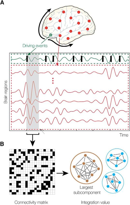
70 Intrinsic Ignition Framework
The Intrinsic Ignition Framework [25] measures the degree of elicited whole-brain integration of
spontaneously occurring events across time. Figure 1 describes the algorithm to obtain the intrinsic
integration across events of each brain area. Driving events are captured by applying the method
of Tagliazucchi and colleagues which measures dynamical neural events for each brain area [32].
75 Events are fixed as a binary signal by transforming the time-series into z-scores, zi (t), and imposing
a threshold θ such that the binary sequence σ(t)=1 if zi (t) > θ, and is crossing the threshold from
below, and σ(t)=0 otherwise. If a brain area has triggered an event, then the neural activity of all
brain areas is measured in the set time window of 4TR. A binary matrix is obtained representing the
connectivity between brain areas exhibiting simultaneous activity. Afterward, the measure of global
80 integration [19] is applied, returning the broadness of communication across the network for each
driving event (i.e the largest subcomponent). Finally, the process is repeated for each spontaneous
neural event, producing the Intrinsic-Driven Mean Integration (IDMI) and the variability as the
standard deviation of the Intrinsic-Driven Integration for each brain area in the whole-brain network.
Comparisons
85 Statistical analyses were carried out in Matlab (R2017a). We used Monte Carlo permutation test
(number of iterations 10,000) to test the differences between conditions. Furthermore, to ensure
that the observed results were not obtained by chance, we applied a surrogate data testing method.
We created the surrogate data using a random permutation at each time point of the original time-
series and measured the ignition in each spontaneous event on the randomized data. We repeated
90 the process 50 times and then, we compared the average randomized data to the original time-series.
4bioRxiv preprint first posted online Jan. 25, 2019; doi: http://dx.doi.org/10.1101/521963. The copyright holder for this preprint
(which was not peer-reviewed) is the author/funder, who has granted bioRxiv a license to display the preprint in perpetuity.
It is made available under a CC-BY-NC-ND 4.0 International license.
Results
Intrinsic-Driven Mean Integration (IDMI)
We first calculated the IDMI in both states (resting-state and meditation) for each group (controls
and meditators). Figure 2A shows the IDMI for each group and brain state, while Figure 2C
95 shows the IDMI for each group and each brain area. The IDMI captures the spatial diversity
as differences in average intrinsic ignition profiles across the different nodes. The brain activity of
meditators during resting-state showed the highest values of the IDMI compared to the control group
(pbioRxiv preprint first posted online Jan. 25, 2019; doi: http://dx.doi.org/10.1101/521963. The copyright holder for this preprint
(which was not peer-reviewed) is the author/funder, who has granted bioRxiv a license to display the preprint in perpetuity.
It is made available under a CC-BY-NC-ND 4.0 International license.
[11]. In addition, a study that applied graph theoretical analysis [33] characterized the degree of the
hierarchical organization during meditation. This study revealed that some nodes had the highest
integration degree during rest but the lowest during meditation, and vice versa. Our work extends
125 these findings by exploring the brain activity during meditation by characterizing the dynamical
complexity in terms of how local information is broadcasted across the whole-brain.
Here, we have characterized the dynamical complexity underlying resting-state and medita-
tion in healthy controls and experienced meditators as evidenced by the level of intrinsic ignition.
Specifically, in meditators but not in controls, we observed a significant increase of intrinsic igni-
130 tion during resting-state compared to meditation (Figure 2A). In addition, during resting-state,
meditators showed the maximal variability of intrinsic ignition (i.e metastability) across the whole
network, revealing a state of maximum network switching (Figure 2B).
Our results showing an increase of intrinsic ignition during rest compare to meditation are con-
sistent with recent studies on information propagation across the brain. Irrmischer and colleagues
135 found a shift from more complex brain dynamics during rest to a state of reduced information prop-
agation during meditation, importantly, only in meditators [34]. Furthermore, Gard and colleagues
demonstrated using graph theory that yoga and meditation practitioners showed greater network
integration than controls during rest [35]. In addition, the increase of metastability in meditators
during resting-state is congruent with the increase of the temporal complexity of oscillations during
140 rest in meditators observed in the previously mentioned study [34]. Moreover, studies applying
a dynamical functional connectivity approach found that individuals with high trait mindfulness,
transitioned more frequently between brain states at rest [13, 36].
To sum up, our investigations suggest that the dynamics underlying meditation differs from the
dynamics underlying resting-state. Even more, the degree of expertise in meditation of the subjects
145 significantly changes the dynamical complexity underlying both resting-state and meditation. Fu-
ture longitudinal studies (before and after an intervention in the same participants) will be needed
to investigate the necessary time-training time necessary to alter the dynamical complexity induced
by meditation.
6bioRxiv preprint first posted online Jan. 25, 2019; doi: http://dx.doi.org/10.1101/521963. The copyright holder for this preprint
(which was not peer-reviewed) is the author/funder, who has granted bioRxiv a license to display the preprint in perpetuity.
It is made available under a CC-BY-NC-ND 4.0 International license.
Figure Legends
Figure 1: Measuring intrinsic ignition. (A) Events were captured applying a threshold method [32] (see green area).
For each event elicited (gray area), the activity in the rest of the network was measured in the time-window of 4TR
(see red area). (B) A binarized matrix was obtained which represents the connectivity between brain areas where
activity was simultaneous. (C) Applying the global integration measure [19], we obtained the largest subcomponent.
Repeating the process for each driving event, we calculated the mean and the variability of the Intrinsic-Driven
Integration for each brain area across the whole-brain network.
7bioRxiv preprint first posted online Jan. 25, 2019; doi: http://dx.doi.org/10.1101/521963. The copyright holder for this preprint
(which was not peer-reviewed) is the author/funder, who has granted bioRxiv a license to display the preprint in perpetuity.
It is made available under a CC-BY-NC-ND 4.0 International license.
Figure 2: (A) Mean of the Intrinsic-Driven Integration (IDMI) for each group during resting-state and meditation
state. The IDMI was higher in meditators than in controls during resting-state and lower in meditators during
meditation. No significant differences were observed in controls between conditions. Furthermore, we show the
box plot from the surrogate IDMI data (on the bottom in green). The randomized data were significantly smaller
than the original time-series, showing the robust statistical comparisons. (B) Both controls and meditators showed
higher local metastability across the whole-brain during resting-state compared to meditation. However, the effect
was significantly larger for meditators. Furthermore, the metastability in resting-state was significantly higher for
experienced meditators than for controls. P-values are based on Monte-Carlo simulation after Bonferroni correction,
* represents pbioRxiv preprint first posted online Jan. 25, 2019; doi: http://dx.doi.org/10.1101/521963. The copyright holder for this preprint
(which was not peer-reviewed) is the author/funder, who has granted bioRxiv a license to display the preprint in perpetuity.
It is made available under a CC-BY-NC-ND 4.0 International license.
European Union’s Horizon 2020 FET Flagship Human Brain Project 785907 HBP SGA2, by the
160 Catalan Research Group Support 2017 SGR 1545 and by the Foundation Marato TV3 2016.
Conflict of Interest Statement
The authors declare that the research was conducted in the absence of any commercial or financial
relationships that could be construed as a potential conflict of interest.
References
165 [1] Matthieu Ricard, Antoine Lutz, and Richard J. Davidson. Mind of the Meditator. Scientific
American, 311(5):38–45, oct 2014.
[2] Judson a Brewer, Patrick D Worhunsky, Jeremy R Gray, Yi-Yuan Tang, Jochen Weber, and
Hedy Kober. Meditation experience is associated with differences in default mode network
activity and connectivity. Proceedings of the National Academy of Sciences of the United
170 States of America, 108(50):20254–9, dec 2011.
[3] Lisa A Kilpatrick, Brandall Y Suyenobu, Suzanne R Smith, Joshua A Bueller, Trudy Good-
man, J David Creswell, Kirsten Tillisch, Emeran A Mayer, and Bruce D Naliboff. Impact
of Mindfulness-Based Stress Reduction training on intrinsic brain connectivity. NeuroImage,
56(1):290–8, may 2011.
175 [4] Brett Froeliger, Eric L. Garland, Rachel V. Kozink, Leslie A. Modlin, Nan Kuei Chen, F. Joseph
McClernon, Jeffrey M. Greeson, and Paul Sobin. Meditation-state functional connectivity
(msFC): Strengthening of the dorsal attention network and beyond. Evidence-based Comple-
mentary and Alternative Medicine, 2012, 2012.
[5] Wendy Hasenkamp, Christine D Wilson-Mendenhall, Erica Duncan, and Lawrence W Barsalou.
180 Mind wandering and attention during focused meditation: A fine-grained temporal analysis of
fluctuating cognitive states. NeuroImage, 59:750–760, 2012.
[6] Véronique A. Taylor, Véronique Daneault, Joshua Grant, Geneviève Scavone, Estelle Breton,
Sébastien Roffe-vidal, Jérôme Courtemanche, Anaı̈s S. Lavarenne, Guillaume Marrelec, Habib
Benali, and Mario Beauregard. Impact of meditation training on the default mode network
185 during a restful state. Social Cognitive and Affective Neuroscience, 8(1):4–14, 2013.
[7] Kathleen A. Garrison, Dustin Scheinost, R. Todd Constable, and Judson A. Brewer. BOLD
signal and functional connectivity associated with loving kindness meditation. Brain and
Behavior, 4(3):337–347, 2014.
9bioRxiv preprint first posted online Jan. 25, 2019; doi: http://dx.doi.org/10.1101/521963. The copyright holder for this preprint
(which was not peer-reviewed) is the author/funder, who has granted bioRxiv a license to display the preprint in perpetuity.
It is made available under a CC-BY-NC-ND 4.0 International license.
[8] William R Marchand. Neural mechanisms of mindfulness and meditation: Evidence from
190 neuroimaging studies. World journal of radiology, 6(7):471–9, jul 2014.
[9] Yi-Yuan Tang, Britta K. Hölzel, and Michael I. Posner. The neuroscience of mindfulness
meditation. Nature Reviews Neuroscience, 16(4):213–225, mar 2015.
[10] Rajanikant Panda, Rose D Bharath, Neeraj Upadhyay, Sandhya Mangalore, Srivas Chennu,
and Shobini L Rao. Temporal Dynamics of the Default Mode Network Characterize Meditation-
195 Induced Alterations in Consciousness. Frontiers in human neuroscience, 10:372, 2016.
[11] Benjamin W. Mooneyham, Michael D. Mrazek, Alissa J. Mrazek, Kaita L. Mrazek, Dawa T.
Phillips, and Jonathan W. Schooler. States of Mind: Characterizing the Neural Bases of
Focus and Mind-wandering through Dynamic Functional Connectivity. Journal of Cognitive
Neuroscience, 29(3):495–506, mar 2017.
200 [12] Sunghyon Kyeong, Joohan Kim, Dae Jin Kim, Hesun Erin Kim, and Jae-Jin Kim. Effects
of gratitude meditation on neural network functional connectivity and brain-heart coupling.
Scientific Reports, 7(1):5058, 12 2017.
[13] Hilary A. Marusak, Farrah Elrahal, Craig A. Peters, Prantik Kundu, Michael V. Lombardo,
Vince D. Calhoun, Elimelech K. Goldberg, Cindy Cohen, Jeffrey W. Taub, and Christine A.
205 Rabinak. Mindfulness and dynamic functional neural connectivity in children and adolescents.
Behavioural Brain Research, 336:211–218, jan 2018.
[14] Gustavo Deco, Enzo Tagliazucchi, Laufs Helmut, Sanjuán Ana, and Kringelbach Morten. Novel
intrinsic ignition method measuring local-global integration characterises wakefulness and deep
sleep. Sciences Advances, 2017.
210 [15] R. Matthew Hutchison, Thilo Womelsdorf, Elena A. Allen, Peter A. Bandettini, Vince D.
Calhoun, Maurizio Corbetta, Stefania Della Penna, Jeff H. Duyn, Gary H. Glover, Javier
Gonzalez-Castillo, Daniel A. Handwerker, Shella Keilholz, Vesa Kiviniemi, David A. Leopold,
Francesco de Pasquale, Olaf Sporns, Martin Walter, and Catie Chang. Dynamic functional
connectivity: Promise, issues, and interpretations. NeuroImage, 80:360–378, oct 2013.
215 [16] Gustavo Deco, Viktor K. Jirsa, and Anthony R. McIntosh. Emerging concepts for the dynam-
ical organization of resting-state activity in the brain. Nature Reviews Neuroscience, 12(1):43–
56, jan 2011.
[17] Olaf Sporns. Network attributes for segregation and integration in the human brain. Current
Opinion in Neurobiology, 23:162–171, 2013.
10bioRxiv preprint first posted online Jan. 25, 2019; doi: http://dx.doi.org/10.1101/521963. The copyright holder for this preprint
(which was not peer-reviewed) is the author/funder, who has granted bioRxiv a license to display the preprint in perpetuity.
It is made available under a CC-BY-NC-ND 4.0 International license.
220 [18] Elena A Allen, Eswar Damaraju, Sergey M Plis, Erik B Erhardt, Tom Eichele, and Vince D
Calhoun. Tracking whole-brain connectivity dynamics in the resting state. Cerebral cortex
(New York, N.Y. : 1991), 24(3):663–76, mar 2014.
[19] Gustavo Deco, Giulio Tononi, Melanie Boly, and Morten L Kringelbach. Rethinking segrega-
tion and integration: contributions of whole-brain modelling. Nature reviews. Neuroscience,
225 16(7):430–439, 2015.
[20] Enzo Tagliazucchi and Helmut Laufs. Decoding Wakefulness Levels from Typical fMRI Resting-
State Data Reveals Reliable Drifts between Wakefulness and Sleep. Neuron, 82(3):695–708,
2014.
[21] Enzo Tagliazucchi, Robin Carhart-Harris, Robert Leech, David Nutt, and Dante R. Chialvo.
230 Enhanced repertoire of brain dynamical states during the psychedelic experience. Human Brain
Mapping, 35(11):5442–5456, nov 2014.
[22] Selen Atasoy, Gustavo Deco, Morten L. Kringelbach, and Joel Pearson. Harmonic Brain Modes:
A Unifying Framework for Linking Space and Time in Brain Dynamics. The Neuroscientist,
24(3):277–293, jun 2018.
235 [23] Selen Atasoy, Leor Roseman, Mendel Kaelen, Morten L. Kringelbach, Gustavo Deco, and
Robin L. Carhart-Harris. Connectome-harmonic decomposition of human brain activity reveals
dynamical repertoire re-organization under LSD. Scientific Reports, 7(1):17661, 12 2017.
[24] Beatrice M. Jobst, Rikkert Hindriks, Helmut Laufs, Enzo Tagliazucchi, Gerald Hahn, Adrián
Ponce-Alvarez, Angus B. A. Stevner, Morten L. Kringelbach, and Gustavo Deco. Increased
240 Stability and Breakdown of Brain Effective Connectivity During Slow-Wave Sleep: Mechanistic
Insights from Whole-Brain Computational Modelling. Scientific Reports, 7(1):4634, dec 2017.
[25] Gustavo Deco and Morten L. Kringelbach. Hierarchy of Information Processing in the Brain:
A Novel ’Intrinsic Ignition’ Framework. Neuron, 94(5):961–968, jun 2017.
[26] Yan Chao-Gan and Zang Yu-Feng. DPARSF: A MATLAB Toolbox for ”Pipeline” Data Anal-
245 ysis of Resting-State fMRI. Frontiers in systems neuroscience, 4:13, 2010.
[27] B Biswal, F Z Yetkin, V M Haughton, J S Hyde, F Zerrin Yetkin, V M Haughton, and J S
Hyde. Functional connectivity in the motor cortex of resting human brain using echo-planar
MRI. Magnetic resonance in medicine : official journal of the Society of Magnetic Resonance
in Medicine / Society of Magnetic Resonance in Medicine, 34(4):537–541, 1995.
250 [28] M.J. Lowe, B.J. Mock, and J.A. Sorenson. Functional Connectivity in Single and Multislice
Echoplanar Imaging Using Resting-State Fluctuations. NeuroImage, 7(2):119–132, feb 1998.
11bioRxiv preprint first posted online Jan. 25, 2019; doi: http://dx.doi.org/10.1101/521963. The copyright holder for this preprint
(which was not peer-reviewed) is the author/funder, who has granted bioRxiv a license to display the preprint in perpetuity.
It is made available under a CC-BY-NC-ND 4.0 International license.
[29] X. Shen, F. Tokoglu, X. Papademetris, and R.T. Constable. Groupwise whole-brain parcella-
tion from resting-state fMRI data for network node identification. NeuroImage, 82:403–415,
nov 2013.
255 [30] Mark Jenkinson, Peter Bannister, Michael Brady, and Stephen Smith. Improved optimiza-
tion for the robust and accurate linear registration and motion correction of brain images.
NeuroImage, 17(2):825–41, oct 2002.
[31] Chao-Gan Yan, R Cameron Craddock, Xi-Nian Zuo, Yu-Feng Zang, and Michael P Milham.
Standardizing the intrinsic brain: towards robust measurement of inter-individual variation in
260 1000 functional connectomes. NeuroImage, 80:246–62, oct 2013.
[32] Enzo Tagliazucchi, Pablo Balenzuela, Daniel Fraiman, and Dante R Chialvo. Criticality in
large-scale brain FMRI dynamics unveiled by a novel point process analysis. Frontiers in
physiology, 3:15, 2012.
[33] Tun Jao, Chia-Wei Li, Petra E. Vértes, Changwei Wesley Wu, Sophie Achard, Chao-Hsien
265 Hsieh, Chien-Hui Liou, Jyh-Horng Chen, and Edward T. Bullmore. Large-Scale Functional
Brain Network Reorganization During Taoist Meditation. Brain Connectivity, 6(1):9–24, 2
2016.
[34] Mona Irrmischer, Simon J. Houtman, Huibert D. Mansvelder, Michael Tremmel, Ulrich Ott,
and Klaus Linkenkaer-Hansen. Controlling the Temporal Structure of Brain Oscillations by
270 Focused Attention Meditation. Human Brain Mapping, jan 2018.
[35] Tim Gard, Maxime Taquet, Rohan Dixit, Britta K Hölzel, Yves-Alexandre de Montjoye,
Narayan Brach, David H Salat, Bradford C Dickerson, Jeremy R Gray, and Sara W Lazar.
Fluid intelligence and brain functional organization in aging yoga and meditation practitioners.
Frontiers in aging neuroscience, 6:76, 2014.
275 [36] Julian Lim, James Teng, Amiya Patanaik, Jesisca Tandi, and Stijn A.A. Massar. Dynamic
functional connectivity markers of objective trait mindfulness. NeuroImage, 176:193–202, aug
2018.
12You can also read