Effects of endothelial progenitor cells transplantation on hyperlipidemia associated kidney damage in ApoE knockout mouse model
←
→
Page content transcription
If your browser does not render page correctly, please read the page content below
Gong et al. Lipids in Health and Disease   (2020) 19:53
https://doi.org/10.1186/s12944-020-01239-1

 RESEARCH                                                                                                                                           Open Access

Effects of endothelial progenitor cells
transplantation on hyperlipidemia
associated kidney damage in ApoE
knockout mouse model
Piyun Gong1†, Zhongwen Zhang2,3†, Dongmei Zhang4, Zhiwei Zou1, Qian Zhang1, Huimei Ma1, Jingxiu Li5,
Lin Liao2,3* and Jianjun Dong1*

  Abstract
  Background: Hyperlipidaemia causes kidney damage over the long term. We investigated the effect of the
  administration of endothelial progenitor cells (EPCs) on the progression of kidney damage in a mouse model of
  hyperlipidaemia.
  Methods: Apolipoprotein E-knockout (ApoE−/−) mice were treated with a high-cholesterol diet after spleen
  resection. Twenty-four weeks later, the mice were divided into two groups and intravenously injected with PBS or
  EPCs. Six weeks later, the recruitment of EPCs to the kidney was monitored by immunofluorescence. The lipid,
  endothelial cell, and collagen contents in the kidney were evaluated by specific immunostaining. The protein
  expression levels of transforming growth factor-β (TGF-β), Smad2/3, and phospho-Smad3 (p-smad3) were detected
  by western blot analysis.
  Results: ApoE−/− mice treated with a high-fat diet demonstrated glomerular lipid deposition, enlargement of the
  glomerular mesangial matrix, endothelial cell enlargement accompanied by vacuolar degeneration and an area of
  interstitial collagen in the kidney. Six weeks after EPC treatment, only a few EPCs were detected in the kidney
  tissues of ApoE−/− mice, mainly in the kidney interstitial area. No significant differences in TGF-β, p-smad3 or
  smad2/3 expression were found between the PBS group and the EPC treatment group (TGF-β expression, PBS
  group: 1.06 ± 0.09, EPC treatment group: 1.09 ± 0.17, P = 0.787; p-smad3/smad2/3 expression: PBS group: 1.11 ± 0.41,
  EPC treatment group: 1.05 ± 0.33, P = 0.861).
  Conclusions: Our findings demonstrate that hyperlipidaemia causes basement membrane thickening,
  glomerulosclerosis and the vascular degeneration of endothelial cells. The long-term administration of EPCs
  substantially has limited effect in the progression of kidney damage in a mouse model of hyperlipidaemia.
  Keywords: Hyperlipidemia, Endothelial progenitor cells, Cell transplantation, Kidney damage

* Correspondence: liaolin@sdu.edu.cn; cwc_ll@sdu.edu.cn
†
 Piyun Gong and Zhongwen Zhang contributed equally to this work.
2
 Department of Endocrinology and Metabology, the First Affiliated Hospital
of Shandong First Medical University, Jinan 250014, China
1
 Department of Endocrinology, Qilu Hospital of Shandong University,
Shandong University, Jinan, Shandong 250012, China
Full list of author information is available at the end of the article

                                        © The Author(s). 2020 Open Access This article is licensed under a Creative Commons Attribution 4.0 International License,
                                        which permits use, sharing, adaptation, distribution and reproduction in any medium or format, as long as you give
                                        appropriate credit to the original author(s) and the source, provide a link to the Creative Commons licence, and indicate if
                                        changes were made. The images or other third party material in this article are included in the article's Creative Commons
                                        licence, unless indicated otherwise in a credit line to the material. If material is not included in the article's Creative Commons
                                        licence and your intended use is not permitted by statutory regulation or exceeds the permitted use, you will need to obtain
                                        permission directly from the copyright holder. To view a copy of this licence, visit http://creativecommons.org/licenses/by/4.0/.
                                        The Creative Commons Public Domain Dedication waiver (http://creativecommons.org/publicdomain/zero/1.0/) applies to the
                                        data made available in this article, unless otherwise stated in a credit line to the data.
Gong et al. Lipids in Health and Disease   (2020) 19:53                                                           Page 2 of 9

Background                                                       performed splenectomy before EPC transplantation. The
Hyperlipidaemia-associated kidney damage, a progressive          spleens of sixteen ApoE−/− mice and eight C57BL/6 J
disease characterized by fatty degeneration of the kidney,       mice were excised as previously described [8, 9]. In brief,
enlargement of the glomerular mesangial matrix and ac-           mice were anaesthetized with an intraperitoneal dose of
cumulated extracellular matrix, leads to chronic kidney          0.8% pentobarbital sodium (10 mg/kg body weight). A
disease and progressive kidney failure [1, 2]. Existing treat-   10–15 mm incision was made on the left abdomen, and
ments for hyperlipidaemia-associated kidney damage are           the splenic arteries and venous supply were ligated, after
not satisfactory; for example, medication cannot cure or         which the spleen was removed. The animal was allowed
reverse chronic kidney damage, and maintenance dialysis          to recover for 6–7 days before further treatment was
can result in nausea, low blood pressure, and restless leg       performed.
syndrome [2]. Thus, the need to find effective approaches
to restore kidney function and eventually reduce the pro-        EPC culture
gression of kidney damage is critical.                           Bone marrow-derived EPCs were isolated from male
   Endothelial cells play a vital role in the maintenance of     C57BL/6 J mice as previously described [10–12]. In
a normal and healthy vasculature and the endothelial             short, mononuclear cells were obtained under sterile
bed [3]. Hyperlipidaemia induces endothelial cell injury         conditions from the long bones of the mice by flushing
accompanied by vacuolar degeneration, and the loss of            with PBS and then purified by the density gradient
glomerular endothelium predisposes patients to the acti-         method (Sigma-Aldrich, St. Louis, MO, USA). Then, the
vation of platelets, causing slight aneurysmal dilatation        cells were cultured in endothelial cell basal medium-2
of the tubular kidney capillaries, resulting in thickening       (EBM-2) supplemented with reagents from an MV Bul-
of the basement membrane, the formation of a dual-               letKit (Lonza, Walkersville, MD, USA). Three days later,
track sign, and eventual glomerulosclerosis [1, 2, 4].           the medium was replaced with new medium after the
Endothelial progenitor cells (EPCs), a type of bone              nonadherent cells had been removed. After 7 days of cul-
marrow-derived progenitor cell, contribute to endothe-           ture, EPCs were identified by double fluorescent staining
lial repair and vasculogenesis [5, 6], and the administra-       for Bandeiraea simplicifolia lectin 1 (BS-1 lectin) and
tion of EPCs might be an effective approach for                  DiI-labelled acetylated low-density lipoprotein (DiI-
hyperlipidaemia associated with kidney damage. Never-            acLDL) (Sigma-Aldrich) (Fig. 1 a-d).
theless, the potential therapeutic effects of EPCs on
hyperlipidaemia-associated kidney damage have not                Preparation of lentivirus vectors and EPC infection
been addressed. A previous study in an acute kidney in-          Recombinant lentivirus (Lenti) carrying transgenic EGFP
jury model found that administration of EPCs enhanced            was purchased from GeneChem (Shanghai, China). EPCs
microvascular endothelial regeneration and protected             at passage 2 were transfected with lentivirus as previ-
against kidney fibrosis [5]; however, Silvestre found that       ously described [12]. Before injection, EPCs carrying
EPC treatment augmented the lesion burden when EPCs              EGFP accounted for more than 90% of the total EPCs
were transferred from young to old ApoE −/− mice [7].            (data not shown).
These therapies are still controversial and inconclusive.
Thus, we performed this experiment to investigate the            Experimental design
effects of EPC treatment on hyperlipidaemia-related kid-         Splenectomized ApoE−/− mice were treated with a high-
ney disease in an ApoE−/− mouse model.                           fat, high-cholesterol diet (34% sucrose, 21% anhydrous
                                                                 milk fat/butter fat and 0.2% cholesterol) for 24 weeks.
Materials and method                                             Then, the sixteen mice were randomly divided into two
Animals                                                          groups and intravenously injected with 1 × 10⁶ EPCs car-
Sixteen 6- to 8-week-old male ApoE−/− C57BL/6 J mice             rying EGFP or the same volume of sterile PBS (200 μl).
(C57BL/6 J black mice) and eight C57BL/6 J mice were             According to the previously reported method [8], the
purchased from Vital River (Peking, China). All animal ex-       effects of EPCs should be examined in mice that are
periments were performed in accordance with the Regula-          returned to a normal chow diet after a high-fat/high-
tion of Animal Care Management of the Ministry of                cholesterol diet. EPC injection is proposed as a clinical
Public Health, People’s Republic of China (document No.          treatment, and this protocol mimics patients’ dietary
55, 2001). All animal care and study protocols were ap-          habits, i.e., once patients start a treatment, their diet
proved by the Ethics Committee of Shandong University.           should be controlled during the treatment. Therefore,
                                                                 after EPC treatment, animals were returned to a normal
Splenectomy surgery                                              chow diet for the remaining experiments. Eight male
To improve the efficiency of EPC transplantation, we             C57BL/6 J mice treated with a normal chow diet were
followed the protocol by Tousoulis et al. [8] and                used as the normal group.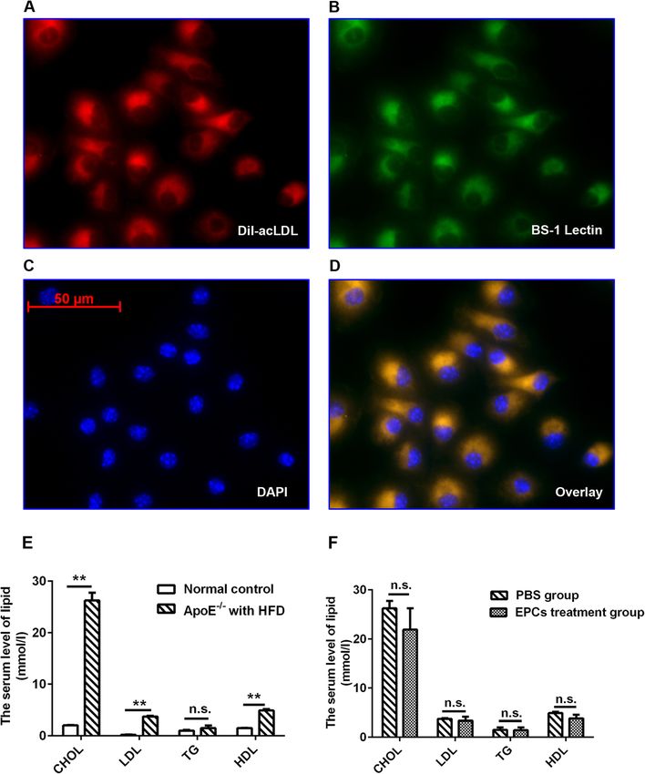
Gong et al. Lipids in Health and Disease       (2020) 19:53                                                                                  Page 3 of 9

 Fig. 1 Characterization of EPCs. The serum level of lipid in the normal control mice and ApoE−/− mice after PBS and EPC treatment. Expanded
 adherent cells could take up (b) BS-1 lectin and bind to (a) DiI-acLDL as shown by the red and green fluorescence. c Nuclei were stained with
 DAPI (blue). Merged images showed that most cells were (d) dual-positive. Dual positive cells were defined as EPCs. The serum level of
 cholesterol (CHOL), low density lipoprotein (LDL), triglyceride (TG) and high-density lipoprotein (HDL) in the normal control mice, ApoE−/− mice
 fed on a high-fat diet with PBS or EPC treatment (e-f, **P < 0.01; n.s., non-significant; HFD, high fat diet). Bars represent mean ± standard deviation

Immunohistochemical analysis                                                  the kidney tissues. The accumulation of glomerular mesan-
After 24 weeks of high-fat diet treatment, the twenty-four                    gial matrix was assessed by periodic acid-Schiff (PAS)
mice were sacrificed after anesthetization with the intraper-                 (Loogene, Beijing, China) staining. The accumulation of
itoneal injection of 0.8% pentobarbital sodium as before.                     EPCs in kidney tissues was evaluated by immunofluores-
The serum levels of cholesterol, triglyceride, low-density                    cence analysis as previously described [9]. The peritubular
lipoprotein and high-density lipoprotein were determined                      capillary density was determined with anti-CD31 antibody
using spectrophotometry. Kidney tissues were collected                        (Abcam, Inc., Cambridge, MA, USA) [9]. The reaction was
after saline perfusion and used to prepare paraffin or fro-                   visualized by staining with 3,3′-diaminobenzidine (DAB)
zen sections as described previously [8]. Frozen sections                     (Abcam, Inc., Cambridge, MA, USA). Images were taken
were stained with Oil red O stain (Sigma-Aldrich, Santa                       with an Olympus light microscope (Olympus, Tokyo,
Clara, CA, USA) to evaluate lipid deposition in the kidney                    Japan). Four to five fields for each mouse were visualised in
tissues. Paraffin sections were stained with Masson’s tri-                    a blinded manner and evaluated using Image-Pro Plus
chrome to evaluate areas of collagen fibre proliferation in                   Software (Media Cybernetics, Silver Spring, MD).
Gong et al. Lipids in Health and Disease (2020) 19:53 Page 4 of 9
Capillary density take up BS-1 lectin and bind DiI-acLDL, indicating that
Each renal tissue sample was examined with light mi- the isolated cells were EPCs. Six weeks after EPC trans-
croscopy at 400× magnification after staining for CD31 plantation, only a few EGFP (+) (labelled putative) EPCs
expression (brown). The density of the peritubular capil- were detected in the kidney tissues of ApoE−/− mice,
laries (PTCs) was calculated using a previously published mainly in the kidney interstitial area (Fig. 2).
with adaptations [13]. Within the endothelial cell- To investigate whether long-term treatment with EPCs
stained area, we randomly chose ten fields from each increased the peritubular capillary density, we deter-
slide and viewed them under a 400× microscope. Each mined the peritubular capillary density (CD31-positive
field had an area of 0.065 mm2. The average number of staining) in ApoE−/− mice fed a high-fat diet. Fig. 1e
capillaries/0.065 mm2 was used to estimate capillary shows that the consumption of a high-fat diet for 24
density [11]. weeks resulted in higher serum cholesterol (P < 0.01),
low-density lipoprotein (P < 0.01) and high-density lipo-
Western blot analysis protein (P < 0.01) levels in the ApoE−/− mice than in the
Western blot analysis was performed as previously de- normal group, indicating that the hyperlipidaemia model
scribed [8]. The following primary antibodies were used: had been successfully constructed. There was no evi-
rabbit polyclonal anti-β-actin (Neomark, Fremont, CA), dence that EPCs influenced the serum levels of lipids in
rabbit polyclonal anti-Smad2/3 antibody (Cell Signaling the ApoE−/− group (Fig. 1f). As shown in Fig. 3, kidney
Technology, Inc., Boston, USA), rabbit polyclonal anti- tubular capillaries exhibited slight aneurysmal dilation,
phospho-Smad3 (Ser423/425; Cell Signaling Technology, and endothelial cells were enlarged, accompanied by
Inc., Boston, USA), and anti-TGF-β antibody (Cell Sig- vacuole degeneration, in ApoE−/− mice fed a high-fat
naling Technology, Inc., Boston, USA). diet. There was an obvious reduction in peritubular ca-
pillary density in the ApoE−/− mice fed a high-fat diet
Statistical analysis compared to control mice, and the capillary density
Data are expressed as the Mean ± Standard deviation, showed a distinct reduction in tubular atrophy and
and statistical analyses were performed with SPSS 25.0 interstitial expansion. However, disappointingly, when
software (SPSS, Inc., Armonk, NY, USA). Comparisons ApoE−/− mice were treated with EPCs, no significant dif-
between groups were analysed via unpaired Student’s t- ference in the peritubular capillary density was detected
test. Differences for which P < 0.05 were considered sta- (PBS group: 27.05 ± 3.23; EPC treatment group: 30.21 ±
tistically significant. 8.91, P = 0.529).
Results Effect of EPCs on dyslipidaemia-induced pathological
Contribution of EPCs to angiogenesis in the kidneys of changes in the kidney
ApoE−/− mice To assess the effects of EPCs on the pathological effects
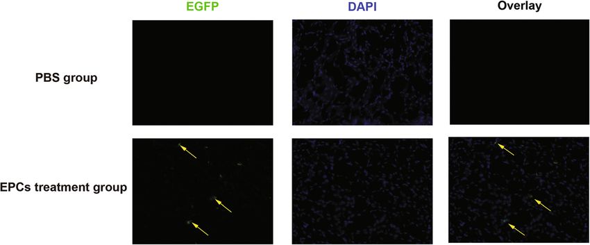
To examine the recruitment of EPCs to the kidney tis- of hyperlipidaemia on the kidney, we examined the lipid
sue, EPCs were transfected with lentivirus encoding burden (the positive area by Oil red O staining) and the
EGFP. Before transplantation, isolated cells were identi- accumulation of glomerular mesangial matrix (the posi-
fied by double fluorescent staining for BS-1 lectin and tive area by PAS staining) by immunostaining. As shown
DiI-acLDL. As shown in Fig. 1 a-d, isolated cells could in Fig. 4, lipid droplets filled the glomerular capillary
Fig. 2 Localization of EPCs in the kidney tissue of ApoE−/− mice. Representative images of EPC incorporation on kidney tissues 6 weeks after PBS
and EPC treatment. Nuclei were stained with DAPI (blue). Arrows and arrowheads identify EPCs (green)Gong et al. Lipids in Health and Disease (2020) 19:53 Page 5 of 9
Fig. 3 Effect of EPCs transplantation on the kidney capillary density of ApoE−/−mice. a CD31 staining in the cortical interstitium of the mice 6
weeks after EPCs treatment. b Quantitative analysis of capillary density in PBS group and EPCs treatment group (n = 4; NS, non-significant). Arrows
refer to the endothelium of the luminal side of the cortical interstitium. Bars represent mean ± standard deviation
lumen in the mouse model of hyperlipidaemia, whereas (Masson’s trichrome, PBS group: 20.11 ± 5.84%, EPC
no glomerular staining was observed in mice fed a nor- treatment group: 21.07 ± 3.16%, P = 0.782; TGF-β ex-
mal diet. There was no significant difference in positive pression, PBS group: 1.06 ± 0.09, EPC treatment group:
Oil red O staining between the PBS group and EPC 1.09 ± 0.17, P = 0.787; p-smad3/smad2/3 expression: PBS
treatment group (PBS group: 5.37 ± 2.42%; EPC treat- group: 1.11 ± 0.41, EPC treatment group: 1.05 ± 0.33, P =
ment group: 5.78 ± 2.38%, P = 0.844). Compared to the 0.861, Fig. 4b and Fig. 5a-b).
normal group, ApoE−/− mice fed a high-fat diet showed
an increased mesangial area and glomerular surface area Discussion
and thickening of the Bowman’s capsule and basement Although EPC treatment has been heralded as a promis-
membranes, as evidenced by the increased positively ing therapeutic strategy for patients with peripheral
stained area determined by PAS staining and Masson’s vascular disease [10], the effect of EPC treatment has
trichrome staining. However, there were no statistically not been studied previously in an animal model of
significant differences in these parameters between the hyperlipidaemia. Previous animal studies focused on uni-
PBS and EPC treatment groups (PBS group: 9.99 ± lateral ureteral obstruction-induced kidney fibrosis and
2.33%; EPC treatment group: 11.02 ± 3.34%, P = 0.683). acute kidney ischaemia rather than hyperlipidaemia-
associated kidney damage [11, 14–16]. Nevertheless,
Effect of EPCs on kidney remodelling in the mouse model hyperlipidaemia-associated kidney damage is present in
of hyperlipidaemia most clinical situations in which the mobilization of
Most forms of progressive kidney disease eventually de- EPCs is planned. The current study is the first to evalu-
velop into kidney fibrosis and remodelling. TGF-β and ate the effect of the long-term administration of EPCs
p-smad3 are the most commonly used markers of kidney on the progression of kidney damage in a mouse model
differentiation, development and interstitial fibrosis. of hyperlipidaemia. In this study, we found that the ad-
Thus, we examined the protein expression levels of ministration of EPCs failed to exert an obvious thera-
TGF-β, p-smad3 and smad 2/3 in the mouse kidneys. peutic effect on the progression of kidney damage.
Additionally, Masson’s trichrome staining was used to These data raise the possibility that EPCs are ineffective
evaluate the collagen content of the mouse kidneys. in the treatment of hyperlipidaemia-associated kidney
Compared to the normal group, ApoE−/− mice treated damage.
with a high-fat diet exhibited an increased collagen con- ApoE, a constituent of very low-density lipoprotein
tent in the glomeruli, tubules and interstitium (Fig. 4a). synthesized in the liver and other tissues, serves as a
However, no significant difference in collagen content or high-affinity ligand for ApoE receptors and is thus re-
TGF-β, p-smad3 or smad2/3 expression was found be- sponsible for the cellular uptake of lipoprotein particles
tween the PBS group and EPC treatment group [17]. Tomiyama-Hanayama et al. showed that eight-Gong et al. Lipids in Health and Disease (2020) 19:53 Page 6 of 9 Fig. 4 Masson’s trichrome, PAS and Oil Red O staining of kidney tissue from ApoE−/− mice after PBS and EPC treatment. a Staining of the collagen, glycogen and lipid in the normal group, PBS group and EPCs treatment group. Arrowheads indicate positive staining areas. b Quantitative analysis of a (n = 4; NS, non-significant). Bars represent mean ± standard deviation Fig. 5 The effects of EPCs treatment on the protein expressions of smad 2/3 and TGF-β in the kidney tissue of ApoE−/− mice (a) Relative p-smad 3, smad 2/3 and TGF-β protein expressions measured by western blot in 3 groups of mice. b Quantitative analysis of a (n = 3; NS, non-significant). Data were standardized to β-actin levels after being quantified by phosphor imaging. Representative blots were shown in the upper panel. Bars represent mean ± standard deviation
Gong et al. Lipids in Health and Disease   (2020) 19:53                                                                             Page 7 of 9

week-old ApoE−/− mice fed a high-fat diet for four weeks       tissues of ApoE−/− mice. The exact mechanism by which
exhibited striking lipid deposition and foam cell forma-       EPCs ameliorate kidney fibrosis has not been fully eluci-
tion in their glomeruli [18]. ApoE−/− mice at 36 weeks of      dated. As reported by Shuai L et al., kidney fibrosis was
age showed more severe renal injury than 24-week-old           improved through the transplantation of bone marrow-
mice [19]. Consistent with previous studies, we found          derived EPCs, which increased the capillary density and
that in 6- to 8-week-old ApoE−/− mice fed a high-fat diet      restored angiogenic activity [33]. In this study, we found
for 24 weeks, the mesangial area and glomerular surface        that very few transplanted EPCs gathered in the kidney
area were increased, kidney tubular capillaries exhibited      lesions of hyperlipidaemic mice and that their role in
slight aneurysmal dilation, and endothelial cells were en-     promoting angiogenesis was very limited. Therefore, the
larged, accompanied by vacuole degeneration.                   expression levels of TGF-β and p-smad3 were not sig-
   Hyperlipidaemia leads to clogged arteries, which can        nificantly different in ApoE−/− mice treated with EPCs
ultimately cause cardio-cerebrovascular diseases [20–          compared to control mice.
22]. Further studies have shown that hyperlipidaemia              Our study has several limitations. First, we only stud-
can also cause direct injury to the kidneys, leading to        ied a single therapeutic dose of EPCs on hyperlipidemia
lipid kidney toxicity [23–25]. Therefore, despite the rela-    associated kidney damage, additional experiments are re-
tive paucity of data on the therapeutic effects of EPCs        quired to explore the effect of different therapeutic dose
on hyperlipidaemia-associated kidney damage, prior ath-        of EPCs on hyperlipidemia associated kidney damage.
erosclerosis studies provide ample support for the use of      Second, the process of EPCs homing and mobilizing to
EPCs in animal models of hyperlipidaemia. In a mouse           the injured site is very complex, further studies are
model of hypertension-hypercholesterolemia, the admin-         needed to illuminate the inadequate kidney repair
istration of EPCs reduced hepatic lipid accumulation           achieved by EPCs in ApoE−/− mice.
and consequently alleviated hypertension and dyslipidae-
mia [26]. In contrast, Silvestre et al. found that EPC         Conclusions
treatment aggravated the atherosclerosis burden when           Our findings demonstrate that hyperlipidaemia causes
EPCs were transferred from young to old ApoE−/− mice           basement membrane thickening, glomerulosclerosis and
[7]. Therefore, the results of prior atherosclerosis studies   the vascular degeneration of endothelial cells. The long-
conflict in terms of both the potential benefits and ad-       term administration of EPCs substantially had a limited
verse outcomes of EPC treatment. Circulation [27] noted        effect in the progression of kidney damage in a mouse
that this controversy is mainly attributed to the very lim-    model of hyperlipidaemia. We assessed a new method to
ited recruitment of stem cells to the lesion site. Al-         improve renal fibrosis, and although its effectiveness was
though stem cells were injected into the injured site,         limited, our data have implications for other researchers.
stem cells that gradually aged with increasing treatment       Additionally, we are carrying out further studies to valid-
time did not exhibit therapeutic efficacy. This is why al-     ate our findings and delineate their causes.
though the clinical use of stem cell therapies is effective
over the short term, outcomes indicate that stem cell          Abbreviations
therapies are not effective over the long term.                EPCs: Endothelial progenitor cells; ApoE: Apolipoprotein E knockout; TGF-
                                                               β: Transforming growth factor-β; p-smad3: Phospho-Smad3; EBM-
   Prior animal studies of the effects of EPCs on kidney-      2: Endothelial cell basal medium-2; BS-1 lectin 1: Bandeiraea simplicifolia
associated diseases are somewhat limited, with one prior       lectin 1; DiI-acLDL: DiI-labeled acetylated low-density lipoprotein;
study showing that the transplantation of EPCs in a mur-       Lenti: Lentiviruses; PAS: Periodic acid-schiff; PTCs: Peritubular capillaries;
                                                               ECM: Extracellular matrix
ine model of unilateral ureteral obstruction had a protect-
ive effect [11]. Similarly, the long-term administration of    Acknowledgements
EPCs was found to increase the kidney microvasculature,        We are grateful for the support by the key laboratory of cardiovascular
protecting the stenotic kidney in experimental renovascu-      remodeling and function research.
lar disease [28, 29]. Nevertheless, these were acute injury
models that may not recapitulate the type of effects of        Authors’ contributions
                                                               LL and JJD conception and design of the study; PYG, ZWZha, ZWZo, QZ,
hyperlipidaemia-mediated kidney damage.                        HMM and JXL generation, collection, assembly, analysis and/or interpretation
   Most forms of progressive kidney disease eventually         of data; LL and JJD drafting or revision of the manuscript. All authors read
develop into kidney fibrosis, and TGF-β and p-smad3,           and approved the final manuscript.

the most important fibrotic factors, can increase degrad-
                                                               Funding
ation of the extracellular matrix (ECM), inhibit ECM           This work was funded by National Natural Science Foundation of China
synthesis and contribute to cellular epithelial to mesen-      Grants (No.81570742,81670757,81770822,81800732), Natural Science
chymal trans differentiation [30–32]. In this study, we        Foundation of Shandong Province (No. ZR2016HQ26, ZR2017LH025), the Key
                                                               Research & Development Plan of Shandong Province (No. 2018GSF118176),
found that administration of EPCs had no influence on          Shandong Provincial Medicine and Health Science and Technology
the expression of TGF-β and p-smad3 in the kidney              Development Program (No.2017WS461).
Gong et al. Lipids in Health and Disease (2020) 19:53 Page 8 of 9
Availability of data and materials plaques in ApoE−/− mice. Stem Cell Res Ther. 2015;6:36. https://doi.org/10.
The datasets generated during and/or analyses during the current study are 1186/s13287-015-0026-0.
available from the corresponding author on reasonable request. 13. Ohashi R, Kitamura H, Yamanaka N. Peritubular capillary injury during the
progression of experimental glomerulonephritis in rats. J Am Soc Nephrol.
Ethics approval and consent to participate 2000;11(1):47–56 https://pubmed.ncbi.nlm.nih.gov/10616839/.
The study was approved by the Ethics Committee of Qilu Hospital of 14. Xue J, Qin Z, Li X, Cao P, Jia R. Protective effects of ischemic
Shandong University. preconditioning-mediated homing of endothelial progenitor cells on renal
acute ischemia and reperfusion injury in male rats. Ann Transplant. 2017;22:
66–74. https://doi.org/10.12659/AOT.901738.
Consent for publication 15. Liang CJ, Shen WC, Chang FB, Wu VC, Wang SH, Young GH, et al.
All the authors have agreed to publish this article. Endothelial progenitor cells derived from wharton's jelly of human umbilical
cord attenuate ischemic acute kidney injury by increasing vascularization
Competing interests and decreasing apoptosis, inflammation, and fibrosis. Cell Transplant. 2015;
The authors declare that they have no competing interests. 24:1363–77. https://doi.org/10.3727/096368914x681720.
16. Patschan D, Schwarze K, Tampe B, Zeisberg M, Patschan S, Muller GA.
Author details Endothelial Colony forming cells (ECFCs) in murine AKI - implications for
1 future cell-based therapies. BMC Nephrol. 2017;18:53. https://doi.org/10.
Department of Endocrinology, Qilu Hospital of Shandong University,
Shandong University, Jinan, Shandong 250012, China. 2Department of 1186/s12882-017-0471-3.
Endocrinology and Metabology, the First Affiliated Hospital of Shandong 17. Jawień J, Nastałek P, Korbut R. Mouse models of experimental
First Medical University, Jinan 250014, China. 3Department of Endocrinology atherosclerosis. J Physiol Pharmacol. 2004;55(3):503–17 https://pubmed.ncbi.
and Metabology, Shandong Provincial Qianfoshan Hospital, Cheeloo College nlm.nih.gov/15381823/.
of Medicine, Shandong University, Jinan 250014, China. 4Department of 18. Tomiyama-Hanayama M, Rakugi H, Kohara M, Mima T, Adachi Y, Ohishi M,
Cardiovascular Medicine, Ninth Hospital of Xi’an, Xi’an 710054, China. 5Quality Katsuya T, Hoshida Y, Aozasa K, Ogihara T, Nishimoto N. Effect of interleukin-
control office, People’s Hospital of Gaoqing, Zibo 256300, China. 6 receptor blockage on renal injury in apolipoprotein E-deficient mice. Am J
Physiol Renal Physiol. 2009;297:F679–84. https://doi.org/10.1152/ajprenal.
Received: 21 November 2019 Accepted: 13 March 2020 90680.2008.
19. Wen M, Segerer S, Dantas M, Brown PA, Hudkins KL, Goodpaster T, et al.
Renal injury in apolipoprotein E-deficient mice. Lab Invest. 2002;82:999–
1006.
References
1. Davignon J. Apolipoprotein E and atherosclerosis: beyond lipid effect. 20. Munshi RP, Joshi SG, Rane BN. Development of an experimental diet model
Arterioscler Thromb Vasc Biol. 2005;25:267–9. https://doi.org/10.1161/01.ATV. in rats to study hyperlipidemia and insulin resistance, markers for coronary
0000154570.50696.2c. heart disease. Indian J Pharmacol. 2014;46:270–6. https://doi.org/10.4103/
2. Cases A, Coll E. Dyslipidemia and the progression of renal disease in chronic 0253-7613.132156.
renal failure patients. Kidney Int Suppl. 2005:S87–93. https://doi.org/10.1111/ 21. Eirin A, Zhu XY, Ebrahimi B, Krier JD, Riester SM, van Wijnen AJ, et al.
j.1523-1755.2005.09916.x. Intrarenal delivery of Mesenchymal stem cells and endothelial progenitor
3. Kang DH, Kanellis J, Hugo C, Truong L, Anderson S, Kerjaschki D, et al. Role cells attenuates hypertensive cardiomyopathy in experimental Renovascular
of the microvascular endothelium in progressive renal disease. J Am Soc hypertension. Cell Transplant. 2015;24:2041–53. https://doi.org/10.3727/
Nephrol. 2002;13:806–16. https://doi.org/10.1089/089277902753619654. 096368914x685582.
4. Hickson LJ, Eirin A, Lerman LO. Challenges and opportunities for stem cell 22. Herz J, Sabellek P, Lane TE, Gunzer M, Hermann DM, Doeppner TR. Role of
therapy in patients with chronic kidney disease. Kidney Int. 2016;89:767–78. neutrophils in exacerbation of brain injury after focal cerebral ischemia in
https://doi.org/10.1016/j.kint.2015.11.023. hyper-lipidemic mice. Stroke. 2015;46:2916–25. https://doi.org/10.1161/
5. Ishida Y, Kimura A, Kuninaka Y, Inui M, Matsushima K, Mukaida N, et al. strokeaha.115.010620.
Pivotal role of the CCL5/CCR5 interaction for recruitment of endothelial 23. Gervais M, Pons S, Nicoletti A, Cosson C, Giudicelli JF, Richer C. Fluvastatin
progenitor cells in mouse wound healing. J Clin Invest. 2012;122:711–21. prevents renal dysfunction and vascular NO deficit in apolipoprotein E-
https://doi.org/10.1172/ jci43027. deficient mice. Arterioscler Thromb Vasc Biol. 2003;23:183–9. https://doi.org/
6. Wang R, Zhang K, Li S, Tong Z, Li G, Zhao Z, et al. Apolipoprotein (a) 10.1161/01.atv.0000051404.84665.49.
impairs endothelial progenitor cell-mediated angiogenesis. DNA Cell Biol. 24. Zhang X, Urbieta-Caceres VH, Eirin A, Bell CC, Crane JA, Tang H, et al.
2013;32:243–51. https://doi.org/10.1089/dna.2013.1963. Humanin prevents intra-renal microvascular remodeling and inflammation
7. Kundu N, Domingues CC, Chou C, Ahmadi N, Houston S, Jerry DJ, Sen S. in hypercholesterolemic ApoE deficient mice. Life Sci. 2012;91:199–206.
Use of p53-silenced endothelial progenitor cells to treat ischemia in https://doi.org/10.1016/j.lfs.2012.07.010.
diabetic peripheral vascular disease. J Am Heart Assoc. 2017;6. https://doi. 25. Sastre C, Rubio-Navarro A, Buendia I, Gomez-Guerrero C, Blanco J, Mas S,
org/10.1161/JAHA.116.005146. et al. Hyperlipidemia-associated renal damage decreases Klotho expression
8. Tousoulis D, Briasoulis A, Vogiatzi G, Valatsou A, Kourkouti P, Pantopoulou A, in kidneys from ApoE knockout mice. PLoS One. 2013;8:e83713. https://doi.
et al. Effects of direct infusion of bone marrow-derived progenitor cells and org/10.1371/journal.pone.0083713.
indirect mobilization of hematopoietic progenitor cells on atherosclerotic 26. Georgescu A, Alexandru N, Andrei E, Dragan E, Cochior D, Dias S. Effects of
plaque and inflammatory process in atherosclerosis. Int J Cardiol. 2013;168: transplanted circulating endothelial progenitor cells and platelet
4769–74. https://doi.org/10.1016/j.ijcard.2013.07.229. microparticles in atherosclerosis development. Biol Cell. 2016;108:219–43.
9. Lima LC, Porto ML, Campagnaro BP, Tonini CL, Nogueira BV, Pereira TM, https://doi.org/10.1111/boc.201500104.
et al. Mononuclear cell therapy reverts cuff-induced thrombosis in 27. Haghighat A, Weiss D, Whalin MK, Cowan DP, Taylor WR. Granulocyte
apolipoprotein E-deficient mice. Lipids Health Dis. 2012;11:96. https://doi. colony stimulating factor and granulocyte macrophage colony-stimulating
org/10.1186/1476-511x-11-96. factor exacerbate atherosclerosis in apolipoprotein E-deficient mice.
10. Asahara T, Murohara T, Sullivan A, Silver M, van der Zee R, Li T, et al. Circulation. 2007;115:2049–54. https://doi.org/10.1161/circulationaha.106.
Isolation of putative progenitor endothelial cells for angiogenesis. Science 665570.
(New York, NY). 1997;275:964–nmu. https://doi.org/10.1126/science.275.5302. 28. Chade AR, Zhu X, Lavi R, Krier JD, Pislaru S, Simari RD, et al. Endothelial
964. progenitor cells restore renal function in chronic experimental renovascular
11. Ma YY, Sun D, Li J, Yin ZC. Transplantation of endothelial progenitor cells disease. Circulation. 2009;119:547–57. https://doi.org/10.1161/circulationaha.
alleviates renal interstitial fibrosis in a mouse model of unilateral ureteral 108.788653.
obstruction. Life Sci. 2010;86:798–807. https://doi.org/10.1016/j.lfs.2010.03. 29. Chade AR, Zhu XY, Krier JD, Jordan KL, Textor SC, Grande JP, et al.
013. Endothelial progenitor cells homing and renal repair in experimental
12. Zhang Z, Dong J, Lobe CG, Gong P, Liu J, Liao L. CCR5 facilitates endothelial renovascular disease. Stem cells (Dayton, Ohio). 2010;28:1039–47. https://doi.
progenitor cell recruitment and promotes the stabilization of atherosclerotic org/10.1002/stem.426.Gong et al. Lipids in Health and Disease (2020) 19:53 Page 9 of 9
30. Sun YB, Qu X, Li X, Nikolic-Paterson DJ, Li J. Endothelial dysfunction
exacerbates renal interstitial fibrosis through enhancing fibroblast Smad3
linker phosphorylation in the mouse obstructed kidney. PLoS One. 2013;8:
e84063. https://doi.org/10.1371/journal.pone.0084063.
31. Qu X, Jiang M, Sun YB, Jiang X, Fu P, Ren Y, et al. The Smad3/Smad4/CDK9
complex promotes renal fibrosis in mice with unilateral ureteral obstruction.
Kidney Int. 2015;88:1323–35. https://doi.org/10.1038/ki.2015.235.
32. Feng M, Tang PM, Huang XR, Sun SF, You YK, Xiao J, Lv LL, Xu AP, Lan HY.
TGF-beta mediates renal fibrosis via the Smad3-Erbb4-IR long noncoding
RNA Axis. Mol Ther. 2018;26:148–61. https://doi.org/10.1016/j.ymthe.2017.09.
024.
33. Shuai L, Li X, He Q, Dang X, Chen H, Zhou P, et al. Angiogenic effect of
endothelial progenitor cells transfected with telomerase reverse
transcriptase on peritubular micro vessel in five out of six subtotal
nephrectomy rats. Ren Fail. 2012;34:1270–80. https://doi.org/10.3109/
0886022x.2012.723592.
Publisher’s Note
Springer Nature remains neutral with regard to jurisdictional claims in
published maps and institutional affiliations.You can also read