Increased Incidence and Plasma-Biofilm Formation Ability of SCCmec Type IV Methicillin-Resistant Staphylococcus aureus (MRSA) Isolated From ...
←
→
Page content transcription
If your browser does not render page correctly, please read the page content below
ORIGINAL RESEARCH
                                                                                                                                              published: 26 March 2021
                                                                                                                                       doi: 10.3389/fcimb.2021.602833

                                             Increased Incidence and
                                             Plasma-Biofilm Formation Ability of
                                             SCCmec Type IV Methicillin-Resistant
                                             Staphylococcus aureus (MRSA)
                                             Isolated From Patients With Bacteremia
                            Edited by:
                         Haider H. Dar,
                                             Masakaze Hamada 1, Tetsuo Yamaguchi 1*, Ayami Sato 1,2, Daisuke Ono 3, Kotaro Aoki 1,
University of Pittsburgh, United States
                                             Chiaki Kajiwara 1, Soichiro Kimura 1, Tadashi Maeda 4, Masakazu Sasaki 5,
                       Reviewed by:          Hinako Murakami 5, Yoshikazu Ishii 1 and Kazuhiro Tateda 1,5
                      Md Suhail Alam,
            University of Notre Dame,        1 Department of Microbiology and Infectious Diseases, Toho University School of Medicine, Tokyo, Japan, 2 Department of

                         United States       Surgery, Toho University Sakura Medical Center, Chiba, Japan, 3 Department of Infectious Diseases and Infection Control,
                        Pankaj Kumar,        Saitama Medical Center, Saitama Medical University, Saitama, Japan, 4 Department of General Medicine and Emergency
      Jamia Hamdard University, India        Care, Toho University Omori Medical Center, Tokyo, Japan, 5 Department of Clinical Laboratories, Toho University Omori
               *Correspondence:              Medical Center, Tokyo, Japan
                 Tetsuo Yamaguchi
 tetsuo.yamaguchi@med.toho-u.ac.jp
                                             In Japan, Staphylococcal cassette chromosome mec (SCCmec) type IV methicillin-
                  Specialty section:         resistant Staphylococcus aureus (MRSA) is an increasingly prominent cause of
        This article was submitted to        bacteremia, but the virulence of most of these strains is unclear. We aimed to
    Molecular Bacterial Pathogenesis,
              a section of the journal
                                             investigate the relationship between the molecular characteristics and the ability to form
                   Frontiers in Cellular     biofilms in the presence of blood plasma (plasma-biofilms) of MRSA strains isolated from
          and Infection Microbiology         bloodstream infections. In this study, the molecular characteristics and biofilms of MRSA
      Received: 11 September 2020            strains isolated from blood cultures between 2015 and 2017 were analyzed by PCR-
       Accepted: 12 February 2021
         Published: 26 March 2021            based assays, crystal violet staining, and confocal reflection microscopy methods. Among
                                 Citation:   the 90 MRSA isolates, the detection rate of SCCmec type II clones decreased from 60.7
    Hamada M, Yamaguchi T, Sato A,           to 20.6%. The SCCmec type IV clone replaced the SCCmec type II clone as the dominant
Ono D, Aoki K, Kajiwara C, Kimura S,
      Maeda T, Sasaki M, Murakami H,
                                             clone, with a detection rate increasing from 32.1 to 73.5%. The plasma-biofilm formation
Ishii Y and Tateda K (2021) Increased        ability of the SCCmec type IV clone was higher than the SCCmec type II clone and even
         Incidence and Plasma-Biofilm         higher in strains harboring the cna or arcA genes. Plasma-biofilms, mainly composed of
 Formation Ability of SCCmec Type IV
  Methicillin-Resistant Staphylococcus       proteins, were formed quickly and strongly. Our study demonstrated the increased
                 aureus (MRSA) Isolated      plasma-biofilm formation ability of SCCmec type IV strains.
       From Patients With Bacteremia.
Front. Cell. Infect. Microbiol. 11:602833.   Keywords: Staphylococcus aureus, Staphylococcal cassette chromosome mec (SCCmec) type IV, methicillin-
       doi: 10.3389/fcimb.2021.602833        resistant Staphylococcus aureus (MRSA), biofilm, plasma, bloodstream infection

Frontiers in Cellular and Infection Microbiology | www.frontiersin.org              1                                        March 2021 | Volume 11 | Article 602833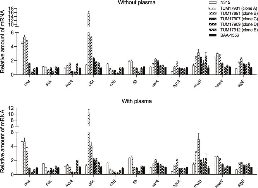
Hamada et al.                                                                                                     CA-MRSA Biofilm Formation in Plasma

INTRODUCTION                                                                 which convert soluble monomeric fibrinogen into self-
                                                                             polymerizing insoluble fibrin and activate a coagulation cascade.
Methicillin-resistant Staphylococcus aureus (MRSA) was first                      The plasma-biofilm is a recently proposed concept and refers
reported in 1961 (Jevons, 1961). Over the subsequent decades,                to the biofilm formed in the presence of plasma (Besser et al.,
hospital-associated MRSA (HA-MRSA) has spread, remaining of                  2019). In recent years, some reports have suggested that S. aureus
great concern as a cause of infection in immunocompromised                   uses the fibrin and fibrinogen recruited by coagulase to form the
hospital patients, affecting hospitals and healthcare facilities             biofilm scaffold in the host’s blood (Gronnemose et al., 2017;
worldwide. In the 1990s, community-associated MRSA (CA-                      Besser et al., 2019). We noticed differences in the plasma-biofilm
MRSA) emerged as another apparent threat (Udo et al., 1993).                 formation ability between different clones in our previous study
CA-MRSA exhibits hyper-virulence and has been reported to                    (Sato et al., 2019). It is assumed that if the ability of each strain to
circulate in communities through the infection and/or                        form plasma-biofilm differs in vivo, MRSA strains with a high
colonization of healthy individuals, particularly children and               level of plasma-biofilm formation ability would exhibit high
young adults.                                                                virulence in the blood.
    Outbreaks of CA-MRSA have been reported worldwide                            In this study, we investigated the molecular characteristics of
(DeLeo et al., 2010); especially prevalent in the United States,             MRSA strains isolated from blood culture samples between 2015
and approximately 80% of S. aureus isolates from skin and soft               and 2017, a period during which MRSA isolation was on the
tissue infections (SSTIs) are MRSA (Miller et al., 2005). Most of            increase in our medical facility. To assess the pathogenicity of
them are of a single CA-MRSA genotype called the USA300                      MRSA in the bloodstream, the biofilms of each MRSA strain
clone (ST8/Staphylococcal cassette chromosome mec (SCCmec)                   formed in the presence of plasma were compared by the crystal
type IV), which causes not only SSTIs but also lethal infections             violet (CV) staining method (Merritt et al., 2005) and confocal
such as necrotizing pneumonia and sepsis (Nimmo, 2012;                       reflection microscopy (CRM) method (Yawata et al., 2010). This
Prosperi et al., 2013). Furthermore, the USA300 clone that has               study aimed to clarify the relationship between the molecular
spread throughout community settings also invades hospital-                  characteristics and plasma-biofilm formation ability of MRSA
associated settings and has become as prominent a pathogen as                strains isolated from bacteremia cases.
HA-MRSA, causing nosocomial infections.
    Japan is an HA-MRSA-endemic country; however, the
methicillin resistance ratio of S. aureus has dropped from                   MATERIALS AND METHODS
nearly 70% in 2000 to less than 50% today (Japan Nosocomial
Infections Surveillance1). We previously clarified that the                   Bacterial Strains
methicillin resistance ratio of S. aureus isolates from SSTIs in             Between January 2007 and December 2017, S. aureus strains
communities is approximately 20%, which is significantly lower                were isolated from blood culture samples from 796 cases at the
than in the United States (Yamaguchi et al., 2015). Furthermore,             Clinical Microbiology Laboratory at the Toho University Omori
most of the CA-MRSA strains are SCCmec type IV but are                       Medical Center in Tokyo, Japan. If multiple S. aureus isolates
neither USA300 clones nor Panton-Valentine Leucocidin (PVL)-                 were present in various samples from the same patient, only one
producing strains. The virulence of other SCCmec type IV clones,             isolate was selected; however, if isolates from the same patient
except specific clones such as the USA300 clone, is not clear.1               showed different antimicrobial susceptibility, each was treated as
    Among the cases in which S. aureus has been isolated from                a separate case.
blood culture samples at the Toho University Omori Medical                       Between 2015 and 2017, MRSA strains were isolated from
Center in Tokyo, Japan, the methicillin resistance rate has                  blood cultures from 98 cases (33 in 2015, 31 in 2016, and 34 in
dropped from 66.7% in 2007 to 33.3% in 2013. However, the                    2017), and genetic analyses such as SCCmec typing and virulence
methicillin resistance rate has re-risen since 2015, reaching                gene detection were performed on 90 MRSA strains (28 in 2015,
45.9% in 2017. The same trend has also been observed in other                28 in 2016, and 34 in 2017) that had been stored. Two strains of
institutions in Japan (Miura et al., 2018), thus investigating what          MRSA, ATCC BAA-1556 (FPR3757 strain; USA300 clone) and
is causing the increase in the number of MRSA cases is crucial.              N315 (New York/Japan clone), were positive controls for
    The biofilm formed by bacteria is involved in establishing                virulence gene detection and biofilm formation analysis. This
bacteremia, including device-related infections (Hoiby et al.,               study was approved by the Ethics Committee of the Faculty of
2015), and that bacterial biofilms comprise a variety of                      Medicine, Toho University (approval number A17114), and
components and substances from both the bacteria and the                     pathogen protocols were approved by the Toho University
host (Alhede et al., 2014; Zapotoczna et al., 2016). Especially,             Safety Committee for Pathogens (approval number 18-43-87).
S. aureus possesses a specific virulence factor called coagulase,
which is thought to play a significant role in biofilm formation in            Molecular Characterization
S. aureus bloodstream infections. Coagulase binds to host                    Each isolate was cultured overnight in brain heart infusion (BHI)
prothrombin and forms active staphylothrombin complexes,                     broth (Eiken Chemical Co., Tokyo, Japan), and the cells were
                                                                             harvested by centrifugation at 8,000×g for 1 min. Bacterial
                                                                             genomic DNA was then extracted using a DNeasy Blood &
1
    https://janis.mhlw.go.jp/english/report/index.html                       Tissue Kit (QIAGEN, Valencia, CA, USA) with lysostaphin

Frontiers in Cellular and Infection Microbiology | www.frontiersin.org   2                                    March 2021 | Volume 11 | Article 602833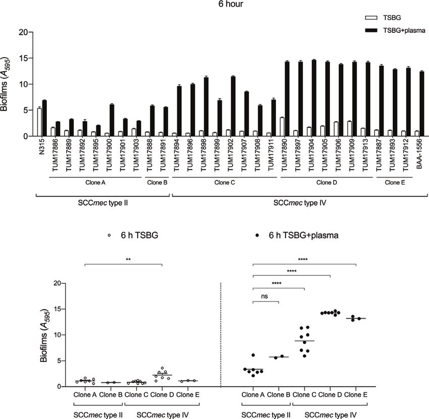
Hamada et al.                                                                                                    CA-MRSA Biofilm Formation in Plasma

(Wako, Osaka, Japan). Genomic DNA was a template for PCR-                    1 mM CaCl2; proteinase K was solubilized in 20 mM Tris-HCl
based screening assays. MRSA SCCmec types were determined                    buffer (pH 7.5) and 100 mM NaCl (Gutierrez et al., 2014).
using a PCR-based assay as previously described (Kondo et al.,
2007; Yamaguchi et al., 2020). The two multiplex PCR sets                    Visualization of Biofilms by CRM
identify the mec gene complex type and ccr gene complex type,                MRSA shaking cultures were diluted to 1:500 with TSBG, and the
and the SCCmec type is determined by the combination pattern                 diluted pre-cultures were diluted to 1:1 with TSBG-containing
of the mec gene complex type and ccr gene complex type. The                  rabbit plasma. Then, 300 ml aliquots were inoculated in 8-well
SCCmec type is classified from I to XIV; however, this method                 coverglass chambers (IWAKI, Tokyo, Japan). After incubation at
can only be used to determine type I to VI. Because this method              35°C for 6 h under aerobic conditions, biofilms that formed at
could not be used to determine the other SCCmec types,                       the bottom of glass chambers were gently washed with PBS, and
including types VII–XIV, isolates for which the SCCmec type                  the 3D structures were visualized with a CRM method (Yawata
could not be identified were treated as non-typeable (NT),                    et al., 2010). A Carl Zeiss Laser Scanning Microscope (LSM 710)
meaning these isolates were SCCmec types VII–XIV, or                         equipped with a 63×/1.40 numerical aperture Plan-Apochromat
new types.                                                                   objective (Carl Zeiss Microscopy GmbH, Jena, Germany) was
   Staphylococcal virulence genes were also detected using a                 used to acquire CRM images. Biofilms were illuminated with a
PCR assay, using reported primers (Jarraud et al., 2002; Diep                514 nm argon laser, and the reflected light was collected through
et al., 2006). The target virulence genes included the PVL-                  a 505–530 nm band-pass filter. An NT 80/20 half mirror was a
encoding gene (lukSF-PV), the toxic shock syndrome toxin-1                   beam splitter. The biomass calculation of biofilm structures
(TSST-1) gene (tst), and the arginine catabolic mobile element               was performed using a COMSTAT2 program 2 (Heydorn
(ACME) gene (arcA). PCR assays to identify microbial surface                 et al., 2000).
componen ts rec ognizing a dhe sive m atrix m olecule
(MSCRAMM) genes were also performed, following described                     Measurement of Biofilm-Related Gene
protocols (Tristan et al., 2003; Vancraeynest et al., 2004). The             Transcription by Reverse-Transcription
MSCRAMM-encoding genes included fnbA, fnbB, fib, clfA, clfB,                  Quantitative PCR (RT-qPCR)
bbp, cna, eno, and ebps.                                                     fnbB, arcA, cna, and bbp were found to be strain-specific
                                                                             virulence factors, and the strains that possessed these genes
Quantification of Biofilms by CV                                               exhibited higher ability of plasma-biofilm formation. Next, we
Staining Assay                                                               compared the transcript levels of the nonspecific factor genes,
For routine cultures, 27 MRSA strains from 2016, BAA-1556 or                 because differences in protein production might influence the
N315, were grown oxically overnight on BHI agar (Becton                      biofilm formation. Target genes included the regulatory genes
Dickinson, Franklin Lakes, NJ, USA) at 35°C. After incubation,               (sarA, agrA, rnaIII, saeR, and sigB), four MSCRAMM-encoding
the colonies were inoculated in BHI broth and then grown                     genes such as fibronectin-binding protein (fnbA) and fibrinogen-
oxically for 12 h at 35°C with shaking at 160 rpm. Shaking                   binding protein (fib, clfA, and clfB), and coagulase (coa) and
cultures were diluted to 1:500 with tryptic soy broth (Becton                staphylokinase (sak) genes involved in the synthesis and
Dickinson, Franklin Lakes, NJ, USA) containing 0.5% (w/v)                    degradation of fibrin. These genes were present in all strains.
glucose (TSBG), and the diluted pre-cultures were diluted 1:1                We had already confirmed the presence of the genes fnbB, arcA,
with TSBG or TSBG containing 14.28% (v/v) “Eiken” rabbit                     cna, and bbp, and we knew that there were strains that did not
plasma (Eiken Chemical Co., Tokyo, Japan). The final plasma                   possess the genes. Even if the transcript levels of these genes were
concentrations were 7.14%. Next, 100 ml aliquots were inoculated             evaluated, it was clear that the strains that did not carry the genes
in 96-well round-bottom polystyrene plates and statically                    would not respond to PCR. Therefore, we excluded the strain-
incubated at 35°C for 1, 2, 6, or 24 h under aerobic conditions.             specific virulence factors (fnbB, arcA, cna, and bbp) from the list
MRSA biofilms formed in 96-well plates were quantified                         of target genes evaluated for transcript levels using RT-qPCR.
following a reported CV staining method (Merritt et al., 2005);              MRSA shaking cultures were diluted to 1:500 with TSBG, and the
the biofilms were washed with phosphate-buffered saline (PBS)                 diluted pre-cultures were diluted to 1:1 with TSBG or TSBG-
and stained with 150 µl of 0.2% (w/v) CV solution. CV-stained                containing rabbit plasma. One milliliter aliquots were then
biofilms were washed with PBS and solubilized with 150 µl of                  inoculated on 12-well flat-bottom polystyrene plates. After
33.3% (v/v) acetic acid. The eluents were transferred to fresh 96-           incubation at 35°C for 1 h under aerobic conditions, 2 ml of
well plates to determine the absorbance at 595 nm (A595).                    RNAprotect (QIAGEN, Valencia, CA, USA) were added to 1 ml
Sometimes, the eluents were diluted to 1:10 with acetic acid.                of static cultures, and then biofilms that formed at the bottom of
    In the biofilm formation tests in the presence of extracellular           the 12-well plates were scraped by vigorous pipetting. Bacterial
matrix (ECM)-degrading agents, 100 mg/ml DNase I (Roche                      suspensions including both non-adherent cells and biofilm cells
Diagnostics, Mannheim, Germany) or proteinase K (Sigma                       were collected into conical tubes. To ensure a sufficient RNA
Aldrich, St. Louis, MO, USA) were added to TSBG or TSBG-                     yield, samples were collected from static cultures in two wells.
containing plasma. After incubation for 6 h in the presence of these
ECM-degrading agents, biofilm formation was measured using the
                                                                             2
CV staining method. DNase I was solubilized in 150 mM NaCl and                   www.comstat.dk

Frontiers in Cellular and Infection Microbiology | www.frontiersin.org   3                                   March 2021 | Volume 11 | Article 602833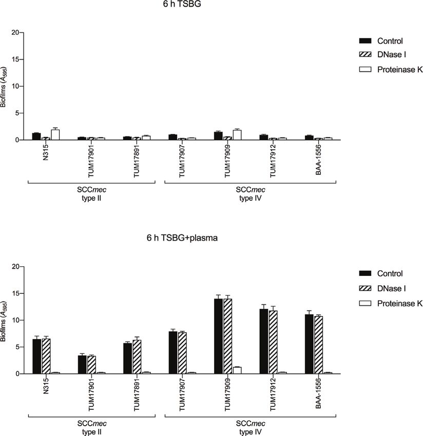
Hamada et al.                                                                                                        CA-MRSA Biofilm Formation in Plasma

   Bacterial pellets after centrifugation were kept at -80°C. The                All SCCmec type IV strains were positive for the fnbA, fib,
pellets were suspended in 0.1 ml of Tris-EDTA buffer                         clfA, clfB, eno, and ebps genes but negative for the bbp gene.
supplemented with 0.2 mg/ml of lysostaphin and then                          SCCmec type IV strains were classified into three types according
incubated at 37°C for 30 min. Extraction of bacterial RNA was                to the presence of the fnbB, cna, and arcA genes. Eight isolates
performed using the RNeasy Mini Kit (QIAGEN, Valencia, CA,                   identified as clone type C (SCCmec type IV, arcA-negative, cna-
USA), following the manufacturer’s protocol. After extraction,               negative, and fnbB-positive isolates) were the most frequently
RNA samples were treated with TURBO DNase (Ambion®,                          detected. Clone type D strains included SCCmec type IV, arcA-
Thermo Fisher Scientific, MA, USA), and RNA concentrations                    negative, cna-positive, and fnbB-negative isolates. Clone type E
in the samples were confirmed using BioSpec-nano (Shimadzu,                   strains included SCCmec type IV, arcA-positive, cna-negative,
Tokyo, Japan). cDNA was acquired using the High Capacity                     and fnbB-positive isolates, characteristics shared by the
cDNA Reverse Transcription Kit (Applied Biosystems ™ ,                       USA300 clone.
Thermo Fisher Scientific, MA, USA) and mixed with a Fast
SYBR Green Master Mix (Applied Biosystems™, Thermo Fisher                    Biofilm Formation
Scientific, MA, USA). Finally, RT-qPCR was performed to                       Molecular characteristic analysis suggested that the SCCmec type
quantify the transcription of biofilm-related genes using the                 IV clone has been the dominant clone causing bacteremia. A
Applied Biosystems 7500 Fast Real-Time PCR System.                           previous report showed that S. aureus forms mature biofilms in
Reported oligonucleotide primers were used for RT-qPCR                       the presence of blood plasma (Sato et al., 2019), so we compared
(Sabet et al., 2006; Burian et al., 2010; Kaito et al., 2011;                the biofilm formation of 18 SCCmec type IV isolates in plasma
Atshan et al., 2013; Ferreira et al., 2013; Schilcher et al., 2016;          with that of 9 SCCmec type II isolates after 6 h. Figure 2A shows
Tranchemontagne et al., 2016). PCR conditions were 95°C for                  the biofilm formation of each isolate, and Figure 2B shows the
20 s, 40 cycles at 95°C for 3 s and 60°C for 30 s. Ct values                 distribution of biofilm formation of clone types A–E. In the
were calculated using the 7500 Fast software version 2.3 (Applied            absence of plasma, the biofilm formation of SCCmec type IV
Biosystems™, Thermo Fisher Scientific, MA, USA). The data                     clones was like that of SCCmec type II clones. In the presence of
were analyzed using the DDCt method. The 16S rRNA gene was                   plasma, the biofilm of each isolate was thicker than in the
an internal standard.                                                        absence of plasma, and SCCmec type IV clones formed more
                                                                             mature biofilms than SCCmec type II clones (Figures 2A, B).
                                                                             Similar results were also observed after 24 h when biofilm
Statistical Analysis                                                         formation in the absence of plasma reached the near-
The experiment was repeated at least twice under the same                    maximum level (Supplementary Figures 1A, B).
conditions. One-way ANOVA followed by Tukey’s multiple                          Clone type D (SCCmec type IV, cna-positive) showed the
comparison test was performed using GraphPad Prism                           highest level of biofilm formation ability among the five clones,
(version 6.0h, GraphPad Software Inc., San Diego, CA, USA).                  regardless of the presence or absence of plasma. Clone type E
P
Hamada et al. CA-MRSA Biofilm Formation in Plasma
TABLE 1 | Molecular characteristics of methicillin-resistant Staphylococcus aureus (MRSA) strains used in this study (isolated in 2016).
SCCmec type Clone type Number of strains Adherence gene Virulence gene
arcA cna fnbB fnbA, fib, clfA, clfB, eno, and ebpS bbp lukSF-PV tst
II N315 a
Reference – – – + – – +
A 7 – – – + – – +/-
B 2 + – – + – – –
IV C 8 – – + + – – +/-
D 7 – + – + – – –
E 3 + – + + – + –
BAA-1556b Reference + – + + – + –
a
This strain is the New York/Japan clone known as hospital-associated MRSA and is used as a reference strain of SCCmec type II.
b
This strain is the USA300 clone known as community-associated MRSA and is used as a reference strain of SCCmec type IV.
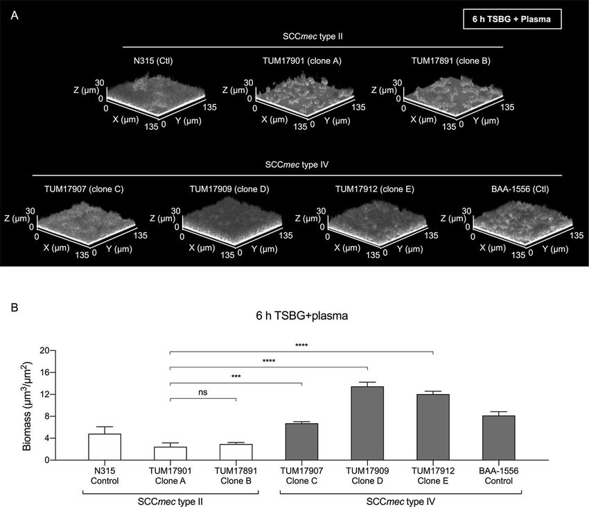
(Figure 2B). Clone type C (SCCmec type IV, fnbB-positive) also staining assay results (Figure 2A), TUM17907 (clone C),
formed more mature biofilms than clone types A and B (SCCmec TUM17909 (clone D), and TUM17912 (clone E) formed more
type II) in the presence of plasma. These results indicate that the mature mat-like biofilm structures than SCCmec type II strains
cna, arcA, and fnbB genes are involved in the biofilm formation including TUM17901 (clone A) and TUM17891 (clone B)
ability of SCCmec type IV clones in plasma. (Figures 3A, B).
To investigate the details of the biofilm formation mechanism
in plasma, one representative strain that formed an average Biofilm Formation in the Presence
biofilm was selected from each clone (clone A, TUM17901; of ECM-Degrading Agents
clone B, TUM17891; clone C, TUM17907; clone D, In previous reports (Cheng et al., 2010; Zapotoczna et al., 2015;
TUM17909; and clone E, TUM17912). Similar to the CV Liesenborghs et al., 2018; Trivedi et al., 2018), S. aureus was
A
B
FIGURE 2 | Quantification of biofilms by crystal violet (CV) staining assay. (A) Amount of biofilm formation of each strain with or without plasma. (B) Comparison of
biofilm formation between clones. Three wells were used for each strain per experiment, and the experiment was conducted three times. Data from the total of nine
experiments were compared between strains. **p < 0.005, ****p < 0.0001; one-way ANOVA followed by Tukey’s multiple comparison test was used in (B). TSBG,
tryptic soy broth containing 0.5% glucose; ns, not significant.
Frontiers in Cellular and Infection Microbiology | www.frontiersin.org 5 March 2021 | Volume 11 | Article 602833Hamada et al. CA-MRSA Biofilm Formation in Plasma
FIGURE 3 | Visualization of biofilms by confocal reflection microscopy (CRM). (A) Biofilm in the presence of plasma (Plasma-biofilm) structures of the strain selected
from each clone. (B) Comparison of the biomass of plasma-biofilms calculated by image analysis. Using a CRM method, the plasma-biofilm structures of each
experimental strain and control strain (BAA-1556 and N315) were observed, and the biomass was measured by image analysis using a COMSTAT2 program. The
biomass of plasma-biofilms was calculated in four fields of view and the means were compared. The results were confirmed by two independent experiments. ***p <
0.001, ****p < 0.0001; one-way ANOVA followed by Tukey’s multiple comparison test was used in (B). TSBG, tryptic soy broth containing 0.5% glucose; ns, not
significant. Fields: 135×135×30 mm (xyz) are indicated.
shown to convert the coagulation factor fibrinogen in blood RT-qPCR. Response regulator saeR, sigma factor sigB,
plasma into fibrin and uses it as ECM in biofilms. Here, to transcriptional regulator sarA, and quorum sensing system
investigate the components of ECM in biofilms of each strain (agrA/rnaIII) were also evaluated. Transcript levels were
with or without plasma, we used DNase I and proteinase K. In measured after 1 h of incubation (for forming biofilm) because
the absence of plasma, the effect of biofilm formation inhibition the plasma-biofilms formed by SCCmec type IV strains were
by DNase I and proteinase K varied among strains. DNase I already thicker than those of SCCmec type II strains after 2 h in
inhibited biofilm formation in all SCCmec type IV strains the CV staining method (Supplementary Figure 2).
(Figure 4A). In contrast, in the presence of plasma, the biofilm There was little difference in transcription levels among the
formation of all strains was inhibited by proteinase K, but not by treatments with and without plasma (Figures 5A, B). Therefore,
DNase I. (Figure 4B). These results indicate the extensive the factors affecting plasma-biofilms could not be clarified in
involvement of extracellular protein components (probably the this experiment. However, the transcript levels of several factors
coagulation factor fibrinogen in plasma) in plasma-biofilm varied among the strains. Transcript levels of the coa gene in
formation, regardless of strain type. three SCCmec type II strains were higher than in four SCCmec
type IV strains. Transcript levels of the clfA gene in TUM17901
Gene Transcription in Biofilm Formation and TUM17891 were higher than in four SCCmec type IV
The transcript levels of coagulase (coa) and staphylokinase (sak) strains and N315. Transcript levels of the saeR gene in three
involved in the synthesis and degradation of fibrin and of SCCmec type II strains were higher than in four SCCmec type IV
MSCRAMMs such as fibronectin-binding protein (fnbA) and strains, indicating the promotion of coa gene transcription via
fibrinogen-binding protein (fib, clfA, and clfB) were measured by saeR. With plasma, transcript levels of the agrA and rnaIII genes
Frontiers in Cellular and Infection Microbiology | www.frontiersin.org 6 March 2021 | Volume 11 | Article 602833Hamada et al. CA-MRSA Biofilm Formation in Plasma
A
B
FIGURE 4 | Biofilm formation tests in the presence of extracellular matrix (ECM)-degrading agents. Biofilm formation was measured by crystal violet staining assay.
(A) The effect of DNase I and proteinase K on biofilm formation in the absence of plasma. (B) The effect of DNase I and proteinase K on biofilm formation in the
presence of plasma. Three wells were used for each strain per experiment, and the experiment was conducted twice. Data from the total of six experiments were
compared between strains. TSBG, tryptic soy broth containing 0.5% glucose; Control, biofilm without treatment.
in TUM17901 and TUM17891 were higher than in four SCCmec bacteremia cases develop during hospitalization, it is thought
type IV strains and N315, indicating the promotion of clfA gene that the CA-MRSA genotype invades hospital-associated
transcription via agrA and rnaIII in the presence of plasma. settings from community settings and spreads bloodstream
infections. We investigated why SCCmec type IV clones are
more likely to cause bloodstream infections by examining their
plasma-biofilms.
DISCUSSION We successfully characterized the relationship between the
molecular characteristics of the strains and their ability to form
Various types of clones have been identified as CA-MRSA in plasma-biofilms. No significant differences in biofilm formation
countries other than the United States, including global clones ability between each clone were observed in the absence of
(ST30/SCCmec type IV) in various parts of the world, European plasma; however, the plasma-biofilm formation ability of
clones (ST80/SCCmec type IV) in Europe, and Taiwanese clones SCCmec type IV strains was increased compared with that
(ST59/SCCmec type IV or V) in Southeast Asia. These strains of SCCmec type II strains in the presence of plasma, with an
share common characteristics such as classification as SCCmec earlier formation of thicker structures (Figures 2, 3). These thick
type IV and positivity for the PVL gene. In Japan, an increasing plasma-biofilms were strongly inhibited by proteinase K,
number of CA-MRSA isolations are unique and different from suggesting that they were robust biofilms based on coagulation
those in other countries. proteins (Figure 4).
In this study, our data clarified that the dominant bacteremia- We confirmed the possession of genes that affect biofilm
causing clone in our hospital changed from SCCmec type II (the formation. The results showed that fnbB, arcA, and cna are
HA-MRSA genotype) to SCCmec type IV (the CA-MRSA strain-specific virulence factors, and that some of the strains do
genotype) during the study period (Figure 1). Since most not possess these genes. In addition, the strains that possessed
Frontiers in Cellular and Infection Microbiology | www.frontiersin.org 7 March 2021 | Volume 11 | Article 602833Hamada et al. CA-MRSA Biofilm Formation in Plasma
A
B
FIGURE 5 | Measurement of biofilm-related gene transcription by reverse-transcription quantitative PCR (RT-qPCR). (A) Transcript levels of each strain after 1 h of
incubation without plasma. (B) Transcript levels of each strain after 1 h of incubation with plasma. Two wells were used for each strain per experiment, and the
experiment was conducted three times. Data from the total of three experiments were compared between strains. The data were analyzed using the DDCt method.
The 16S rRNA gene was used as an internal standard.
these genes exhibited a higher plasma-biofilm formation ability. adheres to the cell wall, and proliferates in the blood vessels,
This suggests that these strain-specific factors influence the which may cause high virulence in bacteremia.
formation of plasma-biofilm. We compared the transcript Although our study was successful, this study had limitations.
levels of the nonspecific genes possessed by all strains, because First, the whole genome sequences of the strains were not
we hypothesized that differences in protein production might analyzed; there might be other factors that affect the formation
influence biofilm formation. However, there was no relationship of plasma-biofilms. Second, our data suggested the possibility
between the transcriptional level of MSCRAMM, which is that strain-specific genes such as cna, arcA, and fnbB are involved
present in most S. aureus strains and the amount of plasma in plasma-biofilm formation. However, further analysis using
biofilm formation (Figure 5). Therefore, the presence of strain- gene knockout and complementation strains are necessary to
specific factors, such as fnbB, arcA, and cna, has a stronger prove these hypotheses. Third, the amounts of plasma-biofilm
effect on formation of plasma biofilm than the expression formation are clearly different between SCCmec type II and type
of MSCRAMM. IV, and the composition may also be different. However, our
In contrast to our prediction, visible floating biofilms were study suggested that the main component is protein, whether it is
observed in SCCmec type II strains but not in SCCmec type IV type II or type IV. Additional experiments, such as proteomic
strains, which exhibited adherent biofilms (Supplementary analysis, are required for determining the composition of the
Figure 3). SCCmec type II clones may form floating biofilms plasma biofilm of each strain, which may lead to the
via the functions of coagulase and fibrinogen-binding protein, development of therapies specific to highly pathogenic clones,
while SCCmec type IV clones may adhere via cna, arcA, or fnbB such as the SCCmec type IV strain.
and form mature adherent biofilms using blood plasma. It is This is the first report to demonstrate that SCCmec type IV
necessary to examine the meaning of these differences in plasma- strains had an increased ability to form plasma-biofilms. The
biofilm formation between SCCmec types. results provide novel evidence regarding plasma-biofilm
The fact that SCCmec type IV clones formed robust plasma- formation that may inform further studies of the virulence of
biofilms suggests their high virulence in bacteremia. Plasma- SCCmec type IV strains. These findings indicate that it is
biofilms were formed quickly and robustly; these traits are likely necessary to prevent SCCmec type IV strains from invading
to facilitate the process by which MRSA enters the bloodstream, hospital-associated settings and to design a strategy for targeting
Frontiers in Cellular and Infection Microbiology | www.frontiersin.org 8 March 2021 | Volume 11 | Article 602833Hamada et al. CA-MRSA Biofilm Formation in Plasma
plasma-biofilm formation in bloodstream infections caused by FUNDING
highly virulent MRSAs.
This work was supported by the JSPS KAKENHI Grant-in-Aid
for Young Scientists to TY [Grant number 19K17938] and the
MEXT Grant-in-Aid for Private University Research Branding
DATA AVAILABILITY STATEMENT Project to KT.
The original contributions presented in the study are included in
the article/Supplementary Material. Further inquiries can be
directed to the corresponding author. ACKNOWLEDGMENTS
We thank Prof. Keiichi Hiramatsu (Juntendo University, Tokyo,
Japan) for providing strain N315. We would like to thank Editage
AUTHOR CONTRIBUTIONS for English language editing.
MH and TY conceived and designed this study. MS and HM
isolated and stored bacterial strains from blood culture samples.
MH, TY, AS, and DO performed the experiments using bacterial SUPPLEMENTARY MATERIAL
strains. MH and TY analyzed the data in consultation with TM,
KA, CK, and SK. MH and TY wrote the paper in consultation The Supplementary Material for this article can be found online at:
with YI and KT. All authors contributed to the article and https://www.frontiersin.org/articles/10.3389/fcimb.2021.602833/
approved the submitted version. full#supplementary-material
Heydorn, A., Nielsen, A. T., Hentzer, M., Sternberg, C., Givskov, M., Ersboll, B. K.,
REFERENCES et al. (2000). Quantification of biofilm structures by the novel computer
Alhede, M., Bjarnsholt, T., Givskov, M., and Alhede, M. (2014). Pseudomonas program COMSTAT. Microbiology 146 (Pt 10), 2395–2407. doi: 10.1099/
aeruginosa biofilms: mechanisms of immune evasion. Adv. Appl. Microbiol. 86, 00221287-146-10-2395
1–40. doi: 10.1016/B978-0-12-800262-9.00001-9 Hoiby, N., Bjarnsholt, T., Moser, C., Bassi, G. L., Coenye, T., Donelli, G., et al.
Atshan, S. S., Shamsudin, M. N., Karunanidhi, A., van Belkum, A., Lung, L. T., (2015). ESCMID guideline for the diagnosis and treatment of biofilm infections
Sekawi, Z., et al. (2013). Quantitative PCR analysis of genes expressed during 2014. Clin. Microbiol. Infect. 21 (Suppl 1), S1–25. doi: 10.1016/j.cmi.2014.10.024
biofilm development of methicillin resistant Staphylococcus aureus (MRSA). Jarraud, S., Mougel, C., Thioulouse, J., Lina, G., Meugnier, H., Forey, F., et al.
Infect. Genet. Evol. J. Mol. Epidemiol. Evol. Genet. Infect. Dis. 18, 106–112. (2002). Relationships between Staphylococcus aureus genetic background,
doi: 10.1016/j.meegid.2013.05.002 virulence factors, agr groups (alleles), and human disease. Infect. Immun. 70,
Besser, M., Terberger, J., Weber, L., Ghebremedhin, B., Naumova, E. A., Arnold, 631–641. doi: 10.1128/iai.70.2.631-641.2002
W. H., et al. (2019). Impact of probiotics on pathogen survival in an innovative Jevons, M. P. (1961). “Celbenin”-resistant Staphylococci. Br. Med. J. 1, 113–114.
human plasma biofilm model (hpBIOM). J. Transl. Med. 17, 243. doi: 10.1186/ Kaito, C., Saito, Y., Nagano, G., Ikuo, M., Omae, Y., Hanada, Y., et al. (2011).
s12967-019-1990-4 Transcription and translation products of the cytolysin gene psm-mec on the
Burian, M., Wolz, C., and Goerke, C. (2010). Regulatory adaptation of mobile genetic element SCCmec regulate Staphylococcus aureus virulence.
Staphylococcus aureus during nasal colonization of humans. PLoS One 5, PLoS Pathog. 7, e1001267. doi: 10.1371/journal.ppat.1001267
e10040. doi: 10.1371/journal.pone.0010040 Kondo, Y., Ito, T., Ma, X. X., Watanabe, S., Kreiswirth, B. N., Etienne, J., et al.
Cheng, A. G., McAdow, M., Kim, H. K., Bae, T., Missiakas, D. M., and (2007). Combination of multiplex PCRs for staphylococcal cassette
Schneewind, O. (2010). Contribution of coagulases towards Staphylococcus chromosome mec type assignment: rapid identification system for mec, ccr,
aureus disease and protective immunity. PLoS Pathog. 6, e1001036. and major differences in junkyard regions. Antimicrob. Agents Chemother. 51,
doi: 10.1371/journal.ppat.1001036 264–274. doi: 10.1128/AAC.00165-06
DeLeo, F. R., Otto, M., Kreiswirth, B. N., and Chambers, H. F. (2010). Liesenborghs, L., Verhamme, P., and Vanassche, T. (2018). Staphylococcus aureus,
Community-associated meticillin-resistant Staphylococcus aureus. Lancet master manipulator of the human hemostatic system. J. Thromb. Haemost. 16,
375, 1557–1568. doi: 10.1016/S0140-6736(09)61999-1 441–454. doi: 10.1111/jth.13928
Diep, B. A., Gill, S. R., Chang, R. F., Phan, T. H., Chen, J. H., Davidson, M. G., et al. Merritt, J. H., Kadouri, D. E., and O’Toole, G. A. (2005). Growing and analyzing
(2006). Complete genome sequence of USA300, an epidemic clone of static biofilms. Curr. Protoc. Microbiol. Chapter 1, Unit 1B 1. doi: 10.1002/
community-acquired meticillin-resistant Staphylococcus aureus. Lancet 367, 9780471729259.mc01b01s00
731–739. doi: 10.1016/S0140-6736(06)68231-7 Miller, L. G., Perdreau-Remington, F., Rieg, G., Mehdi, S., Perlroth, J., Bayer, A. S.,
Ferreira, F. A., Souza, R. R., de Sousa Moraes, B., de Amorim Ferreira, A. M., et al. (2005). Necrotizing fasciitis caused by community-associated methicillin-
Americo, M. A., Fracalanzza, S. E., et al. (2013). Impact of agr dysfunction on resistant Staphylococcus aureus in Los Angeles. N. Engl. J. Med. 352, 1445–
virulence profiles and infections associated with a novel methicillin-resistant 1453. doi: 10.1056/NEJMoa042683
Staphylococcus aureus (MRSA) variant of the lineage ST1-SCCmec IV. BMC Miura, Y., Yamaguchi, T., Nakamura, I., Koyama, S., Tamai, K., Okanda, T., et al.
Microbiol. 13, 93. doi: 10.1186/1471-2180-13-93 (2018). Epidemiological trends observed from molecular characterization of
Gronnemose, R. B., Saederup, K. L., Kolmos, H. J., Hansen, S. W. K., Asferg, C. A., methicillin-resistant Staphylococcus aureus Isolates from blood cultures at a
Rasmussen, K. J., et al. (2017). A novel in vitro model for haematogenous Japanese university hospital 2012-2015. Microb. Drug Resist. 24, 70–75.
spreading of S. aureus device biofilms demonstrating clumping dispersal as an doi: 10.1089/mdr.2017.0008
advantageous dissemination mechanism. Cell Microbiol. 19, e12785. Nimmo, G. R. (2012). USA300 abroad: global spread of a virulent strain of
doi: 10.1111/cmi.12785 community-associated methicillin-resistant Staphylococcus aureus. Clin.
Gutierrez, D., Ruas-Madiedo, P., Martinez, B., Rodriguez, A., and Garcia, P. Microbiol. Infect. 18, 725–734. doi: 10.1111/j.1469-0691.2012.03822.x
(2014). Effective removal of staphylococcal biofilms by the endolysin LysH5. Prosperi, M., Veras, N., Azarian, T., Rathore, M., Nolan, D., Rand, K., et al. (2013).
PLoS One 9, e107307. doi: 10.1371/journal.pone.0107307 Molecular epidemiology of community-associated methicillin-resistant
Frontiers in Cellular and Infection Microbiology | www.frontiersin.org 9 March 2021 | Volume 11 | Article 602833Hamada et al. CA-MRSA Biofilm Formation in Plasma Staphylococcus aureus in the genomic era: a cross-sectional study. Sci. Rep. isolates from rabbits for biofilm formation and MSCRAMMs. Vet. Microbiol. 3:1902. doi: 10.1038/srep01902 103, 241–247. doi: 10.1016/j.vetmic.2004.09.002 Sabet, N. S., Subramaniam, G., Navaratnam, P., and Sekaran, S. D. (2006). Yamaguchi, T., Okamura, S., Miura, Y., Koyama, S., Yanagisawa, H., and Simultaneous species identification and detection of methicillin resistance in Matsumoto, T. (2015). Molecular characterization of community-associated staphylococci using triplex real-time PCR assay. Diagn. Microbiol. Infect. Dis. methicillin-resistant Staphylococcus aureus isolated from skin and pus samples 56, 13–18. doi: 10.1016/j.diagmicrobio.2006.02.013 of outpatients in Japan. Microb. Drug Resist. 21, 441–447. doi: 10.1089/ Sato, A., Yamaguchi, T., Hamada, M., Ono, D., Sonoda, S., Oshiro, T., et al. (2019). mdr.2014.0153 Morphological and biological characteristics of Staphylococcus aureus biofilm Yamaguchi, T., Ono, D., and Sato, A. (2020). Staphylococcal cassette chromosome formed in the presence of plasma. Microb. Drug Resist. 25, 668–676. mec (SCCmec) analysis of MRSA. Methods Mol. Biol. 2069, 59–78. doi: 10.1089/mdr.2019.0068 doi: 10.1007/978-1-4939-9849-4_4 Schilcher, K., Andreoni, F., Dengler Haunreiter, V., Seidl, K., Hasse, B., and Yawata, Y., Toda, K., Setoyama, E., Fukuda, J., Suzuki, H., Uchiyama, H., et al. Zinkernagel, A. S. (2016). Modulation of Staphylococcus aureus biofilm matrix (2010). Monitoring biofilm development in a microfluidic device using by subinhibitory concentrations of clindamycin. Antimicrob. Agents modified confocal reflection microscopy. J. Biosci. Bioeng. 110, 377–380. Chemother. 60, 5957–5967. doi: 10.1128/AAC.00463-16 doi: 10.1016/j.jbiosc.2010.04.002 Tranchemontagne, Z. R., Camire, R. B., O’Donnell, V. J., Baugh, J., and Zapotoczna, M., McCarthy, H., Rudkin, J. K., O’Gara, J. P., and O’Neill, E. (2015). Burkholder, K. M. (2016). Staphylococcus aureus strain USA300 perturbs An essential role for coagulase in Staphylococcus aureus biofilm development acquisition of lysosomal enzymes and requires phagosomal acidification for reveals new therapeutic possibilities for device-related infections. J. Infect. Dis. survival inside macrophages. Infect. Immun. 84, 241–253. doi: 10.1128/ 212, 1883–1893. doi: 10.1093/infdis/jiv319 IAI.00704-15 Zapotoczna, M., O’Neill, E., and O’Gara, J. P. (2016). Untangling the diverse and Tristan, A., Ying, L., Bes, M., Etienne, J., Vandenesch, F., and Lina, G. (2003). Use redundant mechanisms of Staphylococcus aureus biofilm formation. PLoS of multiplex PCR to identify Staphylococcus aureus adhesins involved in Pathog. 12, e1005671. doi: 10.1371/journal.ppat.1005671 human hematogenous infections. J. Clin. Microbiol. 41, 4465–4467. doi: 10.1128/jcm.41.9.4465-4467.2003 Conflict of Interest: The authors declare that the research was conducted in the Trivedi, U., Madsen, J. S., Everett, J., Fell, C., Russel, J., Haaber, J., et al. (2018). absence of any commercial or financial relationships that could be construed as a Staphylococcus aureus coagulases are exploitable yet stable public goods in potential conflict of interest. clinically relevant conditions. Proc. Natl. Acad. Sci. U. S. A. 115, E11771– E11779. doi: 10.1073/pnas.1804850115 Copyright © 2021 Hamada, Yamaguchi, Sato, Ono, Aoki, Kajiwara, Kimura, Maeda, Udo, E. E., Pearman, J. W., and Grubb, W. B. (1993). Genetic analysis of Sasaki, Murakami, Ishii and Tateda. This is an open-access article distributed under community isolates of methicillin-resistant Staphylococcus aureus in the terms of the Creative Commons Attribution License (CC BY). The use, distribution Western Australia. J. Hosp. Infect. 25, 97–108. doi: 10.1016/0195-6701(93) or reproduction in other forums is permitted, provided the original author(s) and the 90100-e copyright owner(s) are credited and that the original publication in this journal is Vancraeynest, D., Hermans, K., and Haesebrouck, F. (2004). Genotypic and cited, in accordance with accepted academic practice. No use, distribution or phenotypic screening of high and low virulence Staphylococcus aureus reproduction is permitted which does not comply with these terms. Frontiers in Cellular and Infection Microbiology | www.frontiersin.org 10 March 2021 | Volume 11 | Article 602833
You can also read