Phyto-mediated silver nanoparticles via Melissa
←
→
Page content transcription
If your browser does not render page correctly, please read the page content below
International Journal of Advanced Biological and Biomedical Research
Available online at http:www.ijabbr.com
Volume 9, Issue 3 (2021) pp. 270-285           DOI: 10.22034/ijabbr.2021.525079.1349

        Original Article

   Phyto-mediated silver nanoparticles via Melissa
     officinalis aqueous and methanolic extracts:
 synthesis, characterization and biological properties
           against infectious bacterial strains
          Fatemeh Dehghan Nayeri1*, Sudabeh Mafakheri2, Maryam
                       Mirhosseini1, Riyaz Sayyed3
    1 Department of Agricultural Biotechnology, Faculty of Agricultural and Natural
         Sciences, Imam Khomeini International University (IKIU), Qazvin, Iran
   2 Department of Horticulture Science, Faculty of Agricultural and Natural Sciences,

              Imam Khomeini International University (IKIU), Qazvin, Iran
     3 Department of Microbiology, PSGVPM'S Arts, Science and Commerce College,

                         Shahada, 425409 Maharashtra, India
                           *Corresponding Author E-mail: nayeri@eng.ikiu.ac.ir
            Received: 13 February 2021, Revised: 13 June 2021, Accepted: 20 July 2021

 ABSTRACT
 Background: The present study was aimed to examine the influence of extraction
 method on the morphology, physico-chemical characteristics and antimicrobial
 properties of AgNPs synthesized from Melissa officinalis.
 Methods: AgNPs were prepared by two extraction methods. The properties of obtained
 nanoparticles were characterized by SEM, UV-Vis, XRD and FTIR techniques.
 Accordingly, SEM images showed different shape, size and morphology of AgNPs using
 two different extracts types.
 Results: The UV-Vis spectroscopy confirmed the formation of AgNPs by observing a
 distinct surface Plasmon resonance band around 450 nm. SEM images showed different
 shape, size and morphology of AgNPs using two different extracts types. AgNPs derived
 from the aqueous extract were rod-shaped with a diameter of 19 to 40 nm whereas
 spherical particles were synthesized by the methanolic extract found smaller with size
 distribution ranging from 13 to 35 nm. The XRD pattern indicated that AgNPs formed
 by the reduction of Ag+ ions using the methanolic extract were crystal-like in nature.
 The functional groups of the methanolic extract involved in synthesis and stabilization
 of AgNPs were investigated by FTIR. In addition, AgNPs containing methanolic extract
 showed higher antioxidant activity.
 Conclusion: In this respect, the biosynthesized particles showed potential bio-
 functionality through which higher antioxidant property and antimicrobial profile
 against infectious strains were observed. By and large, these results have shown that

                                                                                   270 | P a g e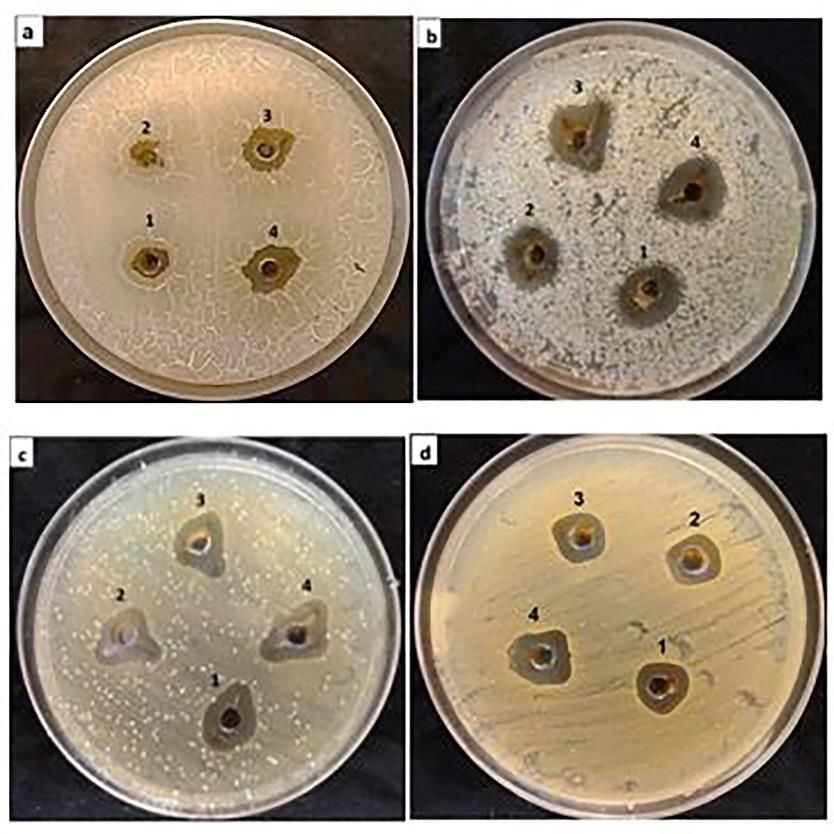
Dehghan Nayeri et al.                          Int. J. Adv. Biol. Biomed. Res. 2021, 9(3):270-285

 using medicinal plants for biosynthesis of AgNPs can boost the quality of prepared
 particles for exhibiting trustworthy functionalities.
 Keywords: Bio-reducing agent; green synthesis; plant extract.

1. Introduction                                NPs      such    as    Berberis     vulgaris
                                               (Berberidaceae)       [10],     Coriandrum
   Nanomaterials are being used as smart
                                               sativum (Apiaceae) [11], Vaccinium
material in almost every field of human
                                               arctostaphylos L. (Ericaceae) [12] and so
activities.   Among       all    types    of
                                               on. Melissa officinalis is a perennial and
nanoparticles,     silver     nanoparticles
(AgNPs( have been used for a wide variety      medicinal herb from the Lamiaceae
                                               family. Due to its lemon-like fragrance, it
of applications in human life [1]. AgNPs
                                               is also known as lemon balm [13].
have more application in the medical field
                                               Different organic compounds present in
as antimicrobials and sterilizers. The
                                               lemon balm extract including protein,
antimicrobial property of AgNPs is well
                                               essential oils, flavonoids, rosmarinic,
known and it is mediated by the silver
                                               caffeic and gallic acids and phenolic
cations released from the AgNO3 (silver
                                               contents are responsible for bio-
nitrate) [2]. These particles have been
                                               reduction of Ag+ ions [14]. Therefore, this
synthesized by different approaches
                                               plant has been used to synthesize bio-
including chemical [1] and biological
                                               AgNPs for reaching higher chemical yields
methods from microorganisms and plants
                                               and bio-activity.
[3].
                                                  This study aimed to examine the
   The chemical methods often generate
                                               impact of aqueous and methanolic
highly toxic compounds that remain
                                               extracts of Melissa officinalis on the size,
adsorbed on the particle surface and pose
                                               shape and morphology of synthesized
adverse effects on human health. This
                                               silver nanoparticles and their biological
warrants the use of safe, biocompatible
                                               properties against infectious microbial
and eco-friendly green biological methods
                                               strains.
for the synthesis of AgNPs [4, 5]. The
green synthesis is based on the use of non-    2. Material and methods
toxic and eco-friendly ingredients
moreover; it is cost-effective, low energy     2.1. Plant extraction
consumption, less complex and less time-       2.1.1. Aqueous extract
consuming process [6].
   Medicinal plants have important roles          The aerial parts of Melissa officinalis
in the development of biochemical              were washed with distilled water and
methods due to the existence of a wide         shade dried for 7 days at 28 °C. The dried
variety of secondary metabolites that          plants were ground into a fine powder.
presented within their tissues [7, 8]. Since   For preparation of the aqueous extract
ancient times, human being has been used       150 mL of sterile distilled water was
medicinal plants for different proposes so     added to 6 g of powdered samples and
that until now the methods for processing      boiled for 10 min. The suspension was left
of medicinal plants for extraction of          for 3 h and then filtered through
trustworthy metabolites have been              Whatman filter paper No. 1. The samples
advanced [7, 9]. In this respect, because of   were centrifuged at 20000 rpm for 15 min
the     development       of   plant-based     and stored at 4 °C until further analysis.
biosynthesis of nanoparticles, a wide
                                               2.1.2. Methanolic extract
range of plant resources have been
explored by researchers to synthesize

                                                                                   271 | P a g e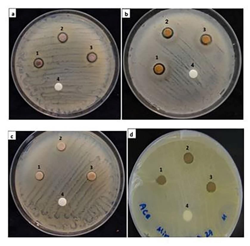
Dehghan Nayeri et al.                         Int. J. Adv. Biol. Biomed. Res. 2021, 9(3):270-285

    For this, about 6 g of powdered sample    after dilution a small aliquot of the sample
was added into a thimble, and extracted in    by distilled water [15].
Soxhlet with methanol for the period until
the solvent in siphon tube of extractor       2.4.2. Scanning Electron Microscopy
becomes colorless. The extracts were          (SEM)
filtered using filter paper and the solvent     The size and shape of AgNPs
was allowed to evaporate from the extract     synthesized by M. officinalis were
in a rotary evaporator to have a syrupy       determined by scanning electron
consistency. The extract was stored at 4 °C   microscopy using high-performance
for further experiments.                      MIRA-3 SEM machine (TESCAN, Brno,
                                              Czech Republic).
2.2.   Biosynthesis          of     silver
nanoparticles                                 2.4.3. X-Ray Diffraction analysis
   The AgNPs were prepared according to          The XRD analysis of synthesized AgNPs
the method of Savithramma et al [30] with     was conducted by using Bruker D8
slight modification. A, 1 mM AgNO3 (silver    Advance X-ray diffractometer (D8-
nitrate) solution was prepared and stored     Advance Bruker, Germany) and the XRD
in amber colored bottle at 4 °C. A 50 mL of   patterns were recorded with a Cu Kα
AgNO3 solution (1 mM) was added into 5        radiation in the range of 20 °C to 280 °C at
mL of lemon balm extracts with constant       40 keV [16].
stirring at 28 °C until the color change as
an indication of the formation of AgNPs.      2.4.4. Fourier Transform               Infrared
The extract content was then centrifuged      spectroscopy
at 14000 rpm for 30 min and the
                                                 The nanoparticles were characterized
reduction of Ag+ ions and the formation of
                                              using the lyophilized samples by
AgNPs was studied by using UV-Vis
                                              potassium bromide pellet technique in
spectroscopy.
                                              the range of 500 to 4000 cm-1 on a Bruker
2.3. Effect of the concentration of           Tensor 27 FTIR spectrometer (Karlsruhe,
extracts on the biosynthesis of silver        Germany). FTIR was used to characterize
nanoparticles                                 the nanoparticles using the lyophilized
                                              samples by potassium bromide pellet
   To study the effect of the different       technique in the range of 500 to 4000 cm-
concentration of extracts, various            1.
volumes of aqueous extract (7, 10, 15 and
20 mL) and methanolic extract (0.1, 0.5, 1,   2.5. DPPH free radical scavenging
and 5 mg /50 mL) were used to prepare         assay
AgNPs.
                                                 Antioxidant activity of AgNPs was
2.4. Characterization of synthesized          quantitatively studied by using DPPH
silver nanoparticles                          method [17]. In this, a 150 μl of 1 mM NPs
                                              solution and ascorbic acid (positive
2.4.1. UV-Vis spectrophotometry               control) were separately mixed with 2.8
   UV–visible spectroscopy was carried        mL of a methanolic solution of DPPH,
out using a UV–Vis spectrophotometer          shaken and incubated in a dark place 4 °C
(Labomed, UV-win5, Germany), in a             for 24 h. After incubation, the absorbance
wavelength range of 300–600 nm. Silver        of the samples (reduction of DPPH
ions reduction was monitored by               radical) was measured by UV–Vis
measuring the UV–Vis spectra of solutions     spectrophotometer at 517 nm against
                                              methanol as blank. The methanolic
                                                                                  272 | P a g e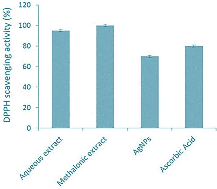
Dehghan Nayeri et al. Int. J. Adv. Biol. Biomed. Res. 2021, 9(3):270-285
solution of DPPH without the sample antimicrobial activity was analyzed by
served as control. The DPPH free radical measuring the diameter of inhibition zone
scavenging activity (percentage of (mm) around the wells.
inhibition) was calculated using the
following formula: 3. Results and discussion
Acontrol − Asample 3.1.1. Extraction and biosynthesis of
Inhibition (%) = × 100
Acontrol AgNPs
The reduction of silver ions into silver
2.6. Antimicrobial assays of the
nanoparticles can be followed by color
biosynthesized AgNPs
change. The fresh extract of M. officinalis
The antimicrobial efficacy of AgNPs was pale yellow in color and after the
was directed against these bacterial and addition of AgNO3 and stirring at 28 °C,
one fungal cultures. Microbial cultures the color of solution changed into pale
namely Staphylococcus aureus PTCC 1113, pink (within 30 min in case of aqueous
Escherichia coli PTCC 1330 and Bacillus extract) and light red (within 15 min in
subtilis NCTC 5398 and fungus case of methanolic extract). The emulsion
Saccharomyces cerevisiae IBRCM 30069 turned dark brown after 24 and 36 h for
were provided from Iranian Biological methanolic and aqueous extracts
Resourece Center IBRC, Iran. Bacterial respectively.
cultures wre mainatianed on LB agar and
fungal culture was preserved on potato 3.1.2. Effect of different concentrations
dextrose agar PDA at 4 °C. of extracts on the biosynthesis of
The antimicrobial assay performed as AgNPs
according to the Kirby–Bauer disc Different volumes of aqueous extract
diffusion [18] and agar well diffusion (7, 10, 15 and 20 mL) and methanolic
methods [19]. In disc diffusion method, extract 0.1, 0.5, 1, and 5 mg /50 mL were
NA and PDA plates were inoculated with used to study the formation of AgNPs.
100 μl of bacterial (1106 cells/mL) and Change in color in the aqueous extract of
fungal spore suspension (1106) lemon balm during biosynthesis of AgNPs
respectively using spread-plating began within 30 min. The color change
technique. After drying the plates, filter occurred first in the lowest concentration
paper discs of 6-mm diameter were (7 mL) followed by color changes in
soaked in 30 and 40 μl of AgNPs solution samples having higher concentrations.
(1 mM), plant extract and distilled water These color changes indicated the
as control and placed on LB agar plates. reduction of silver particles into silver
Plates were incubated at 28 °C for 24-48 nanoparticles. Thus, velocity of the
h. Following the incubation, the formation of AgNPs was inversely
susceptibility of the test organisms was proportional to the concentration of the
determined by measuring diameter of aqueous extract. It began first with lowest
zone of inhibition (mm). concentration and then with increasing
For agar well diffusion test, the concentrations of the aqueous extract.
inoculated plates after drying, were In case of methanolic extract (0.1, 0.5,
punched to make wells of 8-mm diameter, 1, and 5 mg/50 mL) the color changing
and aseptically filled with 20, 30 and 40 μl occurred with higher concentrations of
of biosynthesized 1 mM solution of NPs the extract. The speed of the reaction was
and plant extract. Plates were allowed to increasing with increase of the
diffuse for 2 h at 8 °C followed by concentration of methanolic extract. The
incubation 28 °C for 24-48 h. The concentration 2.5 mg/100 mL responded
273 | P a g e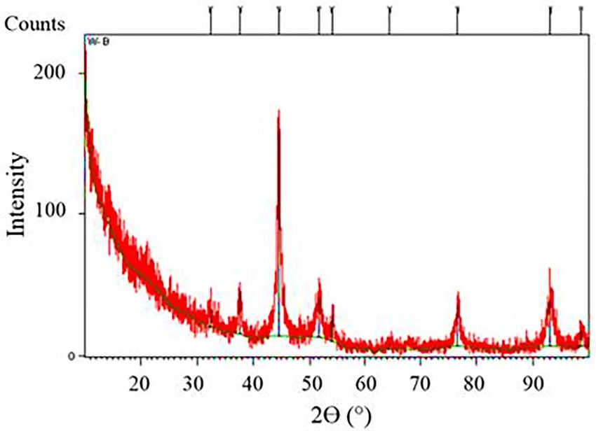
Dehghan Nayeri et al.                         Int. J. Adv. Biol. Biomed. Res. 2021, 9(3):270-285

best compared to 0.5 and 0.25 mg/100 mL        nanoparticles [20]. Biologically, plant
while 0.05 mg/100 mL concentration did         metabolites such as sugars, proteins,
not show any color change indicating no        terpenoids,      polyphenols,     alkaloids,
formation of AgNPs in this lowest              flavonoids and phenolic acids are
concentration.                                 responsible for the reduction and
The results also showed that the size of       stabilization of nanoparticles. It has been
colloidal particles was between 1 to 1000      reported that the OH groups present in
nm. The formation of colloidal solutions       the backbone flavonoids play a critical
from the reduction of silver ions occurs in    role in the reduction of silver ions to
two steps (nucleation and growth)              AgNPs [21]. Since 7 mL aqueous and 2.5
(Figure 1). Reducing and stabilizing           mg/100 mL methanolic extracts resulted
agents are known as the major                  in the faster formation of AgNPs, they
components in the formation of metal           were used for further analysis.

Figure 1. The reducing agents (in plant extract) donate electrons to Ag+ ions lead them
as neutral Ag atoms (Ag0). These atoms due to van der Waal's forces of attractions come
   closer and combine to form cluster of Ag atoms of diameter 1 to 100 nm, called Ag
 nanoparticle. A silver nanoparticle in nano scale may contain about hundreds of atoms
 of silver. If these clusters (nanoparticles) come closer they agglomerate first and then
                 aggregate to form bulky particles, which is not AgNPs [20].
3.2. Characterization of NPs                   recorded by UV-Vis spectroscopy. The
                                               reaction was conducted for 2 h. The UV-
3.2.1. Ultraviolet-visible spectroscopy
                                               Vis absorption spectra of colloidal
   Color change from yellow to brownish-       solutions of AgNPs using M. officinalis
red and dark brown is the first sign of        methanolic extract had absorbance near
nanoparticle production and is due to          450 nm. The broadening peak is a sign of
excitation of the surface Plasmon              the poly-dispersed particles formation
resonance [22]. The biosynthesis of nano       (Figure 2). Hafez et al. [24] produced
particle should be confirmed via physical      AgNPs that showed UV-Vis absorbance at
methods like UV-Vis spectrophotometer,         425 nm and Keshari et al. [25] reported
SEM, XRD and FTIR [23].                        the absorption band of AgNPs at 442 nm.
   The biosynthesis of AgNPs and the           Also, the absorption of the AgNPs was
reduction of Ag+ ions to Ag atoms were         observed near 430 nm [26] and 420 nm

                                                                                  274 | P a g e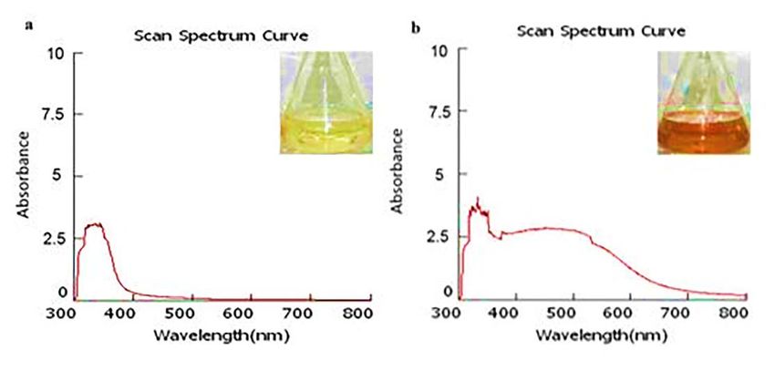
Dehghan Nayeri et al.                           Int. J. Adv. Biol. Biomed. Res. 2021, 9(3):270-285

[27] in the UV–Vis spectrum The                 times [28], fresh and freeze-dried samples
absorbance wavelength depends on the            [29] and particle size [23] .
concentration of plant extract, different

 Figure 2. UV–Vis absorption spectrum of the photosynthesized AgNPs. a) The reaction
  at time zero showed no absorption in 400–500 nm region. b) Spectra represented the
 formation of silver nanoparticles with the help of Melissa officinalis methanolic extract.
      The absorption of the AgNPs was observed near 450 nm which is due to surface
                              Plasmon resonance of AgNPs.
3.2.2. Scanning electron microscope             nanoparticles formed with diameter in
                                                the range of 19-40 nm from the aqueous
   To determine the morphological
                                                extract. Also, the nanoparticles derived
characters nanoparticles synthesized by
                                                from the methanolic extract were
lemon balm extracts, SEM assay was used.
                                                spherical with diameter of 13-35 nm
The SEM images showed rod-shaped
                                                (Figure 3).

Figure 3. SEM images of biosynthesized silver nanoparticles by using different extracts
of Melissa officinalis; a) rod-shaped nanoparticles from aqueous extract and b) spherical
                        shape nanoparticles from methanolic extract.

                                                                                    275 | P a g e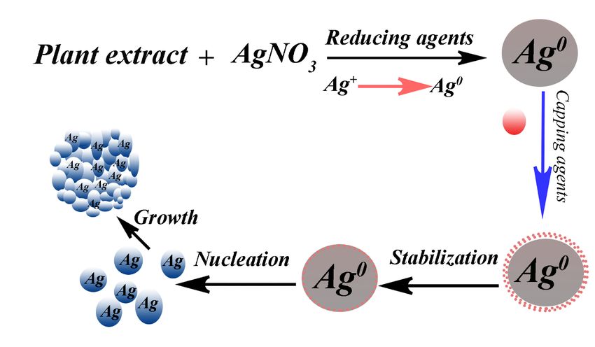
Dehghan Nayeri et al.                          Int. J. Adv. Biol. Biomed. Res. 2021, 9(3):270-285

   It has been reported that the               3.2.3. X-ray diffraction (XRD) analysis
nanoparticles deformation as different            The XRD is one of the most widely used
shapes and diameters is the result of          techniques to characterize the structural
altering the plant extract and/or the
                                               properties of NPs. In order to gain the
reaction conditions depending on the           structural information, the resulting
application [21]. The surface areas and        diffraction patterns obtained from the
the shapes of nanoparticles effect on their    penetration     of    X-rays    into   the
antimicrobial activity [30] due to different   nanomaterials      is   compared      with
effective surface areas and active facets      standards [32]. Figure 4 shows the XRD
[30]. It was suggested that Melissa            pattern of the AgNPs synthesized by using
officinalis scavenged DPPH radical in a        the methanolic extract of lemon balm. The
concentration-dependent manner [31]            AgNPs diffractogram displayed several
that is in agreement with the result of our    sharp intense peaks at 2 theta angles,
study. DPPH assay revealed the sample of       which indicated the crystalline structure
5 mg/50 mL methanolic extract had the          of the AgNPs and confirmed the formation
highest antioxidant activity compared to       of the AgNPs. Four distinct reflections at
the 7 mL aqueous extract.                      37.5° (111), 44.37° (200), 64.56° (220)
   Based on the SEM results, the particles     and 76.58° (311) evidently indicated the
produced by methanolic extract were            formation of the face-centered cubic
spherical and smaller in size in               structure of the AgNPs in the sample.
comparison to the rod-shaped particles
derived from the aqueous extract.
Therefore, further experiments were
conducted using the methanolic sample.

 Figure 4. XRD diffractogram of biosynthesized AgNPs by Melissa officinalis methanolic
                                       extract.
   The XRD outline displayed that the          Additional peaks at 32.25° and 54.62°
AgNPs formed by the reduction of Ag +          were observed on the preparation of
ions by M. officinalis extract are crystal-    AgNPs using M. officinalis methanolic
like in nature. This result is in accordance   extract (Figure 4). These peaks are
with the XRD analysis of Shaik et al. [33].    attributed to the existence of some

                                                                                   276 | P a g e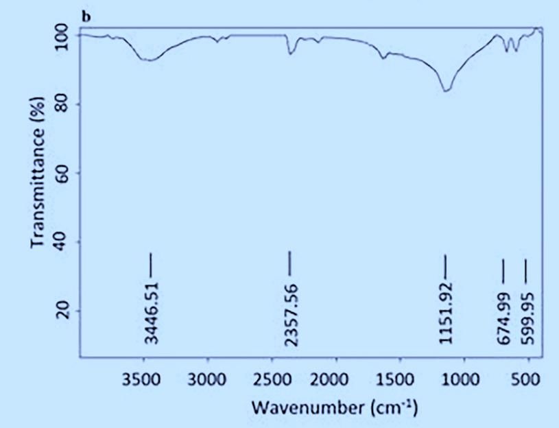
Dehghan Nayeri et al.                          Int. J. Adv. Biol. Biomed. Res. 2021, 9(3):270-285

bioorganic compounds in M. officinalis          stretch, H-bonded corresponded to
leaf broth[34]or related to unreduced and       alcohols and phenols, 2357.56 cm -1
left over residues of AgNO3 in the sample       assigned for single aldehyde, 1151.92 cm-
[35]. It has been suggested that                1 indicates the fingerprint region of C-O

magnesium of the chlorophyll is the             stretching, 674.99 cm-1 could be
center of X-ray diffraction in the              attributed to the presence of C-H bend
bioorganic crystalline phase [36].              alkynes and 674.99 cm-1 and 599.95 cm-1
                                                correspond to halo compound.
3.2.4. Fourier transform infrared                  Figure 5 shows the peaks near
(FTIR) spectroscopic analysis                   3000 cm-1 assigned to O-H stretching and
   Here, FTIR was used to analyze the           aldehyde C-H stretching. The absorption
chemical composition of lemon balm              peaks between 1500 to 2000 cm -1 can be
responsible for reduction of Ag ions. Using     attributed to the presence of C-O
this technique organic functional groups        stretching in carboxyl coupled to the
(COO−, OH, …) attached to the Ag and            amide linkage in amide I. These are the
other chemical residues surface can be          characteristic of the presence of protein
detected. The methanolic extract of aerial      and enzymes in the supernatant and it
parts of lemon balm displayed a number          confirms the extracellular formation of
of absorption peaks, reflecting its complex     AgNPs [37]. Consequently, the occurrence
nature. The results of FTIR analysis            of these peaks in the FTIR spectrum
showed different stretches of bonds             evidently indicates the dual role of the M.
shown at different peaks including              officinalis extract, i.e. both as a green
3446.51 cm-1 could be assigned to O-H           reducing as well as stabilizing agents.

        Figure 5. FTIR spectra of (a) Melissa officinalis methanolic extract and (b)
            biosynthesized AgNPs from Melissa officinalis methanolic extract.
   Interactions between metabolites in          contribute in both reduction                and
the extract and metal ions cause the bio-       stabilization of AgNPs [32].
reduction of metal salts and synthesis of
nanoparticles. The functional groups in         3.3. Antioxidant capacity-DPPH radical
the plant extract act as reducing, capping,     scavenging assay
and stabilizing agents. Negatively charged         The DPPH free radical scavenging
(COO−) and polar (OH and CO) groups             assay was determined by measuring the
presented in the plant extract attach on        ability of plant extracts to capture the
the Ag surface with high tendency and           stable radical DPPH [15, 21]. The initial

                                                                                   277 | P a g e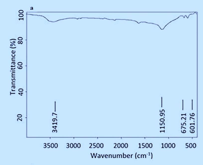
Dehghan Nayeri et al.                         Int. J. Adv. Biol. Biomed. Res. 2021, 9(3):270-285

violet color of DPPH solution changed to       absorbance of DPPH radiation at a
yellow as soon as the AgNPs were added.        wavelength of 517 nm. The color change
This may be because of the presence of         rate in the methanolic extract was also
antioxidant in the medium. However, the        higher than that of the aqueous extract
process of yellowing may slow down and         and AgNPs (Figure 6). The methanolic
the target substance exhibits poor             extract of lemon balm and AgNPs
antioxidant activity or no color change.       exhibited highest antioxidant activity
The inhibitory concentration of the            compared to the aqueous extract.
material is investigated based on the

Figure 6. DPPH radical scavenging activity of two extracts of Melissa officinalis and the
 synthesized AgNPs using methanolic extract. The methanolic extract had the highest
                       antioxidant activity among the samples.
   Various studies have reported the              The antimicrobial effect of spherical
DPPH scavenging activity by AgNPs from         silver nanoparticles was investigated by
plant extracts [24]. The production of iron    agar well diffusion and disc diffusion
NPs using aqueous extracts obtained from       methods. Green synthesis of the AgNPs
26 tree species and antioxidant properties     using this lemon balm extract showed an
of these have been reported [25]. These        effective antimicrobial activity against
researchers interestingly found dried          Gram-positive bacteria such as B. subtilis
leaves (higher concentration) produce          and S. aureus and Gram-negative
extracts     with     higher    antioxidant    bacterium namely E. coli and against
capacities than non-dried leaves (lower        yeast, S. cerevisiae (Figure 7 and 8).
concentration). This is probably due to           In disc diffusion method the diameter
evaporation of water from leaves that          of inhibition zone for B. subtilis, S. aureus,
result in the concentration of antioxidants    E. coli and S. cerevisiae was 5.7, 5.6, 7 and
[25].                                          4 mm respectively, while in agar well
                                               diffusion method it was 10, 10, 11.3 and
3.4. Antimicrobial property of AgNPs           9.25 mm, respectively (Figure 7 and 8).

                                                                                  278 | P a g e
Dehghan Nayeri et al. Int. J. Adv. Biol. Biomed. Res. 2021, 9(3):270-285
Figure 7. Agar well method showing antimicrobial activity of biosynthesized AgNPs at
different volumes; 1) Melissa officinalis methanolic extract, 2) 20, 3) 30 and 4) 40 μl of
biosynthesized AgNPs solution evaluated against a) Saccharomyces cerevisiae, b)
Bacillus subtilis, c) Escherichia coli and d) Staphylococcus aureus.
Figure 8. Disc diffusion method showing inhibition zones produced by biosynthesized
AgNPs at different volumes; 1) Melissa officinalis methanolic extract, 2) 30 and 3) 40 μl
of biosynthesized AgNPs solution plus 4) distilled water evaluated by against a)
Escherichia coli, b) Staphylococcus aureus, c) Bacillus subtilis and d) Saccharomyces
cerevisiae
279 | P a g eDehghan Nayeri et al. Int. J. Adv. Biol. Biomed. Res. 2021, 9(3):270-285
In both methods, Gram-negative the disruption of the cell wall and cell
bacterium, E. coli exhibited the highest membrane that finally leading to bacterial
sensitivity to the AgNPs compared to cell death. Additionally, there are some
other test organisms. The results of the other mechanisms including attack on
present study were similar to those of bacterial surface and membrane through
others [38, 39] and elucidated that AgNPs interaction with sulfur-containing
from methanolic extract of lemon balm proteins [40], disruption of cell
inhibited bacterial and fungal growth. The permeability and respiration, form the
antimicrobial effect of AgNPs was found pits on the cell surface and induce the
to be dependent on the size, shape and proton leakage that causes cell death [41],
concentration of AGNPs, smaller and inhibition of respiratory enzymes of
spherical particles produced higher bacterial cells by combining with the thiol
inhibition compared to larger and non- group [42] as well as cell retention of DNA
spherical particles. This may be due to the replication and preventing cellular
availability of more surface area in division [43] might widely contributed to
smaller and spherical particles [40]. the antibacterial effects of AgNPs. The
Similarly, higher concentrations of AgNPs entrance of AgNPs also produce higher
resulted in more inhibition of the amount of ROS molecules that is linked to
bacterial and fungal growth. Antibacterial deactivation of the respiratory enzymes
property of AgNPs derived from aqueous and disrupt the cellular membrane and
extract of lemon balm leaves against S. damage DNA molecule [44]. Figure 9
aureus and E. coli have been reported by illustrates some possible molecular
Ruíz-Baltazar et al [14]. AgNPs mainly mechanisms might link to antimicrobial
exert their antimicrobial effects through action of AgNPs.
Figure 9. Proposed mechanism of AgNPs formation and antimicrobial activities exerted
by Melissa officinalis. (by Photoshop software).
280 | P a g eDehghan Nayeri et al. Int. J. Adv. Biol. Biomed. Res. 2021, 9(3):270-285
Gram-negative and Gram-positive UV-Vis: Ultraviolet-visible spectroscopy
bacteria were more susceptible to AgNPs
compared to the fungal strain [37]. These Acknowledgment
differences in bactericidal and fungicidal This work was done in Agricultural
effects of the AgNPs are due to the Biotechnology Department of Imam
differences in structural organization of Khomeini International University,
the bacterial and fungal cell walls. The Qazvin, The Islamic Republic of Iran. The
bacteria are evolutionarily prokaryotic authors appreciate all staffs on their good
types and are less complex. Therefore, collaborations.
they are more susceptible to the toxic
effects of AgNPs compared to the Conflict of Interest Statement:
eukaryotic fungi. Fungi being the
The authors declare no competing
eukaryotic organisms, possess superior
interests.
detoxification system that makes them
resistant to higher concentrations of Consent for publications
AgNPs [37].
The authors have read and approved
4. Conclusion the submitted manuscript.
Green production of AgNPs is a low- Availability of data and material
cost and an eco-friendly method and
could provide lots of benefits for The authors declare that all the data is
researchers. Therefore, it is a good embedded in the manuscript.
alternative for the industrial production
of NPs. The biosynthesis of AgNPs using Authors' contributions:
lemon balm extracts and their F.D.N designed the experiment,
antibacterial, antifungal and antioxidant analyzed and interpreted the data and
activities were studied. The nanoparticles drafted the manuscript, M.M performed
produced by the methanolic extract of the experiments, collected the data and
lemon balm resulted in quicker formation prepared figures, S.M. helped in
of AgNPs and exhibited potent performing experiments and R.Z.S revised
antimicrobial and antioxidant activities. and edited the manuscript. All the authors
Due to the antimicrobial properties of the read and approved the final manuscript.
green AgNPs, they can be used as an
antiseptic, sterilant and antimicrobial Ethics approval and consent to
agents. The antioxidant properties of participate:
green AgNPs also make them ideal This article does not contain any
candidate for food industries to eliminate studies with human participants or
the development of infectious pathogens animals performed by any of the authors.
within food cans or similar products.
Funding information:
Abbreviation:
This work was supported by the Imam
AgNO3: Silver nitrate Khomeini International University [grant
AgNPs: Silver nanoparticles number 11590], Qazvin, Iran.
DPPH: 2,2-diphenyl-1-picrylhydrazyl
FTIR: Fourier Transform Infrared Reference
SEM: Scanning Electron Microscope
XRD: X-Ray Diffraction 1. Suganthy N, Ramkumar V S,
UV: Ultraviolet Pugazhendhi A, Benelli G, Archunan G.
(2018). Biogenic synthesis of gold
281 | P a g eDehghan Nayeri et al. Int. J. Adv. Biol. Biomed. Res. 2021, 9(3):270-285
nanoparticles from Terminalia arjuna Journal of Medical Bacteriology, 8(5-6):
bark extract: assessment of safety 8-20.
aspects and neuroprotective potential 9. Mohammadi-Motlagh H-R, Shokohinia
via antioxidant, anticholinesterase, and Y, Mojarrab M, Rasouli H, Mostafaie A.
antiamyloidogenic effects. (2017). 2-Methylpyridine-1-ium-1-
Environmental Science and Pollution sulfonate from Allium hirtifolium: An
Research, 25(11): 10418-10433. anti-angiogenic compound which
2. Khutale G V, Casey A. (2017). Synthesis inhibits growth of MCF-7 and MDA-
and characterization of a MB-231 cells through cell cycle arrest
multifunctional gold-doxorubicin and apoptosis induction. Biomedicine &
nanoparticle system for pH triggered Pharmacotherapy, 93: 117-129.
intracellular anticancer drug release. 10. Behravan M, Panahi A H, Naghizadeh
European Journal of Pharmaceutics and A, Ziaee M, Mahdavi R, Mirzapour A.
Biopharmaceutics, 119: 372-380. (2019). Facile green synthesis of silver
3. Zarayneh S, Sepahi A A, Jonoobi M, nanoparticles using Berberis vulgaris
Rasouli H. (2018). Comparative leaf and root aqueous extract and its
antibacterial effects of cellulose antibacterial activity. International
nanofiber, chitosan nanofiber, journal of biological macromolecules,
chitosan/cellulose combination and 124: 148-154.
chitosan alone against bacterial 11. Ashraf A, Zafar S, Zahid K, Shah M S, Al-
contamination of Iranian banknotes. Ghanim K A, Al-Misned F, Mahboob S.
International Journal of Biological (2019). Synthesis, characterization,
Macromolecules, 118: 1045-1054. and antibacterial potential of silver
4. Khan I, Saeed K, Khan I. (2019). nanoparticles synthesized from
Nanoparticles: Properties, applications Coriandrum sativum L. Journal of
and toxicities. Arabian Journal of Infection and Public Health, 12(2): 275-
Chemistry, 12(7): 908-931. 281.
5. Rasouli H, Popović-Djordjević J, Sayyed 12. Khodadadi S, Mahdinezhad N, Fazeli-
R, Zarayneh S, Jafari M, Fazeli-Nasab B. Nasab B, Heidari M J, Fakheri B, Miri A.
(2020). Nanoparticles: a new threat to (2021). Investigating the Possibility of
crop plants and soil Rhizobia? Green Synthesis of Silver Nanoparticles
Sustainable Agriculture Reviews 41 Using Vaccinium arctostaphlyos
(pp. 201-214): Springer. Extract and Evaluating Its Antibacterial
6. Rasouli H. (2019). Devil's hand conceals Properties. BioMed Research
behind the obscure side of AgNPs: A International, 2021.
letter to the editor. International 13. Abdel-Naime W A, Fahim J R, Fouad M
Journal of Biological Macromolecules, A, Kamel M S. (2016). Botanical studies
125: 510-513. of the leaf of Melissa officinalis L.,
7. Rasouli H, Hosseini-Ghazvini S M-B, Family: Labiatae, cultivated in Egypt.
Khodarahmi R. (2019). Therapeutic Journal of Pharmacognosy and
potentials of the most studied Phytochemistry, 5(6): 98.
flavonoids: highlighting antibacterial 14. de Jesús Ruíz-Baltazar Á, Reyes-López
and antidiabetic functionalities Studies S Y, Larrañaga D, Estévez M, Pérez R.
in natural products chemistry (Vol. 60, (2017). Green synthesis of silver
pp. 85-122): Elsevier. nanoparticles using a Melissa
8. Fazeli Nasab B. (2019). Evaluation of officinalis leaf extract with
Antibacterial Activities of antibacterial properties. Results in
Hydroalcoholic Extract of Saffron Physics, 7: 2639-2643.
Petals on Some Bacterial Pathogens.
282 | P a g eDehghan Nayeri et al. Int. J. Adv. Biol. Biomed. Res. 2021, 9(3):270-285
15. Thaipong K, Boonprakob U, Crosby K, and biopolymer nanocomposites: a
Cisneros-Zevallos L, Byrne D H. (2006). comparative study on physico-
Comparison of ABTS, DPPH, FRAP, and chemical, antimicrobial and anticancer
ORAC assays for estimating antioxidant activity. Bulletin of Materials Science,
activity from guava fruit extracts. 41(2): 1-11.
Journal of food composition and 24. Hafez R A, Abdel-Wahhab M A, Sehab
analysis, 19(6-7): 669-675. A F, El-Din A-Z A K. (2017). Green
16. Bauer A. (1966). Antibiotic synthesis of silver nanoparticles using
susceptibility testing by a standardized Morus nigra leave extract and
single disc method. American Journal of evaluation their antifungal potency on
Clinical Pathology, 45: 149-158. phytopathogenic fungi. Journal of
17. Andrews J M. (2001). The Applied Pharmaceutical Science, 7(02):
development of the BSAC standardized 041-048.
method of disc diffusion testing. 25. Keshari A K, Srivastava R, Singh P,
Journal of Antimicrobial Chemotherapy, Yadav V B, Nath G. (2020). Antioxidant
48(suppl_1): 29-42. and antibacterial activity of silver
18. Bauer A, Kirby W, Sherris J C, Turck M. nanoparticles synthesized by Cestrum
(1966). Antibiotic susceptibility testing nocturnum. Journal of Ayurveda and
by a standardized single disk method. Integrative Medicine, 11(1): 37-44.
American journal of clinical pathology, 26. Salehi S, Shandiz S A S, Ghanbar F,
45(4_ts): 493-496. Darvish M R, Ardestani M S, Mirzaie A,
19. Walther C. (2003). Comparison of Jafari M. (2016). Phytosynthesis of
colloid investigations by single particle silver nanoparticles using Artemisia
analytical techniques-a case study on marschalliana Sprengel aerial part
thorium-oxyhydroxides. Colloids and extract and assessment of their
Surfaces A: Physicochemical and antioxidant, anticancer, and
Engineering Aspects, 217(1-3): 81-92. antibacterial properties. International
20. Jain S, Mehata M S. (2017). Medicinal journal of nanomedicine, 11: 1835.
plant leaf extract and pure flavonoid 27. Pallela P, Ummey S, Ruddaraju L K,
mediated green synthesis of silver Pammi S V N, Yoon S G. (2018). Ultra
nanoparticles and their enhanced Small, mono dispersed green
antibacterial property. Scientific synthesized silver nanoparticles using
Reports, 7(1): 1-13. aqueous extract of Sida cordifolia plant
21. Machado R, Alves-Pereira I, Ferreira R. and investigation of antibacterial
(2018). Plant growth, phytochemical activity. Microb Pathog, 124: 63-69.
accumulation and antioxidant activity 10.1016/j.micpath.2018.08.026
of substrate-grown spinach. Heliyon, 28. Khatami M, Noor F G, Ahmadi S,
4(8): 1-21. Aflatoonian M. (2018). Biosynthesis of
22. Solidum R S, Alguno A C, Ag nanoparticles using Salicornia
Capangpangan R Y. (2018). Controlling bigelovii and its antibacterial activity.
the surface plasmon absorption of Electronic Physician, 10(4): 1-8.
silver nanoparticles via green 29. Moodley J S, Krishna S B N, Pillay K,
synthesis using Pennisetum Govender P. (2018). Green synthesis of
purpureum leaf extract. Paper silver nanoparticles from Moringa
presented at the Key Engineering oleifera leaf extracts and its
Materials. antimicrobial potential. Advances in
23. Palem R R, Ganesh S D, Kroneková Z, Natural Sciences: Nanoscience and
Sláviková M, Saha N, Sáha P. (2018). Nanotechnology, 9(1): 1-10.
Green synthesis of silver nanoparticles
283 | P a g eDehghan Nayeri et al. Int. J. Adv. Biol. Biomed. Res. 2021, 9(3):270-285
30. Raza M A, Kanwal Z, Rauf A, Sabri A N, 37. Panáček A, Kolář M, Večeřová R,
Riaz S, Naseem S. (2016). Size-and Prucek R, Soukupova J, Kryštof V,
shape-dependent antibacterial studies Hamal P, Zbořil R, Kvítek L. (2009).
of silver nanoparticles synthesized by Antifungal activity of silver
wet chemical routes. Nanomaterials, nanoparticles against Candida spp.
6(4): 74. Biomaterials, 30(31): 6333-6340.
31. Kamdem J P, Adeniran A, Boligon A A, 38. Rao N H, N L, Pammi S V, Kollu P, S G, P
Klimaczewski C V, Elekofehinti O O, L. (2016). Green synthesis of silver
Hassan W, Ibrahim M, Waczuk E P, nanoparticles using methanolic root
Meinerz D F, Athayde M L. (2013). extracts of Diospyros paniculata and
Antioxidant activity, genotoxicity and their antimicrobial activities. Mater Sci
cytotoxicity evaluation of lemon balm Eng C Mater Biol Appl, 62: 553-557.
(Melissa officinalis L.) ethanolic 10.1016/j.msec.2016.01.072
extract: Its potential role in 39. Pethakamsetty L, Kothapenta K,
neuroprotection. Industrial Crops and Nammi H R, Ruddaraju L K, Kollu P,
Products, 51: 26-34. Yoon S G, Pammi S V N. (2017). Green
32. Mittal A K, Chisti Y, Banerjee U C. synthesis, characterization and
(2013). Synthesis of metallic antimicrobial activity of silver
nanoparticles using plant extracts. nanoparticles using methanolic root
Biotechnology Advances, 31(2): 346- extracts of Diospyros sylvatica. Journal
356. of Environmental Sciences, 55: 157-163.
33. Shaik M R, Khan M, Kuniyil M, Al- https://doi.org/10.1016/j.jes.2016.04.
Warthan A, Alkhathlan H Z, Siddiqui M 027
R H, Shaik J P, Ahamed A, Mahmood A, 40. Morones J R, Elechiguerra J L,
Khan M. (2018). Plant-extract-assisted Camacho A, Holt K, Kouri J B, Ramírez J
green synthesis of silver nanoparticles T, Yacaman M J. (2005). The
using Origanum vulgare L. extract and bactericidal effect of silver
their microbicidal activities. nanoparticles. Nanotechnology,
Sustainability, 10(4): 1-14. 16(10): 2346.
34. Shankar S S, Ahmad A, Sastry M. 41. Perumalla A, Hettiarachchy N S.
(2003). Geranium leaf assisted (2011). Green tea and grape seed
biosynthesis of silver nanoparticles. extracts—Potential applications in
Biotechnology progress, 19(6): 1627- food safety and quality. Food Research
1631. International, 44(4): 827-839.
35. Chiguvare H, Oyedeji O O, Matewu R, 42. Korshed P A. (2018). The molecular
Aremu O, Oyemitan I A, Oyedeji A O, mechanisms of the antimicrobial
Nkeh-Chungag B N, Songca S P, Mohan properties of laser processed nano-
S, Oluwafemi O S. (2016). Synthesis of particle (PhD thesis): The University of
Silver Nanoparticles Using Buchu Plant Manchester (United Kingdom).
Extracts and Their Analgesic 43. Umadevi M, Rani T, Balakrishnan T,
Properties. Molecules, 21(6): 774. Ramanibai R. (2011). Antimicrobial
36. Jyoti K, Baunthiyal M, Singh A. (2016). activity of silver nanoparticles
Characterization of silver prepared under an ultrasonic field.
nanoparticles synthesized using Urtica International Journal of Pharmaceutical
dioica Linn. leaves and their synergistic Sciences and Nanotechnology, 4: 1491-
effects with antibiotics. Journal of 1496.
Radiation Research and Applied 44. Anees Ahmad S, Sachi Das S, Khatoon
Sciences, 9(3): 217-227. A, Tahir Ansari M, Afzal M, Saquib
Hasnain M, Kumar Nayak A. (2020).
284 | P a g eDehghan Nayeri et al. Int. J. Adv. Biol. Biomed. Res. 2021, 9(3):270-285
Bactericidal activity of silver https://doi.org/10.1016/j.mset.2020.
nanoparticles: A mechanistic review. 09.002
Materials Science for Energy
Technologies, 3: 756-769.
How to cite this article: Fatemeh Dehghan Nayeri, Sudabeh Mafakheri, Maryam
Mirhosseini, Riyaz Sayyed. Phyto- mediated silver nanoparticles via Melissa officinalis
aqueous and methanolic extracts: synthesis, characterization and biological properties
against infectious bacterial strains. International Journal of Advanced Biological and
Biomedical Research, 2021, 9(3), 270-285. Link:
http://www.ijabbr.com/article_245059.htmL
285 | P a g eYou can also read