Snail and Sonic Hedgehog activation in neuroendocrine tumors of the ileum
←
→
Page content transcription
If your browser does not render page correctly, please read the page content below
Endocrine-Related Cancer (2007) 14 865–874

Snail and Sonic Hedgehog activation in
neuroendocrine tumors of the ileum
Volker Fendrich 2,4, Jens Waldmann4, Farzad Esni 2, Annette Ramaswamy 5,
Michael Mullendore1,3, Malte Buchholz 4, Anirban Maitra1,3 and
Georg Feldmann1,3
Departments of 1Pathology, 2Department of Surgery and 3The Sol Goldman Pancreatic Cancer Research Center, Johns Hopkins
University School of Medicine, Baltimore, MD21231, USA
Departments of 4Surgery and 5Pathology, Philipps-Universität Marburg, Baldingerstraße, D-35043 Marburg, Germany
(Correspondence should be addressed to V Fendrich; Email: fendrich@med.uni-marburg.de)

Abstract
The transcription factor Snail represses E-cadherin and induces epithelial–mesenchymal transition, a
process also exploited by invasive cancer cells. Aberrant Hedgehog (Hh) signaling was recently
observed in a variety of epithelial cancers and it has been shown that the Hh target gene Gli1 induces
expression of Snail. In this study, we examined whether Snail and Sonic Hedgehog (SHH) are
expressed in neuroendocrine tumors (NETs) of the ileum. Using immunohistochemistry, we found
expression of Snail in 22 out of 37 (59%) of evaluated NET samples, but not in adjacent normal
tissues. Snail expression was mostly restricted to the invasive front of the tumors. Six of seven liver
metastases analyzed were positive for Snail. Intratumoral expression of SHH was detected in 27 out
of 37 (73%) tumors. As opposed to Snail, cells expressing SHH were found to be distributed more
randomly throughout the tumors. Out of 30 primary NETs, 16 (53%) showed both Snail and SHH
expression. Furthermore, we found downregulation of E-cadherin in Snail-expressing cells by
immunofluorescence. Real-time RT-PCR revealed conservation of the Hh target genes Gli1, Gli2,
and Ptch in the pancreatic carcinoid cell line BON-1, which were downregulated upon Hh inhibition
with cyclopamine. Moreover, Hh inhibition attenuated in vitro cell growth in a dose-dependent
manner. In conclusion, we describe for the first time that Snail and SHH are overexpressed in a large
subset of NETs of the ileum. Aberrant activation of these pathways might be involved in invasion and
metastatic spread in NETs.
Endocrine-Related Cancer (2007) 14 865–874

Introduction                                                            (EMT) during embryonic development. In EMT,
Neuroendocrine tumors (NETs) of the gastrointestinal                    epithelial cells acquire fibroblast-like properties and
(GI) tract are a poorly understood group of lesions that                show reduced intercellular adhesion and increased
encompass a broad category of neoplasms derived                         motility (Zhou & Hung 2005). Numerous obser-
from neuroendocrine cells of the GI mucosa. As a                        vations support the idea that EMT has a central
result of increasing incidence over the past 25 years,                  role in tumor progression. During progression to
NETs are approximately as common as esophageal and                      metastatic competence, carcinoma cells acquire
gastric cancer, occurring in an estimated 38.5 per                      mesenchymal gene-expression patterns and proper-
million in the US population (Maggard et al. 2004). In                  ties. This results in changed adhesive properties
this heterogenous group, the 5-year survival rate lies                  and the activation of proteolysis and motility,
around 60% for NETs of the ileum which comprise the                     which allows the tumor cells to metastasize and
majority of the tumors (Rorstad 2005). The presence of                  establish secondary tumors at distant sites (Thiery
metastases, especially to the liver, is associated with a               & Sleeman 2006). During EMT, the E-cadherin
reduced 5-year survival.                                                promoter is frequently repressed by specific
   The increased motility and invasiveness of                           transcriptional repressors, including Snail, Slug,
cancer cells in the first phase of metastasis are                       and Twist and by subsequent promoter hyper-
reminiscent of epithelial–mesenchymal transition                        methylation. Of the transcriptional repressors, the
Endocrine-Related Cancer (2007) 14 865–874                                                    Downloaded from Bioscientifica.com at 04/26/2020 05:10:14AM
                                                                                                                 DOI:10.1677/ERC-07-0108
                                                                                                                                            via free access
1351–0088/07/014–865 q 2007 Society for Endocrinology Printed in Great Britain      Online version via http://www.endocrinology-journals.org
V Fendrich et al.: Hh and Snail in neuroendocrine tumors

best studied is Snail, a highly unstable protein. It is        Immunostaining
rapidly phosphorylated by glycogen synthase                    For immunolabeling, formalin-fixed and paraffin-
kinase-3b (GSK-3b) and subsequently degraded                   embedded archived tumor samples and corresponding
by the ubiquitin–proteasome pathway. Conversely,               normal tissues were stained as previously described
inhibition of GSK-3b function results in upregula-             (Esni et al. 2004). Concentrations and sources of
tion of Snail by an NF-kB-dependent pathway, loss              primary antibodies are listed in Table 1. Briefly, slides
of E-cadherin expression, and EMT. Additional                  from archived NETs were heated to 60 8C for 1 h,
protein modification further stabilizes Snail protein          deparaffinized using xylene, and hydrated by a graded
and promotes EMT and tumor invasion (Christofori               series of ethanol washes. Antigen retrieval was
2006). Expression of Snail in epithelial tumors                accomplished by microwave heating in 10 mM sodium
increases their aggressiveness, as seen in                     citrate buffer of pH 6.0 for 10 min. For immunohis-
experimentally induced breast tumors, where high               tochemistry, endogenous peroxidase activity was
Snail expression correlates with an increased risk             quenched by 10-min incubation in 3% H2O2. Non-
of tumor relapse and poor survival rates in human              specific binding was blocked with 10% serum.
breast cancer (Moody et al. 2005).                             Sections were then probed with primary antibodies
   Recently, aberrant activation of the Hedgehog (Hh)          overnight at 4 8C. For immunohistochemistry, bound
pathway has been found in the majority of upper                antibodies were detected using the avidin–biotin
aerodigestive tract epithelial cancers of endodermal           complex (ABC) peroxidase method (ABC Elite Kit,
lineage (Berman et al. 2003). In a recent study, Louro         Vector Labs, Burlingame, CA, USA). Final staining
et al. (2002) showed that the Hh target gene Gli1 can          was developed with the Sigma FAST DAB peroxidase
rapidly and directly induce expression of Snail.               substrate kit (Sigma). For immunofluorescence, bound
   The purpose of the present study is to examine the          antibodies were developed with the secondary
in vivo expression of Snail and the Hh ligand Sonic            antibody (Jackson Immuno Research, West Grove,
Hedgehog (SHH) in NETs of the ileum and to clarify             PA, USA) and corresponding Cy3-, Cy2-, and Cy5-
whether the Hh pathway is activated in BON-1 human             conjugated antibodies (Jackson Immuno Research).
carcinoid cells.                                                  Mammary carcinoma samples from our tissue bank,
                                                               which had previously been shown to express high
                                                               levels of Snail, were used as positive controls along
                                                               with each batch of Snail IHC stainings. SHH
Patients and methods                                           expression in crypts of adjacent normal ileum tissues,
Subjects                                                       which has been described previously (van den Brink
                                                               et al. 2002), was used as internal positive control for
A series of 37 NETs of the ileum, including seven liver        SHH immunohistochemistry.
metastases, were obtained from the tissue bank of the
Department of Pathology, Philipps-University of Mar-
burg, Germany. Histological diagnoses were confirmed           Fluorescence microscopy
by an experienced pathologist (A R). Histopathological         Fluorescence confocal microscopy was performed with
diagnosis and grading were done in accordance to               a confocal laser scanning microscope (410LSM, Zeiss)
Klöppel (2004). The immunohistochemistry results for          using 40! (NA 1.2) C-apochromat or 100! (NA 1.4)
Snail1 and SHH were scored as follows: negative, !5%           objectives. The sections were scanned for Cy3-tagged
of cells positive; C, !30% of tumor cells positive; CC,        (excitation 543 nm) and FITC-tagged (excitation
O30% of tumor cells positive.                                  488 nm) markers. Sections (ten sections with images
   The study protocol conformed to the guidelines of           512!512 pixels) were scanned at a pixel size of
the local ethics committee.                                    0.07 mm and step size of 1 mm. Image analysis was

Table 1 Working dilutions of antibodies used for immunohistochemistry and immunofluorescence

Antibody                     Species                   Working dilution    Source

a -E-cadherin                Rat                       1:200               Zymed, San Francisco, CA, USA
a-SHH                        Goat                      1:50                R&D Sytems, Minneapolis, MN, USA
a-b-Catenin                  Rabbit                    1:500               Santa Cruz Biotechnology, Santa Cruz, CA, USA
a-Snail                      Goat                      1:100               Santa Cruz Biotechnology
Chromogranin A               Mouse                     1:100               Santa Cruz Biotechnology

                                                                                      Downloaded from Bioscientifica.com at 04/26/2020 05:10:14AM
                                                                                                                                    via free access
866                                                                                               www.endocrinology-journals.org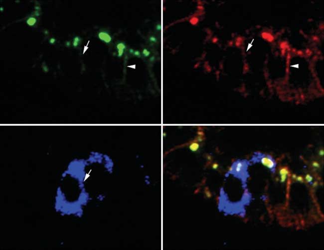
Endocrine-Related Cancer (2007) 14 865–874

performed using MetaMorph series 5.0 software                Strand System for RT-PCR (Invitrogen) at 42 8C for
(Universal Imaging Corp). For specimens not requiring        50 min. For amplification of cDNA specific for Ptch1,
confocal analysis, images were obtained on an inverted       Gli1, Gli2, Gli3, SHH, and PGK-1 Assays-on-De-
motorized microscope (Axiovert 200M, Zeiss) out-             mand (Applied Biosystems, Foster City, CA, USA)
fitted with Cy5, Cy3, FITC, and DAPI filters. Three-         were used together with the QuantiTect Probe PCR
and four-color images were captured using a Zeiss            kit (Qiagen). All PCRs were carried out on a 7300
Axiocam, and pseudocolors assigned using Zeiss               Real-Time PCR System (Applied Biosystems) over
Axiovision software.                                         40 cycles, with denaturation for 15 s at 95 8C and
                                                             combined annealing/extension at 60 8C for 1 min.
Cell culture                                                 Determination of Snail mRNA was performed over
                                                             40 cycles with 15 s of denaturation at 95 8C,
BON-1 pancreatic carcinoid cells (Parekh et al. 1994)        annealing at 61 8C for 20 s, extension at 72 8C for
were grown in DMEM/FK-12 (Invitrogen) supple-                30 s and data acquisition at 78 8C using the
mented with 10% fetal calf serum (FCS; Invitrogen)           Quantitect SYBR Green PCR kit (Qiagen); primer
and 1! Pen/Strep (Biofluids, Camarillo, CA, USA) at          sequences were forward: 5 0 -ggttcttctgcgctactgct-3 0
37 8C in a humidified atmosphere containing 5% CO2.          and reverse: 5 0 -tagggctgctggaaggtaaa-3 0 . Relative
The cell line was routinely tested for mycoplasma            fold mRNA expression levels were determined
infection using the MycoSensor PCR Assay Kit                 using the 2(KDDCt) method (Livak & Schmittgen
(Stratagene, La Jolla, CA, USA).                             2001) with PGK1 (phosphoglycerate kinase 1) as
                                                             housekeeping control.
Proliferation assays
Cells were seeded into 96-well plates at a density of 2000   Statistical analysis
cells in 200 ml full medium/well and incubated overnight.    Kruskal–Wallis analysis was performed using SPSS
Next, they were starved in low-serum medium (0.5%            version 14.0 for Microsoft Windows. Two-tailed t-test
FBS) for 24 h. Then, medium was replaced for 100 ml          was performed using GraphPad Prism for Windows version
low-serum medium supplemented with vehicle, 1.5, 3, or       4.0. P!0.05 was regarded as statistically significant. Prism
6 mM cyclopamine, respectively, for 6 days. Untreated        was also used to generate boxplots in Figs 1E and 2E.
cells served as controls and wells containing medium
only were used for background correction. To determine
viable cell mass, 20 ml of 3-(4,5-dimethylthiazolyl-2)-      Results
2,5-diphenyltetrazolium bromide reagent (Promega)
                                                             Patients
were added per well. Plates were incubated at 37 8C for
another 60 min and optical density read at 490 nm on a       Eighteen males and nineteen females with a median
Wallac 1420 plate reader (Perkin–Elmer, Boston, MA,          age of 63 years (range 34–91 years) at the time of
USA). All experiments were done in triplicate.               surgery were included in this study. All NETs were
                                                             histologically well-differentiated and tested positive
RNA extraction and real-time quantitative                    for chromogranin A by IHC (not shown). Clinical
RT-PCR                                                       characteristics are listed in Table 2.

Cells in the log growth phase were serum starved in 0.5%
                                                             Overexpression of Snail in NETs of the ileum
FCS for 1 day and then incubated with cyclopamine
(6 mM) or ethanol for another 4 days. Next, cells were       Immunohistochemical staining revealed expression of
lysed in the culture dish with 600 ml buffer RLT and         Snail in 22 out of 37 (59%) of NETs tested (Table 2). In
whole RNA extracted using the RNeasy kit (Qiagen)            tumors, in which Snail was found to be expressed, Snail-
with on-column DNA digestion following the standard          positive cells were not distributed homogeneously
protocol provided by the manufacturer.                       throughout the whole tumor. Instead, Snail expression
   Human tissue samples were homogenized with a              was mostly restricted to peripheral areas rendering what
rotor stator homogenizer (Polytron PT1200C, Kine-            appeared to be the invasive front of the tumors, i.e., where
matica, Newark, NJ, USA) in 600 ml lysis buffer RLT          malignant cells invade bordering non-malignant tissue
(Qiagen). Total RNA was then extracted using the             (Fig. 1A and B). Fifteen tumors did not show any Snail-
RNeasy kit (Qiagen) following the standard protocol.         positive cells (1C). Six of seven (86%) liver metastases
   The mRNA was reverse transcribed into cDNA                analyzed were positive for Snail in IHC (Fig. 1D). In
with oligo-dT primers using the Superscript First            normal intestinal epithelium, Snail staining was absent in
                                                                                  Downloaded from Bioscientifica.com at 04/26/2020 05:10:14AM
                                                                                                                                via free access
www.endocrinology-journals.org                                                                                                867
V Fendrich et al.: Hh and Snail in neuroendocrine tumors
Figure 1 IHC staining for Snail in tissue samples from human GI-NETs. (A) Representative example of Snail expressing carcinoid
and adjacent normal tissue lacking Snail expression (lower half of image). (B) In Snail-positive samples, Snail was not expressed
homogenously throughout the whole tumor, but was mostly limited to the invasive front. (C) In some of the examined carcinoid
samples, no Snail expression could be found in the tumor, but only in mesenchymal cells. (D) All liver metastases showed Snail
expression. (E) Steady-state Snail mRNA levels were determined by real-time RT-PCR in a subset of 19 samples. In each group, the
upper and lower hinges of the boxes mark the 25th and 75th percentiles respectively; whiskers denote the ranges.
all samples. Correlation of intratumoral Snail over- small sample number and considerable variation between
expression with clinicopathological features was not different cases, as well as SHH expression in krypts of
analyzed due to the small sample number. non-malignant, normal intestine (Fig. 2E).
These results were confirmed at the mRNA level on Out of 30 primary NETs, 16 (53%) showed both Snail
a subset of 19 samples using quantitative real-time and SHH expressions, whereas six (20%) were negative
RT-PCR. We found increased steady-state Snail for both markers. Eight (27%) primary NETs which
mRNA levels in samples that were classified as ‘C’ expressed SHH showed no Snail staining, but none of the
(PZ0.0043) or ‘CC’ (PZ0.0007) at the protein primaries expressed Snail in the absence of SHH.
level, respectively, when compared with samples with Three of six liver metastases, which expressed Snail,
negative IHC staining (Fig. 1E). lacked expression of SHH as seen in IHC (Table 2). Of
four pairs of primary tumors with matched liver
metastasis tissue samples from the same patients included
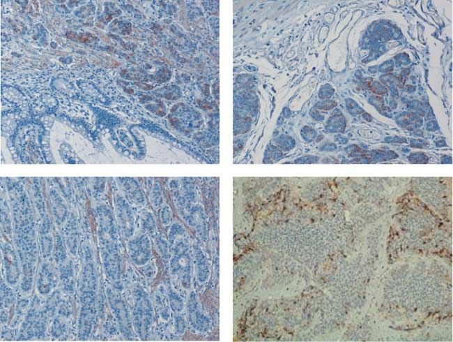
Overexpression of SHH in this series, Snail expression was found in all four
Intratumoral expression of the Hh ligand SHH was metastases, but only in two of four primaries. In case
detected in 27 out of 37 (73%) tumors studied (Table 2). number 30, the primary tumor was classified as ‘C’ for
As opposed to Snail, cells expressing SHH were found to snail by IHC, whereas the matched metastasis is ‘CC’.
be distributed more randomly throughout larger areas of Neither Snail nor SHH protein expression was
the tumors (Fig. 2A). A total of ten tumors did not show detected in non-malignant liver tissue adjacent to
any SHH-positive cells (Fig. 2C). Three of the seven metastatic foci.
(43%) liver metastases also expressed SHH (Fig. 2D). As
expected, SHH expression was also seen in normal
Suppression of E-cadherin expression by Snail
intestinal epithelium (Fig. 2B).
in NETs
SHH mRNA levels tended to be higher in samples that
were classified as positive by IHC, but these differences To examine the influence of Snail on E-cadherin
did not reach statistical significance, possibly due to the expression in NETs, immunofluorescence analysis for
Downloaded from Bioscientifica.com at 04/26/2020 05:10:14AM
via free access
868 www.endocrinology-journals.orgEndocrine-Related Cancer (2007) 14 865–874
Figure 2 IHC staining for SHH in tissue samples from human GI-NETs. Marked overexpression of SHH was seen in the majority of
studied samples (A), whereas some completely lacked expression of SHH (B). SHH expression was also detected in normal
intestinal epithelium (C). Two of four liver metastases also expressed SHH (D). (E) Steady-state SHH mRNA levels as revealed by
real-time RT-PCR. Results are presented as boxplots as described above.
Snail, E-cadherin, and b-catenin was performed in a Discussion
subset of eight randomly selected Snail-positive NETs
The GI tract is the predominant site of origin for NETs,
of the ileum (Fig. 3). E-cadherin was found to be
which were formerly called carcinoids, although they can
downregulated on Snail-expressing cells, while high
occur throughout the body. NETs are generally slow
levels of E-cadherin were expressed abundantly on the
growing but frequently metastasize to the liver, ranking
surfaces of cells lacking Snail expression (Fig. 3A and
second to colorectal carcinoma as a source of isolated
C). b-Catenin was expressed on the membranes of all
cells irrespective of presence or absence of Snail liver metastases (Chen et al. 1998). Molecular data on
protein and was used as control counterstaining in NETs of the GI tract have been accumulating in recent
areas with suppressed E-cadherin expression (Fig. 3B). years, but the genetic basis of endocrine tumor
development and progression is still poorly understood.
EMT occurs during embryonic morphogenesis in
Conservation of Hh pathway in BON-1 cells multicellular organisms, in which embryonic mesench-
The pancreatic carcinoid cell line BON-1 was used for ymal cells are formed and become motile following the
in vitro studies. Quantitative real-time RT-PCR analysis loss of epithelial cell polarity. In recent years, EMT has
revealed expression of the Hh pathway-related genes also been recognized as a potential mechanism for
Gli1, Gli2, and Ptch. Upon Hh inhibition with the small cancer progression (Thiery 2002). Cancer cells under-
molecule-smoothened antagonist cyclopamine, all of going EMT have lost specific target recognition and are
these target genes were found to be downregulated, in usually equipped with autocrine loops of growth
line with preexisting aberrant activation of the Hh signals, mechanisms to evade apoptosis, and the
pathway in this cell line. Interestingly, expression of potential to elicit angiogenesis for independent nutrient
the inhibitory transcription factor Gli3 was not altered supply (Gotzmann et al. 2004). A central event in EMT
upon Hh inhibition (Fig. 4A). is downregulation of E-cadherin that leads to the loss
Furthermore, we found that Hh inhibition with of cell–cell contact and the consecutive progression of
cyclopamine reduced BON1 cell growth in vitro in a the cells toward a malignant phenotype. The transcrip-
dose-dependent manner (Fig. 4B), further supporting our tion factor Snail is one major suppressor of E-cadherin
hypothesis of aberrant Hh activation in this tumor entity. and a strong inducer of EMT. Snail downregulates
Downloaded from Bioscientifica.com at 04/26/2020 05:10:14AM
via free access
www.endocrinology-journals.org 869870
V Fendrich et al.: Hh and Snail in neuroendocrine tumors
Table 2 Clinical characteristics and results of Snail and Sonic Hedgehog (SHH) immunohistochemistry in 33 patients with neuroendocrine tumors of the ileum
Patient no. Age (Years) Sex NET localization Histology Ki-67 (%)a LN Mtxb Liver Mtxc Snail1 expression SHH expression
1 57 M Liver metastasis ND ND ND Negative Negative
2 57 F Ileum carcinoid Well-differentiated ND ND ND CC CC
3 73 M Liver metastasis ND ND ND CC CC
4 61 M Ileum carcinoid Well-differentiated ND C C Negative Negative
5 77 F Ileum carcinoid Well-differentiated ND ND ND CC CC
6 56 M Ileum carcinoid Well-differentiated 30 C C C C
7 72 M Ileum carcinoid Well-differentiated 3 C C CC CC
8 52 M Ileum carcinoid Well-differentiated 1 C K CC CC
9 63 F Ileum carcinoid Well-differentiated 20 C C C CC
10 42 M Ileum carcinoid Well-differentiated 1 C C Negative C
11 68 M Ileum carcinoid Well-differentiated 1 C K CC C
12 91 M Ileum carcinoid Well-differentiated 1 C C Negative C
13 60 F Ileum carcinoid Well-differentiated 5 C C CC C
14 55 F Ileum carcinoid Well-differentiated 5 C C C CC
15 52 F Ileum carcinoid Well-differentiated 1 C C Negative C
16 66 F Liver metastasis 1 C C C Negative
17 57 M Ileum carcinoid Well-differentiated 5 C K Negative CC
18 66 M Ileum carcinoid Well-differentiated 3 C C C C
19 67 F Ileum carcinoid Well-differentiated 1 C C Negative Negative
20 66 F Ileum carcinoid Well-differentiated 2 C C Negative CC
Liver metastasis ND C Negative
21 62 M Ileum carcinoid Well-differentiated 17 C C Negative Negative
22 44 M Ileum carcinoid Well-differentiated 5 C C Negative Negative
23 43 F Ileum carcinoid Well-differentiated 5 C C C C
Liver metastasis ND C C
24 39 M Ileum carcinoid Well-differentiated 2 C C CC CC
25 79 M Ileum carcinoid Well-differentiated 1 C K C CC
26 67 F Ileum carcinoid Well-differentiated 5 C K Negative CC
27 70 M Ileum carcinoid Well-differentiated 1
Downloaded from Bioscientifica.com at 04/26/2020 05:10:14AM
C K CC CC
28 65 F Ileum carcinoid Well-differentiated 1 C C C CC
29 74 F Ileum carcinoid Well-differentiated ND ND ND Negative CC
30 40 M Ileum carcinoid Well-differentiated 4 C C C C
www.endocrinology-journals.org
Liver metastasis ND CC Negative
31 75 F Ileum carcinoid Well-differentiated 1 C K Negative Negative
32 91 M Ileum carcinoid Well-differentiated 10 C C Negative Negative
33 34 F Ileum carcinoid Well-differentiated 1 C C Negative C
Liver metastasis ND C C
ND, no data available.
a
Ki-67 proliferation index in percent.
b
Presence (C) or absence (K) of lymph node metastases at time of diagnosis.
c
Presence (C) or absence (K) of liver metastases at time of diagnosis.
via free accessEndocrine-Related Cancer (2007) 14 865–874
Figure 3 Immunofluorescence staining for (A) E-cadherin, (B) b-catenin, (C) Snail, and (D) merge. E-cadherin expression was
downregulated on cells, in which Snail was expressed (arrow), whereas E-cadherin expression was seen on the surface of cells that
did not stain for Snail (arrowhead). b-Catenin was expressed on surfaces of both Snail-positive and -negative cells. The figure shows
results from one representative of eight performed experiments.
E-cadherin in different types of tumors, e.g., hepato- 2005). Six of seven (86%) liver metastases analyzed in
cellular carcinomas (Jiao et al. 2002), carcinomas from our study were positive for Snail, possibly supporting
the esophagus, cardia, stomach (Rosivatz et al. 2006), our hypothesis that Snail might be involved in the
and colorectal carcinomas (Roy et al. 2005). development of metastases by promoting a more
Our study is the first to show that Snail is expressed invasive phenotype through EMT.
in NETs of the ileum, and to the best of our knowledge Our observation of lack of SHH and Snail expression
this is also the first report of Snail overexpression in an in non-malignant normal liver tissue is in line with
endocrine related cancer at all. We found significant previous reports by others (Sugimachi et al. 2003,
expression of Snail in more than 50% of the NETs we Sicklick et al. 2006), although the latter group detected
investigated. Our results are in line with a meta- Snail mRNA transcripts in adult hepatocytes by in situ
analysis from Zikusoka et al. (2005) of comparative hybridization.
genomic hybridization data, which found that in up to To verify that in GI-NETs expression of Snail is
50% of all carcinoids there is a gain on chromosome associated with loss of E-cadherin, we performed
20q13, the location of Snail. immunofluorescence analysis for Snail, E-cadherin,
As seen in tumors induced in the skin of mice (Cano and b-catenin. In fact, E-cadherin was found to be
et al. 2000), Snail-expressing cells were mostly found downregulated on Snail-expressing cells, while high
at the invasive front of the NETs. At this site, tumor levels of E-cadherin were expressed abundantly on the
cells migrate into and invade the surrounding tissue surfaces of cells lacking Snail expression (Fig. 3), a
either as single cells or in collective clusters. Figure 1 finding commonly related to Snail-induced EMT and
shows the Snail-positive invasive front of a NET of the metastasis (Huber et al. 2005).
ileum, with no Snail staining detectable in normal Genes of the Hh pathway, commonly involved in
intestinal epithelium. The presence of hepatic metas- embryonic patterning and stem cell maintenance in
tases in GI NETs has obvious implications in terms of adult tissues, are frequently mutated in basal cell
patients’ quality of life and overall prognosis. The carcinomas (Athar et al. 2006) and medulloblastomas
5-year survival rate in patients with hepatic metastases (Taylor et al. 2002). Moreover, recent studies by our
from midgut carcinoids is 0–50% as opposed to own group as well as by others described aberrant
75–99% in patients without liver metastases (Rorstad activation of Hh signaling in several malignancies of
Downloaded from Bioscientifica.com at 04/26/2020 05:10:14AM
via free access
www.endocrinology-journals.org 871V Fendrich et al.: Hh and Snail in neuroendocrine tumors
induced by Gli1 in vitro and in skin tumors. In line with
these results, we found expression of SHH and Snail in
53% of primary NETs. Eight primary NETs expressed
SHH without consecutive Snail expression, but no
primary tumor expressed Snail in the absence of SHH.
These results suggest that also in ileum NETs Snail is
induced as a downstream target of SHH or, likewise,
Gli1. Overall, we found expression of SHH in 27 out of
37 (73%) ileum-derived NET samples analyzed
(Table 2). SHH-positive cells were distributed through-
out larger areas of the tumors. As expected, SHH
expression was also seen in normal intestinal epi-
thelium (Nielsen et al. 2004).
Our in vivo findings are in line with the in vitro
expression of Hh-related genes Gli1, Gli2, Gli3, and
Ptch in BON-1 cells (Fig. 4). After treating this cell
line with cyclopamine, we found a significant down-
regulation of the Hh target genes Gli1, Gli2, and Ptch
at the mRNA level, as demonstrated by RT-PCR
(Fig. 4). Furthermore, we found that Hh inhibition with
cyclopamine reduced cell growth in vitro in a dose-
dependent manner (Fig. 4). Although to date cyclopa-
mine cannot be chemically synthesized but has to be
extracted from the corn lily Veratrum californicum
(Cooper et al. 1998) and is thus costly and difficult to
obtain in larger amounts for clinical use, considerable
research effort is currently undertaken to pharma-
cologically target the Hh-signaling pathway using new
small molecule inhibitors, and our results suggest that
the Hh pathway might constitute a potential therapeutic
Figure 4 (A) Expression of Gli1, Gli2, Gli3, and Ptch mRNA in
BON-1 cells treated with solvent only or cyclopamine. Upon
target in NETs. One such synthetic drug has recently
treatment with cyclopamine, RT-PCR revealed downregulation been shown to reduce medulloblastoma growth in a
of all Hh target genes tested. (B) Hh inhibition with cyclopamine transgenic mouse model (Romer et al. 2004).
lead to reduced proliferation of BON-1 cells in a dose-
dependent manner.
In conclusion, our data presented here show that the
sister pathways Snail and Hh are activated in a
considerable subset of GI-NETs, leading to down-
regulation of E-cadherin in line with EMT. Hh activation
the aerodigestive tract (Berman et al. 2003, Thayer
is observed in BON-1 cells and Hh inhibition with
et al. 2003), and particularly high Hh pathway activity
cyclopamine reduces NET cell growth in vitro.
has been found in metastatic lesions of prostate and
pancreatic cancers (Karhadkar et al. 2004, Feldmann
et al. 2007), suggesting that Hh signaling might play a Acknowledgements
role in metastatic tumor spread. Exogenous expression Cyclopamine was donated by Infinity Pharmaceuticals
of the Hh-dependent transcription factor Gli1 in low- (Cambridge, MA, USA). Many thanks to Savita Bisht for
metastatic rodent prostate carcinoma cells caused checking the manuscript and to Malte Buchholz for
enhanced migrationin vitro and development of performing real-time PCRs. This study was supported by
visceral metastases in vivo, correlating with induction the NCI grant R01CA113669 to A M. G F was supported
of Snail expression and E-cadherin repression. by a fellowship grant within the Postdoc-Program of the
All these effects were completely inhibited by German Academic Exchange Service (DAAD). V F was
treatment with the pathway inhibitor cyclopamine, supported by a Research Grant of the University Medical
showing that the Hh pathway indeed contributes to Center Giessen and Marburg. The authors declare that
EMT (Karhadkar et al. 2004). Recently, Li et al. there is no conflict of interest that would prejudice the
(2006) showed in an elegant study that Snail is rapidly impartiality of this scientific work.
Downloaded from Bioscientifica.com at 04/26/2020 05:10:14AM
via free access
872 www.endocrinology-journals.orgEndocrine-Related Cancer (2007) 14 865–874
References Li X, Deng W, Nail CD, Bailey SK, Kraus MH,
Ruppert JM & Lobo-Ruppert SM 2006 Snail
Apelqvist A, Ahlgren U & Edlund H 1997 Sonic Hedgehog induction is an early response to Gli1 that determines
directs specialised mesoderm differentiation in the
the efficiency of epithelial transformation. Oncogene
intestine and pancreas. Current Biology 7 801–804.
25 609–621.
Athar M, Tanq X, Lee JL, Kopelovich L & Kim AL 2006
Livak KJ & Schmittgen TD: 2001 Analysis of relative
Hedgehog signalling in skin development and cancer.
gene expression data using real-time quantitative PCR
Experimental Dermatology 15 667–677.
and the 2(KDeltaDelta C(T)) method. Methods 25
Berman DM, Karhadkar SS, Maitra A et al. 2003 Widespread
402–408.
requirement for hedgehog ligand stimulation in growth of
digestive tract tumours. Nature 425 846–851. Louro ID, Bailey EC, Li X, South LS, McKie-Bell PR,
Cano A, Pérez-Moreno MA, Rodrigo I, Locascio A, Blanco Yoder BK, Huang CC, Johnson MR, Hill AE, Johnson
MJ, del Barrio MG, Portillo F & Nieto MA 2000 The RL et al. 2002 Comparative gene expression profile
transcription factor snail controls epithelial-mesenchymal analysis of Gli1 and c-Myc in an epithelial model
transitions by repressing Ecadherin expression. Nature of malignant transformation. Cancer Research 62
Cell Biology 2 76–83. 5867–5873.
Chen H, Hardacre JM, Uzar A, Cameron JL & Choti MA 1998 Maggard MA, O’Connell JB & Ko CY 2004 Updated
Isolated liver metastases from neuroendocrine tumors: does population-based review of carcinoid tumors. Annals of
resection prolong survival? Journal of the American College Surgery 240 117–122.
of Surgeons 187 88–92. Moody SE, Perez D, Pan TC, Sarkisian CJ, Portocarrero CP,
Christofori G 2006 New signals from the invasive front. Sterner CJ, Notorfrancesco KL, Cardiff RD & Chodosh LA
Nature 44 444–450. 2005 The transcriptional repressor Snail promotes mam-
Cooper MK, Porter JA, Young KE & Beachy PA 1998 mary tumor recurrence. Cancer Cell 8 197–209.
Teratogen-mediated inhibition of target tissue response to Nielsen CM, Williams J, van den Brink GR, Lauwers GY &
SHH signaling. Science 280 1603–1607. Roberts DJ 2004 Hh pathway expression in human gut
Devereux TR, Anna CH, Foley JF, White CM, Sills RC & tissues and in inflammatory gut diseases. Laboratory
Barrett JC 1999 Mutation of beta-catenin is an early event Investigation 84 1631–1642.
in chemically induced mouse hepatocellular carcinogen- Parekh D, Ishizuka J, Townsend CM Jr, Haber B,
esis. Oncogene 18 4726–4733. Beauchamp RD, Karp G, Kim SW, Rajaraman S,
Esni F, Stoffers DA, Takeuchi T & Leach SD 2004 Origin of Greeley G Jr & Thompson JC 1994 Characterization
exocrine pancreatic cells from nestin-positive precursors of a human pancreatic carcinoid in vitro: morphology,
in developing mouse pancreas. Mechanism of Develop- amine and peptide storage, and secretion. Pancreas 9
ment 121 15–25. 83–90.
Feldmann G, Dhara S, Fendrich V, Bedja D, Beaty R, Perl AK, Wilgenbus P, Dahl U, Semb H & Christofori G
Mullendore M, Karikari C, Alvarez H, Iacobuzio-Donahue 1998 A causal role for E-cadherin in the transition from
C, Jimeno A et al. 2007 Blockade of hedgehog signaling adenoma to carcinoma. Nature 392 190–193.
inhibits pancreatic cancer invasion and metastases: a new Romer JT, Kimura H, Magdaleno S et al. 2004 Suppression of
paradigm for combination therapy in solid cancers. Cancer the SHH pathway using a small molecule inhibitor eliminates
Research 67 2187–2196.
medulloblastoma in Ptc1(C/K)p53(K/K) mice. Cancer
Gotzmann J, Mikula M, Eger A, Schulte-Hermann R, Foisner R,
Cell 6 229–240.
Beug H & Mikulits W 2004 Molecular aspects of epithelial
Rorstad O 2005 Prognostic indicators for carcinoid neuro-
cell plasticity: implications for local tumor invasion and
endocrine tumors of the gastrointestinal tract. Journal of
metastasis. Mutation Research 566 9–20.
Surgical Oncology 89 151–160.
Huber MA, Kraut N & Beuq H 2005 Molecular requirements
Rosivatz E, Becker KF, Kremmer E, Schott C, Blechschmidt
for epithelial-mesenchymal transition during tumor
K, Hofler H & Sarbia M 2006 Expression and nuclear
progression. Current Opinion in Cell Biology 17 548–558.
Jiao W, Miyazaki K & Kitajima Y 2002 Inverse correlation localization of Snail, an E-cadherin repressor, in
between E-cadherin and Snail expression in hepatocel- adenocarcinomas of the upper gastrointestinal tract.
lular carcinoma cell lines in vitro and in vivo. British Virchows Archiv 448 277–287.
Journal of Cancer 86 98–101. Roy HK, Smyrk TC, Koetsier J, Victor TA & Wali RK 2005
Karhadkar SS, Bova GS, Abdallah N, Dhara S, Gardner D, The transcriptional repressor Snail is overexpressed in
Maitra A, Isaacs JT, Berman DM & Beachy PA 2004 human colon cancer. Digestive Diseases and Sciences 50
Hedgehog signalling in prostate regeneration, neoplasia 42–46.
and metastasis. Nature 431 707–712. Sicklick JK, Li Y-X, Jayaraman A, Kannangai R, Qi Y,
Klöppel G, Perren A & Heitz PU 2004 The gastroenter- Vivekanandan P, Ludlow JW, Owzar K, Chen W,
opancreatic neuroendocrine cell system and its tumors: Torbenson MS et al. 2006 Dysregulation of the Hedgehog
the WHO classification. Annals of New York Academy of pathway in human hepatocarcinogenesis. Carcinogenesis
Sciences 1014 13–27. 27 748–757.
Downloaded from Bioscientifica.com at 04/26/2020 05:10:14AM
via free access
www.endocrinology-journals.org 873V Fendrich et al.: Hh and Snail in neuroendocrine tumors
Sugimachi K, Tanaka S, Kameyama T, Taguchi K, Aishima van den Brink GR, Hardwick JC, Tytgat GN, Brink MA,
S, Shimada M, Sugimachi K & Tsuneyoshi M 2003 Ten Kate FJ, Van Deventer SJ & Peppelenbosch MP 2001
Transcriptional repressor Snail and Progression of Human Sonic Hedgehog regulates gastric gland morphogenesis in
Hepatocellular Carcinoma. Clinical Cancer Research 9 man and mouse. Gastroenterology 121 317–328.
2657–2664. van den Brink GR, Hardwick JCH, Nielsen C, Xu C, ten Kate
Taylor MD, Liu L, Raffel C, Hui CC, Mainprize TG, Zhang X, FJ, Glickman J, van Deventer SJ, Roberts DJ &
Agatep R, Chiappa S, Gao L, Lowrance A et al. 2002 Peppelenbosch MP 2002 Sonic Hedgehog expression
Mutations in SUFU predispose to medulloblastoma. correlates with fundic gland differentiation in the adult
Nature Genetics 31 306–310. gastrointestinal tract. Gut 51 628–633.
Thayer SP, di Magliano MP, Heiser PW, Nielsen CM, Watkins DN, Berman DM, Burkholder SG, Wang B, Beachy
Roberts DJ, Lauwers GY, Qi YP, Gysin S, Fernandez-del PA & Baylin SB 2003 Hedgehog signalling within airway
Castillo C, Yajnik V et al. 2003 Hedgehog is an early and epithelial progenitors and in small-cell lung cancer.
late mediator of pancreatic cancer tumorigenesis. Nature Nature 422 313–317.
425 851–856. Zhou BP & Hung MC 2005 Wnt, hedgehog and snail: sister
Thiery JP 2002 Epithelial-mesenchymal transitions in tumour pathways that control by GSK-3beta and beta-Trcp in the
progression. Nature Review Cancer 2 442–454. regulation of metastasis. Cell Cycle 4 772–776.
Thiery JP & Sleeman JP 2006 Complex networks orchestrate Zikusoka MN, Kidd M, Eick G, Latich I & Modlin IM 2005
epithelial-mesenchymal transitions. Nature Review. Mol- The molecular genetics of gastroenteropancreatic neuro-
ecular and Cellular Biology 7 131–142. endocrine tumors. Cancer 104 2292–2309.
Downloaded from Bioscientifica.com at 04/26/2020 05:10:14AM
via free access
874 www.endocrinology-journals.orgYou can also read