Rethinking brain-wide interactions through multi-region "network of networks" models - OSF
←
→
Page content transcription
If your browser does not render page correctly, please read the page content below
Rethinking brain-wide interactions through multi-region “network of networks”
models
Matthew G. Perich1,*, Kanaka Rajan1,*
1
  Department of Neuroscience, Icahn School of Medicine at Mount Sinai, New York, NY, USA
* - correspondence: mperich@gmail.com, kanaka.rajan@mssm.edu

Abstract
The neural control of behavior is distributed across many functionally and anatomically distinct brain regions even in small
nervous systems. While classical neuroscience models treated these regions as a set of hierarchically isolated nodes, the
brain comprises a recurrently interconnected network in which each region is intimately modulated by many others.
Uncovering these interactions is now possible through experimental techniques that access large neural populations from
many brain regions simultaneously. Harnessing these large-scale datasets, however, requires new theoretical approaches.
Here, we review recent work to understand brain-wide interactions using multi-region "network of networks" models and
discuss how they can guide future experiments. We also emphasize the importance of multi-region recordings, and posit
that studying individual components in isolation will be insufficient to understand the neural basis of behavior.

Highlights
   ● Brains can be viewed as a composition of anatomically-distinct modules called regions
   ● Brain regions interact in complex ways to form a “network of networks”
   ● Untangling this network of networks could lead to new views of functional modularity

Introduction
Animal behavior - whether hunting, running, sleeping, or building a nest - arises from precisely coordinated activity
spanning the entire brain. Rather than operate as a uniform network of neurons, the brain organizes throughout development
into a large number of distinct, but interconnected, brain regions. Most animals demonstrate such anatomical modularity
within the brain [1], and neuroscience researchers seeking to understand fundamental computational principles of the brain
have thus adopted a similarly compartmentalized perspective (Figure 1a). In this view, “low level” sensory regions relay
information about the external world to “high level” regions coordinating behavior, that in turn instruct low-level output or
motor regions. However, such a simplified hierarchical view of the brain cannot account for the myriad, complex ways in
which different regions are connected (Figure 1b), with a large amount of recurrent connections between many low level
and high level brain regions. Due to this architecture, brain regions should not be considered as a discrete set of independent
nodes. Instead, they form a complex and heterogeneous “network of networks” to turn sensory information from the world
into thought and action. In the following sections, we consider the challenges this organization poses for studying and
understanding brain function, and highlight modeling approaches that can enable researchers to untangle the complex web
of multi-region interactions. We conclude that a multi-region view of the brain is critical to understand neural computation,
and that future experimental work should aim to record from numerous regions and diverse cell types, rather than denser
sampling of a spatially-localized population.

Understanding behavior generated by interacting brain regions
What are the implications of this highly recurrent organization for neuroscientists studying the neural basis of behavior? To
understand the computations performed by a given set of neurons, we must be able to observe not only the outputs of these
neurons, but also the inputs driving them. Many researchers historically aimed to characterize a brain region by recording
outputs (e.g., action potentials in V1) in response to known external inputs (e.g., gratings of light presented to the retina).
However, the complex connections between brain regions pose a problem; there are few, if any, cells in the brain that
respond solely to external, measurable stimuli. Most of the nervous system, even retinal cells [4], are modulated by
descending inputs related to brain states such as arousal [5]. Given the recurrent organization of the brain, each cell should
be viewed not just through the lens of external or environmental stimuli, but from the perspective of the other cells in the
brain that provide input.
Figure 1: A. A hierarchical model of the various sensory and motor brain regions underlying whisking in rodents. Adapted
from [2]. B. A detailed map of the known neural connections linking the brain regions underlying whisking in rodents. S2,
secondary somatosensory cortex; Clau, claustrum; NBM, nucleus basalis magnocellularis; VPM, medial venteroposterior
nucleus; Pom, medial posterior nucleus; LD, laterodorsal nucleus; RT, reticular nucleus; Amg, amygdala; PPTg,
pedunculopontine tegmental nucleus and the laterodorsal tegmental nucleus; TMN, tuberomammillary nucleus; LC, locus
coeruleus; GP, globus pallidus; EPN, entopeduncular nucleus; STN, subthalamic nucleus; SNc, substantia nigra pars
compacta; SNr, substantia nigra pars reticulata; IO, inferior olive; PN, pontine nucleus; NRTP, nucleus reticularis tegmenti
pontis; DMN, deep mesencephalic nucleus; DR, dorsal raphe nucleus; AMB, ambiguus nucleus; APT, anterior pretectal
nucleus; KF, Kölliker-Fuse nucleus and parabrachial complex; RN, red nucleus; PAG, periaqueductal gray; PrV, principal
trigeminal nucleus; SpVo, spinal trigeminal nucleus pars oralis; SpVi, spinal trigeminal nucleus pars interpolaris; SpVc,
spinal trigeminal nucleus pars caudalis; MeV, mesencephalic trigeminal nucleus; NXII, hypoglossal nucleus. Adapted from
[3].

Fully characterizing a neuron of interest requires knowledge of both its spiking output as well as the current state of the
surrounding cells. Studying individual regions of the brain in isolation will thus inevitably reach a limit. Fortunately, modern
experimental techniques enable unprecedented access to the brain through the ability to observe and manipulate thousands
of neurons simultaneously, often with cellular-level resolution [6]. These tools allow neuroscientists to extend beyond the
reference points of behavioral output or sensory input and study the activity of a particular brain region with respect to its
interactions with other brain regions. However, these large-scale datasets carry their own challenges. Most existing
approaches to study interactions between brain regions have addressed simple scenarios of two interacting regions [7–11].
As the number of recorded brain regions increases, the number of possible pairwise interactions between these regions
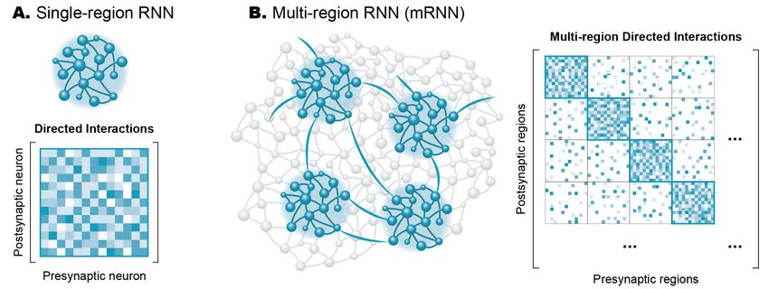
increases quadratically, even without considering the possibility of long-range or indirect interactions. To fully leverage the
power of modern experimental datasets, we require new theoretical and computational tools to quantitatively model the
interactions within the complex circuitry in the brain.

Recurrent neural network models of neural activity
The field of artificial neural networks emerged from neuroscience, attempting to mimic the computational power of the
brain. The units in these models are analogous to neurons, integrating information from many sources through weighted
connections, and output rules governed by nonlinearities. By studying a population of these units, an observer or readout
unit can extract information and make decisions about the nature of the inputs driving the network. Recurrent neural
networks (RNNs) are a special class of artificial neural network models where each unit both outputs to and receives input
from all other units in the network (Figure 2a). This connectivity is described by a weight matrix (which we refer to as the
“directed interaction” matrix) relating all presynaptic units to their postsynaptic targets.

Figure 2: A. Schematic of a recurrent neural network (top) and its associated directed interaction matrix (bottom) relating
each presynaptic unit in the columns to each postsynaptic unit in the rows. B. Schematic of a multi-region RNN where
four distinct brain regions are sampled out of all of the possible neurons in the brain. This mRNN can be summarized
using a multi-region directed interaction matrix summarizing both within-region (shown along the diagonal) and between-
region (shown on the off-diagonal) interactions.

RNN models of a single brain region have emerged as a powerful tool to study neural computation, elucidating potential
means by which neural circuits can generate the desired dynamics. Typically, chaotic RNNs are trained to produce desired
activity patterns based on a specific pattern of external input [12,13]. Single-region RNN models have been applied to
understand neural population dynamics underlying flexible timing of behavior [14–16], sequence generation [17], or the
movement of the arm for reaching [18]. Since the precise connectivity and dynamics of the model are known, researchers
can peek inside the “black box” that transforms the known inputs to the behavioral output to reverse-engineer the properties
of the network [19]. For example, analysis of the single-region RNNs can unveil latent dynamical structure such as attractors
[20] and fixed points [21]. This view provides a means to generate or test potential hypotheses to feedback into experimental
studies.

Recent work introduced a new class of data-driven RNN models [22]. Rather than treat the RNN as a black box to generate
some output, these models are directly constrained by experimentally-recorded neural data. During training, an initially
random connectivity matrix (J) is modified such that the time-varying activity of each RNN unit tracks a target experimental
neuron [22,23]. Like the single-region RNN models described above, these data-driven RNNs also provide access to a
critical component that is difficult or impossible to obtain experimentally: a "directed interaction" matrix (Figure 2a)
describing the functional weights linking pairs of neurons. Since the models are fit directly to observed time-series
recordings, they provide a unique means to analyze the dynamics underlying the experimental data. Data-driven RNN
models have been used to study cellular and synaptic mechanisms enabling short-term memory [24], as well as circuit
mechanisms of sequence propagation [22].

Scaling to multi-region "network of networks" models
RNNs also provide a platform to study how the structural modularity inherent in brain regions enables behavior. Beyond
single-region RNNs, more complex RNNs can be built with distinct submodules representing different brain regions. This
has given rise to a new class of models—multi-region RNNs (mRNNs)—that describes the activity of many brain regions
simultaneously. Such models have been applied to show the necessity of different cortical regions to generate perceptual
decisions [25,26], select motor plans [27,28] or actions [29] in response to visual cues, generate robust dynamics for motor
output [30], and to study interactions between cortical and subcortical regions [31,32]. The data-driven RNN models
described previously can also readily scale to model large, multi-region datasets. Rather than consider each region
individually, mRNNs fit a single model that describes interactions within the entire dataset simultaneously [23]. This
approach gives access to the directed interactions governing the dynamics both within each region and between all pairs of
regions. These interactions can be conceptualized as an extension of the directed interaction matrix, where within-region
connections are summarized along the diagonal of the matrix, and between-region interactions appear off-diagonal (Figure
2b).

Once trained, the mRNN models provide unique access to study the brain-wide interactions of real neural circuits. By
analyzing the mRNN connectivity matrices, researchers can infer the strength and characteristics of directed, functional
interactions within the circuit. Importantly, these matrices provide both a magnitude and a direction of interaction—the
asymmetry in the weight matrix provides unique weights for outgoing and incoming projections between any pair of brain
regions. For example, the model may capture dense, full-rank interactions within a given region due to the many short-range
connections, but identify sparser connections between two regions with a small number of highly weighted connections
(schematized in Figure 2B). An mRNN was recently applied to whole-brain recordings from larval zebrafish to identify
putative pathways between habenula and the raphe nucleus that could enable behavioral state changes in response to
inescapable stress [23].

Most regions throughout the brain are engaged when animals perform a task [33], yet the reasons for this widespread
activation are not fully understood. The mRNN models can be a powerful tool to understand whole-brain coordination of
behavior. For example, mRNN models can be used to identify which brain regions (or combined subcomponents of brain
regions) are necessary and sufficient to describe the neural dynamics relevant for a given behavior, i.e., the least complex
model. The models thus provide insight into which brain regions might be critically mediating a particular behavior and
which might be passive observers or responders. This approach is particularly intriguing as a means to explore comparative
evolution. For a similar class of behaviors, are the same regions or circuits engaged [34]? What mechanisms or patterns are
conserved across species or during development? The mRNN models can also be used to probe the experimental data
directly to test predictions for simulated lesion studies or other manipulations to the circuit that may be prohibitively
challenging or unethical to perform in behaving animals. Lastly, the views afforded by mRNN models can allow researchers
to go beyond anatomical designations into brain regions. By applying clustering algorithms to the directed interaction
matrices, for example, researchers may be able to uncover functional multi-region circuits or sub-circuits within a single
region that underlie the experimental data.

Interplay between theorists and experimentalists can improve mRNNs and guide future experiments
Besides describing existing experimental data, mRNN models can be powerful tools to generate testable hypotheses for
future experiments. By parsing the multi-region interactions inherent in a dataset, researchers can identify which regions or
pathways can best explain the behavioral output. Once identified, the causal relevance of these regions for behavior can be
tested with experimental tools such as optogenetic activation or ablation. Furthermore, the mRNN models can make
predictions for as-yet-unobserved pathways linking regions, whether direct or indirect. For example, in the larval zebrafish
model of behavioral state transitions discussed above, the mRNN model identified a change in interactions from the raphe
nucleus to the habenula for which no known anatomical link is known [23].
Data-driven mRNN models are directly constrained by experimental data. Thus, the quality and reliability of the models
depends on the amount and diversity of data available. Any experimentally measurable features can be leveraged to improve
mRNN models. Connectomics data can be used to build a prior into the mRNN about which pathways are most likely to be
directly connected [35–37]. Predetermined cell types could be incorporated into the model to constrain excitatory or
inhibitory projections [38,39]. Information about the release of neurotransmitters such as dopamine [40,41] could also be
incorporated to help account for long-range or slow-timescale signaling that simultaneously influences the activity of many
brain regions. Additionally, non-neuronal data such as glial recordings [42] or behavioral data [43,44] could be incorporated
into mRNN models to provide additional constraints and help account for common inputs between neurons [45].

While more data, whether neural or behavioral, is always advantageous, effectively modeling interactions between brain
regions requires sampling broadly across as many brain regions as possible. Experimental neuroscientists should thus seek
to sample more diverse populations from different regions in the brain, even if it requires more sparse sampling from each
region. Behaviorally-relevant neural dynamics in many brain regions are typically surprisingly low-dimensional [46,47],
and large-scale population dynamics can be accurately reconstructed even with sparse samples of the local population
[48,49]. Since behavior arises from coordinated activity of nearly all regions in the brain [33,50,51], we argue that recording
from many distinct regions - even with the inevitable tradeoff of smaller numbers of neurons per region - will provide more
insight than recording ever larger numbers of neurons from any single region.

Structural and functional modularity of multi-region circuits
Data-driven mRNNs seek to model the functional interactions between neurons, and thus bears some similarity to the field
of functional connectivity [45]. While mRNNs are subject to some of the same limitations, they also offer tangible
advantages to traditional approaches such as generalized linear models [7,52,53], Granger causality [54], or dynamical
causal modeling [55]. All of these methods provide correlational estimates of interactions and cannot accurately describe
causal mechanisms due to confounders like common input or recurrent connectivity [56]. However, recurrence is built into
the mRNN models, potentially allowing for more reasonable estimates of directed interactions when the data are
appropriately constrained. It is important to emphasize that in all of these methods "interactions" need not correspond to
direct synaptic connections. Functional interactions can arise through a multitude of indirect pathways including multi-
synapse relays, ephaptic coupling [57], or even through widespread neurotransmitter release [40]. Since data from
throughout the brain is fit directly, the mRNN models can meaningfully incorporate these effects during training.

The application of modern analytical tools such as mRNN models to large-scale neuroscientific datasets has the potential
to transform how we view neural computation. Brain regions captured by mRNN models reflect the structural modularity
of the brain. However, rather than focusing on compartmentalized, hierarchical, and discrete models of cognitive function
and behavior, we can leverage these models to develop new, more integrated views of how neural circuits interact to give
rise to behavior. Such models can help us to move beyond "brain regions" as the basis of neural function and instead view
the brain as a more complex "network of networks" comprising functional building blocks spanning numerous regions.
Brain-wide mRNN models can be a first step toward defining these new functional subdivisions.

References
1. Krubitzer LA, Seelke AMH: Cortical evolution in mammals: the bane and beauty of phenotypic variability.
    Proc Natl Acad Sci U S A 2012, 109 Suppl 1:10647–10654.
2. Circuits in the Rodent Brainstem that Control Whisking in Concert with Other Orofacial Motor Actions.
    Neuroscience 2018, 368:152–170.
3. Bosman LWJ, Houweling AR, Owens CB, Tanke N, Shevchouk OT, Rahmati N, Teunissen WHT, Ju C, Gong W,
    Koekkoek SKE, et al.: Anatomical pathways involved in generating and sensing rhythmic whisker movements.
    Front Integr Neurosci 2011, 5:53.
4. Schröder S, Steinmetz NA, Krumin M, Pachitariu M, Rizzi M, Lagnado L, Harris KD, Carandini M: Arousal
    Modulates Retinal Output. Neuron 2020, 107:487–495.e9.
5. Engel TA, Steinmetz NA: New perspectives on dimensionality and variability from large-scale cortical
    dynamics. Curr Opin Neurobiol 2019, 58:181–190.
6. Boyden ES, Zhang F, Bamberg E, Nagel G, Deisseroth K: Millisecond-timescale, genetically targeted optical
control of neural activity. Nat Neurosci 2005, 8:1263–1268.
7. Perich MG, Gallego JA, Miller LE: A Neural Population Mechanism for Rapid Learning. Neuron 2018, 100:964–
976.e7.
8. Semedo JD, Zandvakili A, Machens CK, Yu BM, Kohn A: Cortical Areas Interact through a Communication
Subspace. Neuron 2019, 102:249–259.e4.
9. Perich MG, Conti S, Badi M, Bogaard A, Barra B, Wurth S, Bloch J, Courtine G, Micera S, Capogrosso M, et al.:
Motor cortical dynamics are shaped by multiple distinct subspaces during naturalistic behavior. bioRxiv 2020,
doi:10.1101/2020.07.30.228767.
10. Kaufman MT, Churchland MM, Ryu SI, Shenoy KV: Cortical activity in the null space: permitting preparation
without movement. Nat Neurosci 2014, 17:440–448.
11. Veuthey TL, Derosier K, Kondapavulur S, Ganguly K: Single-trial cross-area neural population dynamics during
long-term skill learning. Nat Commun 2020, 11:4057.
12. Sussillo D, Abbott LF: Generating coherent patterns of activity from chaotic neural networks. Neuron 2009,
63:544–557.
13. DePasquale B, Cueva CJ, Rajan K, Escola GS, Abbott LF: full-FORCE: A target-based method for training
recurrent networks. PLoS One 2018, 13:e0191527.
14. Remington ED, Narain D, Hosseini EA, Jazayeri M: Flexible Sensorimotor Computations through Rapid
Reconfiguration of Cortical Dynamics. Neuron 2018, 98:1005–1019.e5.
15. Wang J, Narain D, Hosseini EA, Jazayeri M: Flexible timing by temporal scaling of cortical responses. Nat
Neurosci 2018, 21:102–110.
16. Sohn H, Narain D, Meirhaeghe N, Jazayeri M: Bayesian Computation through Cortical Latent Dynamics.
Neuron 2019, 103:934–947.e5.
17. Orhan AE, Ma WJ: A diverse range of factors affect the nature of neural representations underlying short-term
memory. Nat Neurosci 2019, 22:275–283.
18. Pandarinath C, O’Shea DJ, Collins J, Jozefowicz R, Stavisky SD, Kao JC, Trautmann EM, Kaufman MT, Ryu SI,
Hochberg LR, et al.: Inferring single-trial neural population dynamics using sequential auto-encoders. Nat
Methods 2018, 15:805–815.
19. Maheswaranathan N, Williams AH, Golub MD, Ganguli S, Sussillo D: Reverse engineering recurrent networks
for sentiment classification reveals line attractor dynamics. Adv Neural Inf Process Syst 2019, 32:15696–15705.
20. Laje R, Buonomano DV: Robust timing and motor patterns by taming chaos in recurrent neural networks. Nat
Neurosci 2013, 16:925–933.
21. Mante V, Sussillo D, Shenoy KV, Newsome WT: Context-dependent computation by recurrent dynamics in
prefrontal cortex. Nature 2013, 503:78–84.
22. Rajan K, Harvey CD, Tank DW: Recurrent Network Models of Sequence Generation and Memory. Neuron
2016, 90:128–142.
23. Andalman AS, Burns VM, Lovett-Barron M, Broxton M, Poole B, Yang SJ, Grosenick L, Lerner TN, Chen R,
Benster T, et al.: Neuronal Dynamics Regulating Brain and Behavioral State Transitions. Cell 2019, 177:970–
985.e20.
24. Fisher D, Olasagasti I, Tank DW, Aksay ERF, Goldman MS: A modeling framework for deriving the structural
and functional architecture of a short-term memory microcircuit. Neuron 2013, 79:987–1000.
25. Pinto L, Rajan K, DePasquale B, Thiberge SY, Tank DW, Brody CD: Task-Dependent Changes in the Large-
Scale Dynamics and Necessity of Cortical Regions. Neuron 2019, 104:810–824.e9.
26. Murray JD, Jaramillo J, Wang X-J: Working Memory and Decision-Making in a Frontoparietal Circuit Model.
The Journal of Neuroscience 2017, 37:12167–12186.
27. Michaels JA, Schaffelhofer S, Agudelo-Toro A, Scherberger H: A modular neural network model of grasp
movement generation. bioRxiv 2019, doi:10.1101/742189.
28. Kleinman M, Chandrasekaran C, Kao JC: Recurrent neural network models of multi-area computation
underlying decision-making. bioRxiv 2019, doi:10.1101/798553.
29. Márton CD, Schultz SR, Averbeck BB: Learning to select actions shapes recurrent dynamics in the
corticostriatal system. Neural Netw 2020, 132:375–393.
30. Li N, Daie K, Svoboda K, Druckmann S: Robust neuronal dynamics in premotor cortex during motor planning.Nature 2016, 532:459–464.
31. Jaramillo J, Mejias JF, Wang X-J: Engagement of Pulvino-cortical Feedforward and Feedback Pathways in
Cognitive Computations. Neuron 2019, 101:321–336.e9.
32. Lo C-C, Wang X-J: Cortico–basal ganglia circuit mechanism for a decision threshold in reaction time tasks.
Nature Neuroscience 2006, 9:956–963.
33. Steinmetz NA, Zatka-Haas P, Carandini M, Harris KD: Distributed coding of choice, action and engagement
across the mouse brain. Nature 2019, 576:266–273.
34. Lovett-Barron M, Andalman AS, Allen WE, Vesuna S, Kauvar I, Burns VM, Deisseroth K: Ancestral Circuits for
the Coordinated Modulation of Brain State. Cell 2017, 171:1411–1423.e17.
35. Abbott LF, Bock DD, Callaway EM, Denk W, Dulac C, Fairhall AL, Fiete I, Harris KM, Helmstaedter M, Jain V, et
al.: The Mind of a Mouse. Cell 2020, 182:1372–1376.
36. Scheffer LK, Xu CS, Januszewski M, Lu Z, Takemura S-Y, Hayworth KJ, Huang GB, Shinomiya K, Maitlin-
Shepard J, Berg S, et al.: A connectome and analysis of the adult central brain. Elife 2020, 9.
37. Turner NL, Macrina T, Alexander Bae J, Yang R, Wilson AM, Schneider-Mizell C, Lee K, Lu R, Wu J, Bodor AL,
et al.: Multiscale and multimodal reconstruction of cortical structure and function. bioRxiv 2020,
doi:10.1101/2020.10.14.338681.
38. Wilmes KA, Clopath C: Inhibitory microcircuits for top-down plasticity of sensory representations. Nature
communications 2019, 10.1 (2019): 1-10.
39. Bittner SR, Williamson RC, Snyder AC, Litwin-Kumar A, Doiron B, Chase SM, Smith MA, Yu BM: Population
activity structure of excitatory and inhibitory neurons. PLOS ONE 2017, 12:e0181773.
40. Tian J, Huang R, Cohen JY, Osakada F, Kobak D, Machens CK, Callaway EM, Uchida N, Watabe-Uchida M:
Distributed and Mixed Information in Monosynaptic Inputs to Dopamine Neurons. Neuron 2016, 91:1374–
1389.
41. Starkweather CK, Babayan BM, Uchida N, Gershman SJ: Dopamine reward prediction errors reflect hidden-
state inference across time. Nature Neuroscience 2017, 20:581–589.
42. Mu Y, Bennett DV, Rubinov M, Narayan S, Yang C-T, Tanimoto M, Mensh BD, Looger LL, Ahrens MB: Glia
Accumulate Evidence that Actions Are Futile and Suppress Unsuccessful Behavior. Cell 2019, 178:27–43.e19.
43. Berman GJ, Bialek W, Shaevitz JW: Predictability and hierarchy in Drosophila behavior. Proc Natl Acad Sci U S
A 2016, 113:11943–11948.
44. Calhoun AJ, Pillow JW, Murthy M: Author Correction: Unsupervised identification of the internal states that
shape natural behavior. Nat Neurosci 2020, 23:293.
45. Stevenson IH, Rebesco JM, Miller LE, Körding KP: Inferring functional connections between neurons. Curr
Opin Neurobiol 2008, 18:582–588.
46. Gallego JA, Perich MG, Miller LE, Solla SA: Neural Manifolds for the Control of Movement. Neuron 2017,
94:978–984.
47. Gao P, Ganguli S: On simplicity and complexity in the brave new world of large-scale neuroscience. Curr Opin
Neurobiol 2015, 32:148–155.
48. Gallego JA, Perich MG, Chowdhury RH, Solla SA, Miller LE: Long-term stability of cortical population
dynamics underlying consistent behavior. Nat Neurosci 2020, 23:260–270.
49. Trautmann EM, Stavisky SD, Lahiri S, Ames KC, Kaufman MT, Ryu SI, Ganguli S, Shenoy KV: Accurate
estimation of neural population dynamics without spike sorting. Neuron. 103.2 (2019): 292-308.
50. Musall S, Kaufman MT, Juavinett AL, Gluf S, Churchland AK: Single-trial neural dynamics are dominated by
richly varied movements. Nat Neurosci 2019, 22:1677–1686.
51. Stringer C, Pachitariu M, Steinmetz N, Reddy CB, Carandini M, Harris KD: Spontaneous behaviors drive
multidimensional, brainwide activity. Science 2019, 364:255.
52. Lawlor PN, Perich MG, Miller LE, Kording KP: Linear-nonlinear-time-warp-poisson models of neural activity. J
Comput Neurosci 2018, 45:173–191.
53. Stevenson IH, London BM, Oby ER, Sachs NA, Reimer J, Englitz B, David SV, Shamma SA, Blanche TJ, Mizuseki
K, et al.: Functional connectivity and tuning curves in populations of simultaneously recorded neurons. PLoS
Comput Biol 2012, 8:e1002775.
54. Seth AK, Barrett AB, Barnett L: Granger Causality Analysis in Neuroscience and Neuroimaging. Journal ofNeuroscience 2015, 35:3293–3297.
55. Razi A, Seghier ML, Zhou Y, McColgan P, Zeidman P, Park H-J, Sporns O, Rees G, Friston KJ: Large-scale DCMs
for resting-state fMRI. Netw Neurosci 2017, 1:222–241.
56. Das A, Fiete IR: Systematic errors in connectivity inferred from activity in strongly recurrent networks. Nat
Neurosci 2020, 23:1286–1296.
57. Han K-S, Chen CH, Khan MM, Guo C, Regehr WG: Climbing fiber synapses rapidly and transiently inhibit
neighboring Purkinje cells via ephaptic coupling. Nat Neurosci 2020, doi:10.1038/s41593-020-0701-z.You can also read