Biological Effects of Glucosinolate Degradation Products from Horseradish: A Horse that Wins the Race - MDPI
←
→
Page content transcription
If your browser does not render page correctly, please read the page content below
biomolecules

Article
Biological Effects of Glucosinolate Degradation
Products from Horseradish: A Horse that Wins
the Race
Marijana Popović 1, * , Ana Maravić 2 , Vedrana Čikeš Čulić 3 , Azra Đulović 1 ,
Franko Burčul 4 and Ivica Blažević 1, *
 1   Department of Organic Chemistry, Faculty of Chemistry and Technology, University of Split,
     Ruđera Boškovića 35, 21000 Split, Croatia; azra@ktf-split.hr
 2   Department of Biology, Faculty of Science, University of Split, Ruđera Boškovića 33, 21000 Split, Croatia;
     amaravic@pmfst.hr
 3   Department of Medical Chemistry and Biochemistry, School of Medicine, University of Split, Šoltanska 2,
     2100 Split, Croatia; vcikesc@mefst.hr
 4   Department of Analytical Chemistry, Faculty of Chemistry and Technology, University of Split,
     Ruđera Boškovića 35, 21000 Split, Croatia; franko@ktf-split.hr
 *   Correspondence: mpopovic@ktf-split.hr (M.P.); blazevic@ktf-split.hr (I.B.); Tel.: +385-21-329-434 (I.B.)

 Received: 27 December 2019; Accepted: 20 February 2020; Published: 21 February 2020

 Abstract: Horseradish degradation products, mainly isothiocyanates (ITC) and nitriles, along with
 their precursors glucosinolates, were characterized by GC-MS and UHPLC-MS/MS, respectively.
 Volatiles from horseradish leaves and roots were isolated using microwave assisted-distillation
 (MAD), microwave hydrodiffusion and gravity (MHG) and hydrodistillation (HD). Allyl ITC was
 predominant in the leaves regardless of the isolation method while MAD, MHG, and HD of the
 roots resulted in different yields of allyl ITC, 2-phenylethyl ITC, and their nitriles. The antimicrobial
 potential of roots volatiles and their main compounds was assessed against sixteen emerging food
 spoilage and opportunistic pathogens. The MHG isolate was the most active, inhibiting bacteria at
 minimal inhibitory concentrations (MICs) from only 3.75 to 30 µg/mL, and fungi at MIC50 between
Biomolecules 2020, 10, 343 2 of 15
Glucosinolates (GSLs) are major constituents of horseradish (Armoracia rusticana P.Gaertn., B.Mey.
& Scherb.), a large-leaved, hardy perennial plant of the Brassicaceae family [4,5]. Degradation
of GSLs generates different chemical structures: isothiocyanates (ITCs), thiocyanates, nitriles,
epithionitriles, and oxazolidinethiones [6,7]. GSL degradation may be initiated enzymatically,
thermally, and chemically [8]. Horseradish was studied by many authors and sinigrin (1) was
found as the most dominant GSL in all parts of the plant, followed by gluconasturtiin (5) and
glucobrassicin (6) [9–13], while the major volatiles were allyl- and 2-phenylethyl ITCs [14–16].
ITCs represent naturally occurring bioactive compounds with various known biological
activities [17]. They were previously found to exhibit broad-spectrum antibacterial potential [18,19],
even against multidrug-resistant clinical strains [20]. The latter study suggests ITCs as a potential
phytotherapeutic alternative to conventional antibiotics in the treatment of common infections, like
those of the urinary tract caused by antibiotic-resistant E. coli [20]. Regular consumption of Brassica
vegetables has also been associated with the reduced risk of cancer [21]. Chemopreventive actions
of ITCs are attributed to their ability to modulate different cellular mechanisms such as: phase I and
phase II drug metabolism enzymes, cell cycle arrest, apoptosis and differentiation, antioxidant and
detoxication proteins, as well as proinflammatory and procarcinogen factors [22]. Park et al. showed
that volatiles obtained by steam distillation of horseradish roots have good insecticidal activity [23].
Good anticandidal and antimicrobial activity of horseradish volatiles obtained with hydrodistillation
was revealed in the study by Petrović et al. [15]. Dekić et al. confirmed those findings, shedding a new
light on good cytotoxic and spasmolytic activities of horseradish volatiles obtained by autolysis [16].
Microwave hydrodiffusion and gravity (MHG) and microwave-assisted distillation (MAD) are new,
green extraction and distillation techniques that use microwave irradiation for evenly heating the
in-situ water of the plant material, inducing thermal degradation of GSLs [24–26]. MHG and MAD can
result in different chemical profiles or different yields compared to the conventional distillation and
extraction techniques [26,27].
The aim of this study was to evaluate the influence of different heating treatments on the GSLs
present in the horseradish leaves and roots during microwave irradiation (using MAD and MHG
techniques) and hydrodistillation (using Clevenger type apparatus, HD). GSLs were identified and
quantified by their desulfo counterparts using UHPLC-DAD-MS/MS, while the volatiles produced
after their thermal degradation were analyzed using GC-MS. In order to expand the knowledge
of beneficial effects of horseradish volatiles, biological activities of the volatile isolates and their
main compounds were investigated. Additionally, mixtures containing different ratios of the main
compounds were used to reveal their possible synergistic or antagonistic effects. The antimicrobial
effectiveness against a range of emerging food spoilage and opportunistic pathogens, among which
clinically isolated multidrug-resistant ESKAPE strains, was assessed by the microdilution assay.
Furthermore, the cytotoxic activity of horseradish volatiles and their main compounds was investigated
using the MTT method against human lung (A549) and human bladder (T24) cancer cell lines.
2. Materials and Methods
2.1. Plant Material and Standards
Horseradish (leaves and roots) was collected in March 2019 from the wild-growing population
in Konavle (near Dubrovnik, Croatia; 42◦ 340 53” N 18◦ 130 03” E) during the spring flowering
phenological stage. The voucher specimen (ZOKAR001) is deposited at the Department of Organic
Chemistry, Faculty of Chemistry and Technology, Split, Croatia. 2-Phenylethyl isothiocyanate (PEITC),
3-phenylpropanenitrile (PPCN), and allyl isothiocyanate (AITC) were purchased from Sigma Aldrich
(St. Louis, MO, USA).Biomolecules 2020, 10, 343 3 of 15
2.2. Analysis of Glucosinolates and Volatiles
2.2.1. Isolation of Desulfoglucosinolates
Isolation of desulfoglucosinolates (dGSLs) from 100 mg of dried horseradish roots and leaves
was performed as reported previously [28,29]. Plant material was firstly subjected to extraction in
MetOH/H2 O (70:30 v/v; Gram-Mol d.o.o, Zagreb, Croatia). The supernatant was loaded on mini-column
filled with DEAE-Sephadex A-25 anion-exchange resin (Sigma-Aldrich, St. Louis, MO, USA) and
the columns were then washed to remove the remaining non-polar compounds. To create optimal
conditions for the sulfatase reaction, mini columns were washed with 20 mM NaOAc buffer (Merck,
Darmstadt, Germany) followed by the addition of sulfatase (type H-1 from Helix pomatia; Sigma-Aldrich,
St. Louis, MO, USA). The reaction was left overnight and the dGSLs were eluted the next day with
ultrapure H2 O (Merck Millipore, Burlington, MA, USA).
2.2.2. HPLC-DAD Analysis of Desulfoglucosinolates
dGSLs were analyzed by high-performance liquid chromatography (Hewlett Packard 1090 Series II
UV-visible, Palo Alto, CA, USA) with binary gradient solvent delivery system, autoinjector, diode-array
detector (wavelength range 190–600 nm), and Nucleosil C-18 column (250 mm × 4 mm, 5 µm particle
size, Macherey-Nagel GmbH & Co. KG, Düren, Germany). The flow rate of 0.8 mL/min was applied
for solvent A (H2 O) and solvent B (acetonitrile:H2 O = 30:70 v/v) as followed: 0.5 min 96% A and 4% B,
28 min 14% A and 86% B, 4.0 min 14% A and 86% B, 2.0 min 5% A and 95% B, 13.0 min 5% A and 95%
B, 1.0 min 96% A and 4% B, and 8.0 min 96% A and 4% B. After each run the system was allowed to
equilibrate for 2 min. The column was set at room temperature (25 ◦ C). The signals were recorded at
the 227 nm by DAD detector. Quantification of dGSLs was performed using an external calibration
curve of pure desulfosinigrin (range from 13.56–542.50 µM). For each individual dGSL response factors
(RPF) was taken in accordance to the literature: RPF 1.0 for 1, 2 and 5 [30]; RPF 1.15 for 3, and 0.29 for
6 [31]; arbitrary 1.0 for 4.
2.2.3. UHPLC-MS/MS Analysis of Desulfoglucosinolates
The analysis was performed on UHPLC-DAD-MS/MS (Ultimate 3000RS with TSQ Quantis MS/MS
detector, Thermo Fischer Scientific, Waltham, MA, USA) using Hypersil GOLD column (3.0 µm,
3.0 × 100 mm, Thermo Fischer Scientific, USA). A gradient consisting of solvent A (50 µM NaCl in
H2 O) and solvent B (acetonitrile:H2 O 30:70 v/v) was applied at a flow rate of 0.5 mL/min as follows:
0.14 min 96% A and 4% B; 7.84 min 14% A and 86% B; 8.96 min 14% A and 86% B; 9.52 min 5% A and
95% B; 13.16 min 5% A and 95% B; 13.44 min 96% A and 4% B; 15.68 min 96% A and 4% B. The column
temperature was held at 40 ◦ C and the injection volume was 5 µL. The system was operated in the
positive ion electrospray mode and the electrospray interface was H-ESI operating with a capillary
voltage of 3.5 kV at 350 ◦ C.
2.2.4. Isolation of Volatiles
Microwave-assisted distillation was performed in the Ethos X microwave system (Milestone,
Brøndby, Denmark) for 30 min at 500 W. The plant material (287 g of the leaves and 374 g of the roots) was
partially immersed in water and transferred into a flask inside the microwave oven. Microwaves heated
the in situ water until the cell walls ruptured and the molecules of interest were released. The vapors
carried the volatile compounds through the coolant into the pentane trap, which retains volatiles.
Microwave hydrodiffusion and gravity was also performed in the Ethos X microwave system
for 10 min at 500 W. The plant material (516 g of the leaves and 493 g of the roots) was placed into a
microwave reactor without any solvent addition. The extracts passed through a cooler before reaching
the collection beaker. The volatiles from the aqueous solution were extracted with dichloromethane
(T.T.T. d.o.o., Sveta Nedelja, Croatia).Biomolecules 2020, 10, 343 4 of 15
Hydrodistillation (HD) of plant material (75.49 g of the leaves and 377 g of the roots) was performed
in the Clevenger apparatus (DeottoLab, Zagreb, Croatia) for 2.5 h, as reported previously [32].
2.2.5. GC-MS Analysis of Volatiles
Identification of volatiles from all the isolates was performed by GC-MS as previously described
on both non-polar VF-5MS and polar CP Wax 52 columns (Varian Inc., Lake Forest, CA, USA) with
slight modifications [33]. Briefly, the chromatographic conditions for both columns were as follows:
helium as carrier gas with the flow rate 1 mL/min−1 ; injector temperature 250 ◦ C; volume injected 1 µL
and the split ratio 1:50. For the VF-5MS column temperature was programmed at 60 ◦ C isothermal
for 3 min, increased to 246 ◦ C at a rate of 3 ◦ C min−1 and held isothermal for 25 min, while for CP
Wax 52 column the temperature was 70 ◦ C isothermal for 5 min, then increased to 240 ◦ C at a rate of
3 ◦ C/min−1 and held isothermal for 25 min. For the MS, ionization voltage was set to 70 eV, ion source
temperature at 200 ◦ C and the mass scan range was 40–350 mass units. The individual peaks were
identified by comparison of their retention indices and by computer matching of mass spectra against
the Wiley 7-spectra library database, along with the comparison of mass spectra from the literature
data [34–36]. All the analyses were run in duplicate and the mean values of component percentage
were obtained from both columns.
2.3. Antimicrobial Activity
2.3.1. Bacterial Strains
The antimicrobial effect of horseradish roots MHG, MAD, HD, and their main volatiles, among
them PEITC and PPCN, was assessed on sixteen strains of opportunistic pathogens, including the
emerging food spoilage microorganisms as well as clinical multidrug-resistant ESKAPE bacterial
strains. Susceptibility testing was carried on Gram-positive Listeria monocytogenes ATCC 19111,
Staphylococcus aureus (ATCC 29213 and a methicillin-resistant S. aureus clinical strain MRSA-1),
Enterococcus faecalis ATCC 29212, Streptococcus pyogenes ATCC 19612, and Bacillus cereus food isolate.
Gram-negative strains included Salmonella enterica serovar Typhimurium WDCM 00031, E. coli
(ATCC 25922 and an extended-spectrum-beta-lactamase (ESBL)-producing multiple-resistant clinical
strain), Klebsiella pneumoniae (ATCC 13883 and ESBL-producing multiple-resistant clinical strain),
and Acinetobacter baumannii (ATCC 19606 and a metallo-beta-lactamase-producing multiple-resistant
hospital strain). Multidrug-resistant clinical strains were obtained from the University Hospital
Centre Split, Croatia. Their origin and antibiotic resistance phenotypes were described previously [37].
Antifungal activity was estimated against an environmental isolate of opportunistic pathogenic yeast
Candida albicans and food isolates of food spoilage moulds Penicillium citrinum and Aspergillus niger.
Antibiotic susceptibility testing was carried out using Etest (AB Biodisk, Solna, Sweden) and VITEK
2 system (bioMérieux, Craponne, France). Microorganisms were stored at −80 ◦ C and subcultured
on tryptic soy agar (TSA, Biolife, Milan, Italy) or Sabouraud dextrose agar (SDA, Biolife, Milan, Italy)
before the testing.
2.3.2. Microdilution Assays
The antimicrobial activity was tested using a two-fold broth microdilution assay according to the
guidelines of the Clinical Laboratory Standards Institute [38,39] and the protocol was described in detail
previously [40]. Experiments were carried out in 96-well microtiter plates with serial two-fold dilutions
of horseradish roots extract, distillates, PEITC, and PPCN as follows: hydrodistillate (4 mg/mL) was
tested in a range from 800 to 0.78 µg/mL; MAD (2 mg/mL) in a range from 400 to 0.39 µg/mL; MHG
(600 µg/mL) in a range from 120 to 0.12 µg/mL; PEITC and PPCN (10 mg/mL) in a range from 2000
to 1.95 µg/mL, respectively. AITC susceptibility was previously tested [40]. The mixtures of three
predominant volatiles in horseradish, PEITC, PPCN, and AITC, mixed in different proportions were
also included in testing in order to evaluate synergistic effects. These three main volatiles (10 mg/mL)Biomolecules 2020, 10, 343 5 of 15
were mixed in ratio, 7:2:1, 4:4:2, and 3:2:5, respectively. For easier comparison, the mixed solutions
were then diluted to reach concentrations used for testing as previously noted. The MIC value was
recorded as the lowest concentration showing no visually detectable bacterial growth in the wells and
was the consensus value of the experiment performed in triplicate. For the determination of minimal
bactericidal concentration (MBC), bacteria from the wells corresponding to the MIC, 2 × MIC and
4 × MIC were plated, and the MBC was recorded as the lowest concentration causing ~99.9% killing of
the start inoculum.
2.4. Cytotoxic Activity
In order to determine cytotoxic activity of horseradish volatiles (PEITC,
PPCN, AITC), individual compounds and their 7:2:1 mixture, cell viability assay
(3-(4,5-dimethylthiazol-2-yl)-2,5-diphenyltetrazolium bromide, MTT) was performed as previously
described [40]. Human bladder cancer cell line T24 and human lung cancer cell line A549 (LGC
Standards) were incubated overnight in 96-well plates at a density of 5000 cells/well followed by
incubation with test substances (in triplicate) at concentrations of 1, 5, 10, 50, and 100 µg/mL for 72 h.
The cells were then incubated with 0.5 g MTT/L at 37 ◦ C for 2 h, followed by the removal of the medium
and addition of 10% dimethylsulfoxide (DMSO) for another 10 min at 37 ◦ C. Formazan formation, the
indicator of metabolically active cells, was measured at 570 nm on the microplate reader (BioSan,
Riga, Latvia). For statistical analyses t-test with unequal variances was performed using statistical
software GraphPad Prism 7.0 (San Diego, CA, USA) with the significance set at p < 0.05. The criteria
used to categorize the activity against the tested cell lines was based on IC50 values as follows:
501 µg/mL = inactive [41]. The calculation of IC50 values was performed with the GraphPad
Prism software version 7.0 (San Diego, CA, USA), normalizing the data by three independent
measurements of untreated controls. The combination index value (CI) was calculated in order to
quantify interactions between the compounds in a mixture as synergism (CI < 1) or antagonism
(CI > 1) using a median-effect analysis by CompuSyn software [42,43].
3. Results and Discussion
3.1. Chemical Characterization
Horseradish leaves and roots, collected from a wild-growing population, were analyzed
qualitatively and quantitatively for the presence of GSLs by their desulfo counterparts using HPLC-DAD
and UHPLC-MS/MS. The results are given in Table 1 and Figures S1 and S2 (Supplementary Materials).
Generally, six GSLs were identified by UV spectra, retention time (tR ) and mass spectra with commercial
standards and the literature. All the structures are given in Figure 1.
Table 1. Content of glucosinolates (µmol/g DW) in the roots and the leaves of horseradish.
Glucosinolate tR Roots Leaves
[M + Na]+
(Trivial Name) (min) (µmol/g DW) (µmol/g DW)
Aliphatic
Allyl GSL (1) *
2.0 3.53 ± 0.37 11.43 ± 0.26 302
(Sinigrin)
But-3-enyl GSL (2)
4.4 tr tr 316
(Gluconapin)
Pent-4-enyl GSL (3)
6.0 tr tr 330
(Glucobrassicanapin)
Branched
sec-Butyl GSL (4)
5.4 tr tr 318
(Glucocochlearin)Biomolecules 2020, 10, 343 6 of 15
Table 1. Cont.
Glucosinolate tR Roots Leaves
[M + Na]+
(Trivial Name) (min) (µmol/g DW) (µmol/g DW)
Arylaliphatic
2-Phenylethyl GSL (5)
7.6 7.21 ± 0.25 tr 366
(Gluconasturtiin)
Indole
Indol-3-yl GSL (6)
6.7 0.15 ± 0.08 tr 391
(Glucobrassicin)
Total (µmol/g DW) 10.89 ± 0.70 11.43 ± 0.26
[M + Na]+ ,
sodium adduct of desulfoglucosinolate; DW, dry weight; tr, trace amounts (99.9%). Next to 1, two other
Met derived GSLs, found in traces in all part plant parts, were gluconapin (2) and glucobrassicanapin
(3). Glucocochlearin (4), the only branched GSL, derived from Ile, was also detected in traces in all
plant parts.
The volatiles from horseradish leaves and roots were isolated by HD, MHG, and MAD. The profiles
and yields of the obtained volatiles are shown in Table 2. Regardless of the isolation method or part of
the plant, the main volatiles found in each sample originated from the degradation of 1 and 5 which
were the major GSLs (Table 1). Allyl ITC (AITC), but-3-enenitrile and allyl thiocyanate were identified as
degradation products of 1, while 2-phenylethyl ITC (PEITC) and 3-phenylpropanenitrile (PPCN) were
identified as degradation products of 5. Other volatiles, namely but-3-enyl-, pent-4-enyl- and sec-butyl
ITC, which originated from the degradation of 2, 3, and 4, respectively were also detected in traces.
Degradation volatiles of 6 were not found due to their thermal instability during GC-MS analysis.Biomolecules 2020, 10, 343 7 of 15
Table 2. Isothiocyanates and other volatiles identified in the roots and the leaves of horseradish
by GC-MS.
HD MAD MHG
Compound RI1 RI2
Roots Leaves Roots Leaves Roots Leaves
But-3-enenitrilea,b,c 1272 - 6.58 1.64 1.05 37.16 11.76 37.05
(E)-Hex-2-enal a,b,c 1311 - - 1.12 - 0.12 - 0.11
sec-Butyl isothiocyanate a,c 1360 936 1.04 4.31 0.17 2.76 0.04 0.10
(Z)-Pent-2-en-1-ol a,b,c 1393 - - 0.07 - - - 0.06
Allyl isothiocyanate (AITC) a,b,c 1429 879 46.36 73.45 14.29 54.77 13.81 52.36
(Z)-Hex-3-en-1-ol a,b,c 1452 862 0.30 5.22 0.03 0.74 0.15 1.65
(E)-Hex-2-en-1-ol a,b,c 1474 - - 0.22 - 0.04 - 0.02
Nonanal a,b,c 1481 - - - - 0.41 - -
Allyl thiocyanate a,c 1504 - 1.15 1.76 0.32 0.41 0.26 0.31
But-3-enyl isothiocyanate a,b,c 1514 992 0.51 0.20 0.10 0.08 0.04 0.04
Pent-4-enyl isothiocyanate a,c 1589 1094 0.24 0.32 0.20 0.04 0.04 0.02
Benzeneacetaldehyde a,b,c 1676 - - 0.12 - 0.37 - 0.19
2-Methoxy-3-(1-methylpropyl)pyrazine a,b - 1173 0.37 - 0.10 - 0.06 -
2-Phenylethyl alcohol a,b,c 1914 - - 0.03 - - 0.04 0.06
3-Phenylpropanenitrile (PPCN) a,b,c 2024 1248 15.44 1.69 18.61 0.49 34.44 0.23
Octanoic acid a,b,c 2056 - 0.01 0.15 0.24 0.25 - 0.00
Nonanoic acid a,b,c 2154 - 0.01 - 0.15 0.12 0.70 0.12
(E)-β-Ionone a,b,c - 1493 - - - 1.28 - -
2-Phenylethyl isothiocyanate (PEITC) a,b,c 2197 1513 27.61 2.81 62.82 0.29 30.53 0.07
Decanoic acid a,b,c 2254 - 0.01 0.96 0.15 0.12 0.83 0.77
Undecanoic acid a,b,c 2351 - - 0.57 0.10 0.04 0.44 0.41
Benzoic acid a,b,c 2371 - - - 0.03 0.04 0.13 0.14
Tridecanoic acid a,b,c 2561 - - 0.33 - - 0.13 0.14
Tetradecanoic acid a,b,c 2645 - - 0.74 - - 0.35 0.33
Pentadecanoic acid a,b,c 2744 - - 0.50 - - 0.46 0.40
Total sum (%) 99.66 96.18 98.36 99.55 94.22 94.60
Yield (ng/g) 66.34 138.96 35.43 3.96 7.57 3.39
Isothiocyanates (%) 75.76 81.09 77.58 57.94 44.46 52.59
Nitriles (%) 22.02 3.33 19.66 37.65 46.20 37.28
Others (%) 1.88 11.76 1.12 3.96 3.56 4.73
HD—hydrodistillation using Clevenger type apparatus; MAD—microwave-assisted distillation; MHG—microwave
hydrodiffusion and gravity; Retention indices (RI1 and RI2 ) determined on a CP Wax 52 and VF-5MS capillary
column, respectively; -, not detected; tr, traces (Biomolecules 2020, 10, 343 8 of 15
3.2. Biological Activities
3.2.1. Antimicrobial Effect of Horseradish and Its Main Volatiles
In order to assess the antimicrobial potential of horseradish, root volatiles obtained by HD, MAD,
and MHG, three main constituents (PEITC, PPCN, and AITC), as well as their mixtures in different
ratios were tested using microdilution assay on a range of spoilage and foodborne microorganisms.
The results are given in Tables 3 and 4. In our previous study, antimicrobial effects of L. latifolium
volatile isolates, as well as their main constituent AITC, were tested against a range of spoilage and
foodborne microorganisms [40].
The root MHG (mixed nitrile—ITC type constituents) extract proved to be the most active in
comparison to the tested distillates i.e., HD and MAD (ITC type constituents). MHG inhibited both
laboratory and clinical antibiotic-resistant bacterial strains at very low MIC values ranging from 3.75
to 30 µg/mL. The fungi were inhibited at even lower doses of MIC50 betweenBiomolecules 2020, 10, 343 9 of 15
Table 3. Antimicrobial activity of horseradish (Armoracia rusticana) roots extract, distillates by microdilution assay a .
HD MAD MHG
Species Strain Origin Agent c
MIC MBC b MIC MBC MIC MBC
Gram-positive bacteria
Listeria monocytogenes ATCC 19111 50 200 25 100 7.5 30 ≤1 (S)
Staphylococcus aureus ATCC 29213 25 >100 25 >100 30 >120 0.25 (S)
Staphylococcus aureus Clinical/MRSA 50 >200 12.5 >50 3.75 >15 ≥16 (R)
Enterococcus faecalis ATCC 29212 50 200 50 200 15 60 ≤1 (S)
Streptococcus pyogenes ATCC 19615 50 200 50 200 15 60 ≤1 (S)
Bacillus cereus Food 50 50 25 25 15 15 ≤1 (S)
Gram-negative bacteria
Salmonella Typhimurium WDCM 00031 50 100 25 50 15 30 ≤1 (S)
Escherichia coli ATCC 25922 50 50 25 50 15 30 0.5 (S)
Escherichia coli Clinical 100 200 50 100 15 30 ≤1 (S)
Klebsiella pneumoniae ATCC 13883 100 200 100 100 30 30 0.12 (S)
Klebsiella pneumoniae Clinical 200 400 100 100 30 30 ≥16 (R)
Acinetobacter baumannii ATCC 19606 50 100 25 50 7.5 15 1 (S)
Acinetobacter baumannii Clinical 50 100 25 50 7.5 15 ≥16 (R)
Fungi MIC50 MIC90 MIC50 MIC90 MIC50 MIC90 MIC90
Candida albicans EnvironmentalBiomolecules 2020, 10, 343 10 of 15
Table 4. Antimicrobial activities of 2-phenylethyl ITC (PEITC) and 3-phenylpropanenitrile (PPCN), and mixed solutions of PEITC, PPCN and allyl ITC (AITC), in
different ratios ΨPEITC:PPCN:AITC against tested strains by microdilution assay a .
PEITC PPCN Ψ7:2:1 c Ψ4:4:2 Ψ2:3:5
Species Strain Origin Agent d
MIC MBC b MIC MBC MIC MBC MIC MBC MIC MBC
Gram-positive
bacteria
Listeria monocytogenes ATCC 19111 125 500 125 1000 12.5 50 7.5 30 25 100 ≤1 (S)
Staphylococcus aureus ATCC 29213 31.25 >125 500 2000 25 >100 30 >120 25 >100 0.25 (S)
Staphylococcus aureus Clinical/MRSA 15.6 62.5 500 2000 25 50 7.5 30 50 100 ≥16 (R)
Enterococcus faecalis ATCC 29212 125 500 500 2000 12.5 12.5 15 60 50 200 ≤1 (S)
Streptococcus pyogenes ATCC 19615 62.5 250 500 2000 25 100 15 60 50 200 ≤1 (S)
Bacillus cereus Food 62.5 62.5 500 1000 25 25 15 15 50 50 ≤1 (S)
Gram-negative
bacteria
Salmonella
WDCM 00031 62.5 125 500 1000 25 50 15 30 50 100 ≤1 (S)
Typhimurium
Escherichia coli ATCC 25922 15.6 31.25 500 1000 25 25 30 60 50 50 0.5 (S)
Escherichia coli Clinical 250 500 500 1000 50 100 15 30 100 200 ≤1 (S)
Klebsiella pneumoniae ATCC 13883 250 250 500 1000 50 100 30 30 100 200 0.12 (S)
Klebsiella pneumoniae Clinical 500 500 500 1000 100 100 30 30 200 400 ≥16 (R)
Acinetobacter
ATCC 19606 125 250 250 500 25 50 7.5 15 50 100 1 (S)
baumannii
Acinetobacter
Clinical 31.25 31.25 125 500 12.5 25 7.5 15 25 50 ≥16 (R)
baumannii
Fungi MIC50 MIC90 MIC50 MIC90 MIC50 MIC90 MIC50 MIC90 MIC50 MIC90 MIC90
Candida albicans EnvironmentalBiomolecules 2020, 10, 343 11 of 15
3.2.2. Cytotoxic Activity of Horseradish and Its Main Volatiles
Cytotoxic activity of horseradish leaves and roots volatiles, individual compounds and their
mixture was tested against human lung (A549) and bladder (T24) cancer cell lines. The results for
tested concentrations after 72 h incubation period are shown in Figures 2 and 3, while calculated IC50
values are given in Table S1. A549 was more resistant than T24 cell line, however, the results show
that all horseradish volatiles obtained by different types of isolation have very high cytotoxic activity,
which is consistent with the previous study [41].
Figure 2. Percentage of metabolically active human lung carcinoma cell A549 and bladder cancer cell
T24 lines after 72 h of incubation with different concentrations of volatiles obtained by HD, MAD,
and MHG from the leaves and the roots of horseradish. Calculated IC50 values (µg/mL) are given in
Table S1. Each data point is presented as mean ± SD (n = 3). Lower case letters represent significance
level in comparison to non-treated cell line samples (a, p < 0.001; b, p < 0.01; c, p < 0.05).
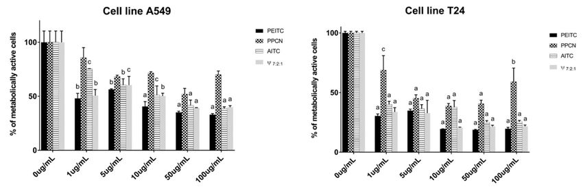
Figure 3. Percentage of metabolically active human lung carcinoma cell A549 and bladder cancer
cell T24 lines after 72h of incubation with the main compounds obtained from horseradish isolates
(2-phenylethyl ITC, 3-phenylpropanenitrile and allyl ITC) and their mixture (7:2:1, respectively).
Calculated IC50 values (µg/mL) are given in Table S1. Each data point is presented as mean ± SD
(n = 3). Lower case letters represent significance level in comparison to non-treated cell line samples
(a, p < 0.001; b, p < 0.01; c, p < 0.05).
The volatiles from the roots, in almost all cases, were more active than the ones from the leaves for
both cell lines (Figure 2, Table S1). HD and MAD from the roots showed the highest activity on A549
having IC50 values of 2.62 and 4.08 µg/mL, respectively. In difference to the root volatiles which areBiomolecules 2020, 10, 343 12 of 15
represented by degradation products originating from 1 and 5, leaves volatiles are represented only by
degradation products of 1. MHG from the leaves showed high cytotoxic activity, while MAD and HD
showed moderate cytotoxic activity with IC50 values of 11.63, 23.47 and 34.22 µg/mL, respectively.
Even better activity was observed when testing leaves and roots volatile isolates against human
bladder cancer cell line (T24). HD, MAD, MHG of all samples largely suppressed cell viability, with IC50
values of 0.57, 0.48, and 1.14 µg/mL, respectively for roots isolates and 7.87, 4.77, and 3.13 µg/mL,
respectively for leaves isolates.
In order to investigate the contribution of the main constituents of the volatiles to the observed
cytotoxic activity, PEITC, PPCN, and AITC were tested individually by MTT test (Figure 3, Table S1).
PEITC showed the highest cytotoxic activity for both cell lines. When comparing the cytotoxic activity
of PEITC, PPCN, and AITC against A549 cell line, PEITC and AITC showed high activity having IC50
values of 6.27 µg/mL and 17.76 µg/mL, respectively. On the other hand PPCN did not reach IC50
value. Thus it can be suggested that the observed root volatiles activities can be attributed to the high
percentages of PEITC and AITC.
To investigate the contribution of AITC and PPCN to the PEITC activity, we tested the mixture of
PEITC:PPCN:AITC in 7:2:1 ratio. This mixture having IC50 of 12.96 µg/mL was two times weaker than
PEITC alone. In order to quantitatively determine interactions between the components a combination
index (CI) method was used and calculated with CompuSyn software. The obtained CI value of 2.39
indicated antagonism of these three components.
PEITC showed the best activity against T24 cell line as well, with IC50 of 0.84 µg/mL, followed
by AITC and PPCN, both showing high activity, with IC50 values of 1.96 µg/mL and 6.52 µg/mL,
respectively. These results suggest that the observed high activities of the volatile isolates can be
attributed to the presence of all three components. The roots volatile isolates, having PEITC as major
constituent, showed better or very similar activity to PEITC indicating some type of interaction between
its constituents (Figure 2 and Table S1). Mixture of PEITC:PPCN:AITC in 7:2:1 ratio was tested as
well showing similar activity after 72 h incubation period (IC50 = 0.95 µg/mL) to PEITC itself. These
observations indicate positive synergistic effect of the main constituents obtained from horseradish
volatile isolates. CI value for T24 cell lines using the same mixture combination was calculated to be
0.61 which indicates synergism.
Tang et al. found that AITC and PEITC induce NAD(P)H:quinone oxidoreductase 1 and elevate
glutathione levels in human bladder cancer cells. They also affect cell-cycle progression; AITC blocks
cells in the G2/M phase while PEITC arrests cells in both G2/M and S phase. PEITC is more potent
than AITC in the induction of apoptosis through the cellular activity of caspase 3/7 [45]. A study
by Tripathi et al. showed that AITC induces replication stress-associated DNA damage response and
slows cell cycle progression through S phase in human lung cancer cells [46], while another study by
Kuang et al. discovered that PEITC induces apoptosis through elevated expression of P53 and P21 or
necrosis, in concentration-dependent manner [47].
4. Conclusions
Different isolation techniques of horseradish roots and leaves isolates resulted in different yields
of AITC, PEITC, and corresponding nitriles in the roots; whilst in the leaves, AITC was predominant
regardless of the isolation method. Horseradish extract and distillates effectively inhibited a range of
opportunistic pathogens, including the emerging food spoilage microorganisms as well as hospital
multidrug-resistant ESKAPE strains. MHG proved to be the most active, inhibiting growth of MRSA,
L. monocytogenes, clinical A. baumannii and fungi. The volatile susceptibility testing clearly showed that
the antimicrobial activity of horseradish roots extract and distillates arises from the combined activity
of its three main volatiles, among which the PEITC was the most active. PEITC was also shown to be
the most contributing to the cytotoxic activity against two cell lines.
Over the past decade, intensive research has been devoted to ITCs as they are recognized
to be responsible for various biological activities (anticancer, antimicrobial, etc.). Their activity isBiomolecules 2020, 10, 343 13 of 15
usually investigated using pure compounds or as a part of volatile mixtures isolated from plants that
commonly contain 2-3 major GSLs as their precursors. On the other hand, the synergistic effects of
their combinations are scarcely studied. Thus, in order to better understand the mechanisms involved
in ITCs interactions, more detailed studies that comprise a higher number of ITCs and their different
proportions in the mixtures are necessary.
Supplementary Materials: The following are available online at http://www.mdpi.com/2218-273X/10/2/343/s1,
Figure S1: Chromatogram of desulfoglucosinolates obtained from the roots and the leaves of horseradish:
d1—desulfosinigrin; d2—desulfogluconapin; d3—desulfoglucobrassicanapin; d4—desulfoglucocochlearin;
—desulfogluconasturtiin; d6—desulfoglucobrassicin, Figure S2: UV-Vis and MS2 spectra at 15V ionization
of 3 main desulfoglucosinolates detected: d1, d5, and d6, Table S1: Calculated IC50 values (µg/mL) for volatiles
obtained by HD, MAD and MHG from the roots and the leaves of horseradish and its main compounds
2-phenylethyl ITC, 3-phenylpropanenitrile, allyl ITC, and their mixture in the proportion similar to the one
obtained by root MAD, 7:2:1, respectively (Ψ7:2:1 ) against human lung cancer cell A549 and bladder cancer cell
T24 lines after 72 h.
Author Contributions: Chemistry investigation: M.P., A.Đ., F.B. and I.B.; Antimicrobial assay: A.M.; Cell culture
and cytotoxic assays: V.Č.Č.; Writing–original draft: M.P., A.M. and I.B.; Writing–review & editing: all authors.
All authors have read and agreed to the published version of the manuscript.
Funding: This research has been fully supported by the Croatian Science Foundation (Grant IP-2016-06-1316).
Acknowledgments: We are also thankful for the scientific-research equipment financed by EU grant “Functional
integration of the University of Split, PMF-ST, PFST and KTFST through the development of the scientific and
research infrastructure” (KK.01.1.1.02.0018).
Conflicts of Interest: The authors declare no conflict of interest.
References
1. Saladino, F.; Bordin, K.; Luciano, F.B.; Franzón, M.F.; Mañes, J.; Meca, G. Antimicrobial activity of the
glucosinolates. In Glucosinolates; Mérillon, J.-M., Ramawat, K.G., Eds.; Springer International Publishing:
Cham, Switzerland, 2017; pp. 249–274.
2. Mazarakis, N.; Snibson, K.; Licciardi, P.V.; Karagiannis, T.C. The potential use of l-sulforaphane for the
treatment of chronic inflammatory diseases: A review of the clinical evidence. Clin. Nutr. 2019, in press.
[CrossRef] [PubMed]
3. Pendleton, J.N.; Gorman, S.P.; Gilmore, B.F. Clinical relevance of the eskape pathogens. Expert Rev. Anti.
Infect. Ther. 2013, 11, 297–308. [CrossRef] [PubMed]
4. Weber, W.W. Seed production in horseradish. J. Hered. 1949, 40, 223–227. [CrossRef]
5. Blažević, I.; Montaut, S.; Burčul, F.; Olsen, C.E.; Burow, M.; Rollin, P.; Agerbirk, N. Glucosinolate structural
diversity, identification, chemical synthesis and metabolism in plants. Phytochemistry 2020, 169, 112100.
[CrossRef]
6. Cole, R.A. Isothiocyanates, nitriles and thiocyanates as products of autolysis of glucosinolates in cruciferae.
Phytochemistry 1976, 15, 759–762. [CrossRef]
7. Hochkoeppler, A.; Palmieri, S. Kinetic properties of myrosinase in hydrated reverse micelles. Biotechnol. Progr.
1992, 8, 91–96. [CrossRef]
8. Blažević, I.; Maleš, T.; Ruščić, M. Glucosinolates of Lunaria annua: Thermal, enzymatic, and chemical
degradation. Chem. Nat. Comp. 2014, 49, 1154–1157. [CrossRef]
9. Wedelsbäck Bladh, K.; Olsson, K.; Yndgaard, F. Evaluation of glucosinolates in nordic horseradish
(Armoracia rusticana). Bot. Lith. 2013, 19, 48–56. [CrossRef]
10. Agneta, R.; Lelario, F.; De Maria, S.; Mollers, C.; Bufo, S.A.; Rivelli, A.R. Glucosinolate profile and distribution
among plant tissues and phenological stages of field-grown horseradish. Phytochemistry 2014, 106, 178–187.
[CrossRef]
11. Ciska, E.; Horbowicz, M.; Rogowska, M.; Kosson, R.; Drabińska, N.; Honke, J. Evaluation of seasonal
variations in the glucosinolate content in leaves and roots of four european horseradish (Armoracia rusticana)
landraces. Pol. J. Food Nutr. Sci. 2017, 67, 301–308. [CrossRef]Biomolecules 2020, 10, 343 14 of 15
12. Agneta, R.; Möllers, C.; Rivelli, A.R.; Evolution, C. Horseradish (Armoracia rusticana), a neglected medical
and condiment species with a relevant glucosinolate profile: A review. Genet. Resour. Crop Evol. 2013,
60, 1923–1943. [CrossRef]
13. Grob, K.; Matile, P. Capillary GC of glucosinolate-derived horseradish constituents. Phytochemistry 1980,
19, 1789–1793. [CrossRef]
14. Masuda, H.; Harada, Y.; Tanaka, K.; Nakajima, M.; Tabeta, H. Characteristic odorants of wasabi
(Wasabia japonica matum), japanese horseradish, in comparison with those of horseradish (Armoracia Rusticana).
ACS Sym Ser. Biotech. Impr. Food Flav. 1996, 637, 67–78.
15. Petrović, S.; Drobac, M.; Ušjak, L.J.; Filipović, V.; Milenković, M.; Niketić, M. Volatiles of roots of wild-growing
and cultivated Armoracia macrocarpa and their antimicrobial activity, in comparison to horseradish, A. rusticana.
Ind. Crops Prod. 2017, 109, 398–403. [CrossRef]
16. Dekić, M.S.; Radulović, N.S.; Stojanović, N.M.; Ranđelović, P.J.; Stojanović-Radić, Z.Z.; Najman, S.;
Stojanović, S. Spasmolytic, antimicrobial and cytotoxic activities of 5-phenylpentyl isothiocyanate, a new
glucosinolate autolysis product from horseradish (Armoracia rusticana P. Gaertn., B. Mey. & Scherb.,
Brassicaceae). Food Chem. 2017, 232, 329–339. [PubMed]
17. Hayes, J.D.; Kelleher, M.O.; Eggleston, I.M. The cancer chemopreventive actions of phytochemicals derived
from glucosinolates. Eur. J. Nutr. 2008, 47, 73–88. [CrossRef]
18. Lin, C.M.; Kim, J.; Du, W.X.; Wei, C.I. Bactericidal activity of isothiocyanate against pathogens on fresh
produce. J. Food Protect. 2000, 63, 25–30. [CrossRef] [PubMed]
19. Wilson, A.E.; Martine, B.; Bindler, F.; Marchioni, E.; Lintz, A.; Ennahar, S. In vitro efficacies of various
isothiocyanates from cruciferous vegetables as antimicrobial agents against foodborne pathogens and
spoilage bacteria. Food Control 2013, 318–324. [CrossRef]
20. Mutters, N.T.; Mampel, A.; Kropidlowski, R.; Biehler, K.; Gunther, F.; Balu, I.; Malek, V.; Frank, U. Treating
urinary tract infections due to MDR E. coli with isothiocyanates—A phytotherapeutic alternative to antibiotics?
Fitoterapia 2018, 129, 237–240. [CrossRef]
21. Van Poppel, G.; Verhoeven, D.T.; Verhagen, H.; Goldbohm, R.A. Brassica vegetables and cancer prevention.
In Advances in Nutrition and Cancer 2; Springer: Boston, MA, USA, 1999; pp. 159–168.
22. Conaway, C.; Yang, Y.M.; Chung, F.L. Isothiocyanates as cancer chemopreventive agents: Their biological
activities and metabolism in rodents and humans. Curr. Drug Metab. 2002, 3, 233–255. [CrossRef]
23. Park, I.K.; Choi, K.S.; Kim, D.H.; Choi, I.H.; Kim, L.S.; Bak, W.C.; Choi, J.W.; Shin, S.C. Fumigant activity of
plant essential oils and components from horseradish (Armoracia rusticana), anise (Pimpinella anisum) and
garlic (Allium sativum) oils against Lycoriella ingenua (Diptera: Sciaridae). Pest Manag. Sci. 2006, 62, 723–728.
[CrossRef] [PubMed]
24. Chemat, F.; Vian, M.A.; Cravotto, G. Green extraction of natural products: Concept and principles. Int. J.
Mol. Sci. 2012, 13, 8615–8627. [CrossRef] [PubMed]
25. Vian, M.A.; Fernandez, X.; Visinoni, F.; Chemat, F. Microwave hydrodiffusion and gravity, a new technique
for extraction of essential oils. J. Chromatogr. A 2008, 1190, 14–17. [CrossRef]
26. Mohamadi, M.; Shamspur, T.; Mostafavi, A. Comparison of microwave-assisted distillation and conventional
hydrodistillation in the essential oil extraction of flowers Rosa damascena Mill. J. Essent. Oil Res. 2013,
25, 55–61. [CrossRef]
27. Dahmoune, F.; Spigno, G.; Moussi, K.; Remini, H.; Cherbal, A.; Madani, K. Pistacia lentiscus leaves as a source
of phenolic compounds: Microwave-assisted extraction optimized and compared with ultrasound-assisted
and conventional solvent extraction. Ind. Crops Prod. 2014, 61, 31–40. [CrossRef]
28. Blažević, I.; Đulović, A.; Čikeš Čulić, V.; Burčul, F.; Ljubenkov, I.; Ruščić, M.; Generalić Mekinić, I.
Bunias erucago L.: Glucosinolate profile and in vitro biological potential. Molecules 2019, 24, 741. [CrossRef]
[PubMed]
29. Grosser, K.; van Dam, N.M. A straightforward method for glucosinolate extraction and analysis with
high-pressure liquid chromatography (HPLC). J. Vis. Exp. 2017, 121, e55425. [CrossRef]
30. Brown, P.D.; Tokuhisa, J.G.; Reichelt, M.; Gershenzon, J. Variation of glucosinolate accumulation among
different organs and developmental stages of Arabidopsis thaliana. Phytochemistry 2003, 62, 471–481. [CrossRef]
31. Wathelet, J.P.; Iori, R.; Leoni, O.; Quinsac, A.; Palmieri, S. Guidelines for glucosinolate analysis in green
tissues used for biofumigation. Agroindustria 2004, 3, 257–266.Biomolecules 2020, 10, 343 15 of 15
32. Blažević, I.; Mastelić, J. Free and bound volatiles of rocket (Eruca sativa Mill.). Flavour Frag. J. 2008, 23, 278–285.
[CrossRef]
33. Bezić, N.; Vuko, E.; Dunkić, V.; Ruščić, M.; Blažević, I.; Burčul, F. Antiphytoviral activity of sesquiterpene-rich
essential oils from four croatian Teucrium species. Molecules 2011, 16, 8119–8129. [CrossRef] [PubMed]
34. Adams, R.P. Identification of Essential Oil Components by Gas Chromatography/Quadrupole Mass Spectroscopy;
Allured Carol Stream: Carol Stream, IL, USA, 2005.
35. Kjær, A.; Ohashi, M.; Wilson, J.M.; Djerassi, C. Mass spectra of isothiocyanates. Acta Chem. Scand. 1963,
17, 2143–2154. [CrossRef]
36. Spencer, G.F.; Daxenbichler, M.E. Gas chromatography-mass spectrometry of nitriles, isothiocyanates and
oxazolidinethiones derived from cruciferous glucosinolates. J. Sci. Food Agric. 1980, 31, 359–367. [CrossRef]
37. Rončević, T.; Vukičević, D.; Ilić, N.; Krče, L.; Gajski, G.; Tonkić, M.; Goić-Barišić, I.; Zoranić, L.; Sonavane, Y.;
Benincasa, M.; et al. Antibacterial activity affected by the conformational flexibility in glycine-lysine based
alpha-helical antimicrobial peptides. J. Med. Chem. 2018, 61, 2924–2936. [CrossRef] [PubMed]
38. CLSI. Reference Method for Broth Dilution Antifungal Susceptibility Testing of Yeasts, 3rd ed.; Approved Standard;
CLSI document M27-A3; CLSI document M27-A3; Clinical and Laboratory Standards Institute: Wayne, PA,
USA, 2008.
39. CLSI. Performance Standards for Antimicrobial Susceptibility Testing, 26th ed.; CLSI Supplement M100S; Clinical
and Laboratory Standards Institute: Wayne, PA, USA, 2016.
40. Blažević, I.; Đulović, A.; Maravić, A.; Čikeš Čulić, V.; Montaut, S.; Rollin, P. Antimicrobial and cytotoxic
activities of Lepidium latifolium L. Hydrodistillate, extract and its major sulfur volatile allyl isothiocyanate.
Chem. Biodivers. 2019, 16, e1800661. [CrossRef] [PubMed]
41. Al-Gendy, A.A.; Nematallah, K.A.; Zaghloul, S.S.; Ayoub, N.A. Glucosinolates profile, volatile constituents,
antimicrobial, and cytotoxic activities of Lobularia libyca. Pharm. Biol. 2016, 54, 3257–3263. [CrossRef]
42. Chou, T.-C. Theoretical basis, experimental design, and computerized simulation of synergism and
antagonism in drug combination studies. Pharmacol. Rev. 2006, 58, 621–681. [CrossRef]
43. Compusyn. Available online: http://www.combosyn.com (accessed on 4 February 2019).
44. Choi, K.-D.; Kim, H.-Y.; Shin, I.-S. Antifungal activity of isothiocyanates extracted from horseradish
(Armoracia rusticana) root against pathogenic dermal fungi. Food Sci. Biotechnol. 2017, 26, 847–852. [CrossRef]
45. Tang, L.; Zhang, Y. Dietary isothiocyanates inhibit the growth of human bladder carcinoma cells. J. Nutr.
2004, 134, 2004–2010. [CrossRef]
46. Tripathi, K.; Hussein, U.K.; Anupalli, R.; Barnett, R.; Bachaboina, L.; Scalici, J.; Rocconi, R.P.; Owen, L.B.;
Piazza, G.A.; Palle, K. Allyl isothiocyanate induces replication-associated DNA damage response in NSCLC
cells and sensitizes to ionizing radiation. Oncotarget. 2015, 6, 5237–5252. [CrossRef]
47. Kuang, Y.F.; Chen, Y.H. Induction of apoptosis in a non-small cell human lung cancer cell line by
isothiocyanates is associated with p53 and p21. Food Chem. Toxicol. 2004, 42, 1711–1718. [CrossRef]
[PubMed]
© 2020 by the authors. Licensee MDPI, Basel, Switzerland. This article is an open access
article distributed under the terms and conditions of the Creative Commons Attribution
(CC BY) license (http://creativecommons.org/licenses/by/4.0/).You can also read