Ductal Carcinoma In Situ of the Breast: An Update with Emphasis on Radiological and Morphological Features as Predictive Prognostic Factors - MDPI
←
→
Page content transcription
If your browser does not render page correctly, please read the page content below
cancers
Review
Ductal Carcinoma In Situ of the Breast: An Update
with Emphasis on Radiological and Morphological
Features as Predictive Prognostic Factors
Lucia Salvatorelli 1, *, Lidia Puzzo 1 , Giada Maria Vecchio 1 , Rosario Caltabiano 1 ,
Valentina Virzì 2 and Gaetano Magro 1
 1    Department of Medical and Surgical Sciences and Advanced Technologies, G.F. Ingrassia, Azienda
      Ospedaliero-Universitaria “Policlinico Vittorio Emanuele”, Anatomic Pathology, School of Medicine,
      University of Catania, 95123 San Giovanni Galermo, Italy; lipuzzo@unict.it (L.P.);
      giadamariavecchio@gmail.com (G.M.V.); rosario.caltabiano@unict.it (R.C.); g.magro@unict.it (G.M.)
 2    U.F. Radiodiagnostica Casa di cura Regina Pacis, 93017 San Cataldo, Italy; valentinavirzi@gmail.com
 *    Correspondence: lucia.salvatorelli@unict.it

 Received: 9 February 2020; Accepted: 2 March 2020; Published: 6 March 2020

 Abstract: Ductal carcinoma in situ (DCIS) shows overlapping epidemiology with invasive ductal
 carcinoma of the breast, sharing similar risk factorssuch as age, mammographic density, family history,
 and hormonal therapy as well as genetic factors such as BRCA1/BRCA2, histotypes, and molecular
 subtypes such as luminal A and B, HER2 enriched, and basal-type, thus suggesting its potential
 precursor role. A small percentage of patients with a history of DCIS die without a documented
 intermediate diagnosis of invasive breast carcinoma (IBC). The increased risk of death is usually
 associated with ipsilateral recurrence such as IBC. The slightly variable incidence of DCIS in different
 countries is mainly due to a different diffusion of mammographic screening and variability of the
 risk factors. The majority of DCIS lesions are not palpable lesions, which can be only radiologically
 detected because of the association with microcalcifications. Mammography is a highly sensitive
 diagnostic procedure for detecting DCIS with microcalcifications, while magnetic resonance imaging
 (MRI) is considered more sensitive to detect DCIS without calcifications and/or multifocal lesions.
 The aim of the present overview was to focus on the clinical, radiological, and pathological features
 of DCIS of the breast, with an emphasis on the practical diagnostic approach, predictive prognostic
 factors, and therapeutic options.

 Keywords: DCIS; diagnosis; mammography; morphological features; immunohistochemical
 profile; prognosis

1. Definition
      Ductal carcinoma in situ (DCIS) is a non-obligate precursor of invasive breast carcinoma. The great
interest for this pre-invasive lesion lies in the fact that its early diagnosis and appropriate treatment
are crucial to prevent the development of an invasive cancer that can be potentially lethal. DCIS is a
segmental disease arising from a terminal duct lobular unit with the potential to progress within the
duct system up to the lactiferous ducts and nipple. DCIS is a malignant proliferation of ductal epithelial
cells that grow with different endoluminal architectural patterns, but restricted to the ductal-lobular
system, and thus without documented stromal invasion. This means that either the basal membrane or
the layer of myoepithelial cells isstill preserved, preventing the possibility of the neoplastic cells to
metastasize. Although a unifying term, DCIS is used inthis highly heterogenous diseasein terms of
extension, morphology, biology, and prognosis [1].

Cancers 2020, 12, 609; doi:10.3390/cancers12030609                                www.mdpi.com/journal/cancers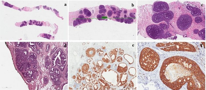
Cancers 2020, 12, 609                                                                               2 of 11

2. Epidemiology
      It is widely accepted that most cases of DCIS are now diagnosed through breast cancer screening
programs and 20% of all breast carcinomas are “in situ” lesions [2]. As most DCIS are surgically treated,
it is not surprising that approximately 20–25% of surgical breast samples evaluated by pathologists
refer to this disease [3,4]. The incidence of DCIS has increased over the last three decades, ranging from
1.87 cases per 100,000 person-years in the first years of the 1970s to 32.5 cases per 100,000 person-years
in 2005 [5]. Breast carcinoma is the most frequent cancer among women in different age groups, with
40% of cases diagnosed in the 0–49 age group, 35% of cases in the 50–69 age group, and 22% of cases in
the oldest age group >70 years. It has been calculated that one case of DCIS is usually identified per
1000 screening mammograms.

3. Clinical Features
     Most DCIS lesions are diagnosed mammographically (70–90%), being rarely detected clinically.
Only a few cases of DCIS (2–3%) present as a small-sized (≤1 cm) palpable mass, or with nipple changes
(discharge or Paget’s disease). Notably a small percentage (up 5%) of DCIS lesions is incidentally
detected at the histological examination of breast tissue evaluated for other reasons.

4. Radiologic Features
      Most DCIS lesions are not usually palpable lesions, being only radiologically detected.
Mammography is a highly sensitive diagnostic procedure for detecting DCIS. Several studies show
that the diagnosis of DCIS mainly depends on the detection of microcalcifications on mammographic
screening (70–90% of cases) [4,6,7], while only a smaller percentage of cases is diagnosed as a palpable
mass or a nipple discharge or ulceration (Paget’s disease). In 2002, it was estimated that about 1/1300
screening mammographies was consistent with the diagnosis of DCIS, histologically proven by needle
core biopsy [8]. Microcalcifications alone are likely the most reliable mammographic indicators of
DCIS in women younger than 50 years, whereas parenchymal abnormalities, especially architectural
distortions, are the alarming features more evident in women older than 50 years, due to the variation
in overall breast density at this age [9]. Briefly, two types of microcalcifications are recognized: fine
and coarse microcalcifications, with a clustered (at least five microcalcifications in a small volume
of tissue: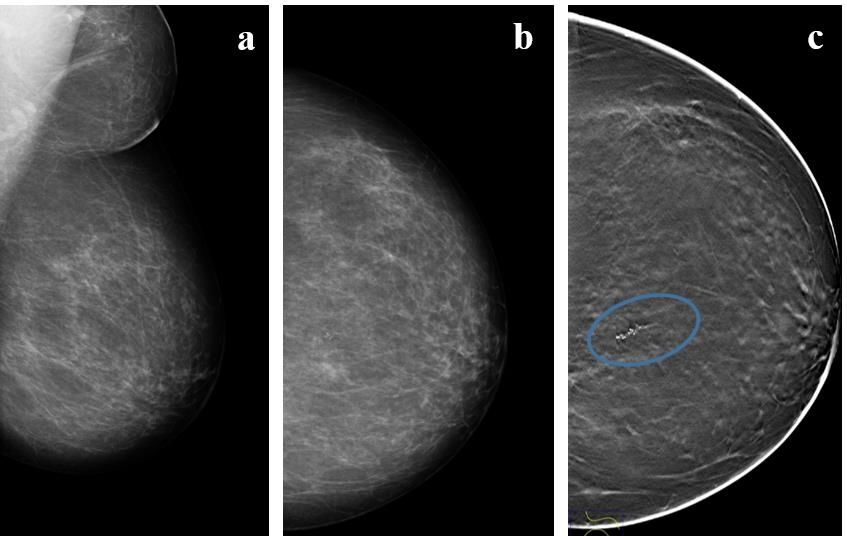
Cancers 2020, 12, 609 3 of 11
Cancers
Cancers 2020,
2020, 12,
12, xx 33 of
of 11
11
Figure
Figure1.1.
Figure Radiologic
1.Radiologic
Radiologic features
features
features of
of aa low-grade
low-grade
of a low-grade ductal
ductal carcinoma
ductal carcinoma carcinoma in situ
situ (DCIS)
in lesion.
in situ (DCIS) (DCIS) lesion.
lesion. (a,b)
(a,b) Mammograms (a,b)
Mammograms
Mammograms
show show
show aa heterogeneously
a heterogeneously heterogeneously dense
denseIn
dense right breast. right
thebreast.
right upperIn
breast. In the
the upper
outer upper outer
outer
quadrant quadrant
quadrant
(UOQ) (UOQ)
(UOQ)
there there
there
is a small
is
is aa opacity
oval small
small oval
with opacity
oval obscuredwith
opacity with obscured
obscured
margins; margins; (c)
(c) ultrasound
margins; shows
(c) ultrasound ultrasound
an irregularshows
mass an
shows an irregular
irregular
with mass
mass
indistinct with
with
margins,
indistinct
indistinct margins,
hypoechoic margins, hypoechoic
hypoechoic
echo pattern echo
echo
without pattern
pattern
posterior without
without posterior
features. posterior features.
features.
Figure
Figure2.
Figure 2.
2. AA
A5757 year-old
57year-old woman
year-oldwoman
womanwithwith high-grade
with high-grade DCIS.
high-gradeDCIS. (a,b)
DCIS. (a,b) Mammograms
(a,b)Mammograms
Mammogramsand and (c)
and (c) Tomosyinthesis
(c)Tomosyinthesis
Tomosyinthesis
imaging
imaging of
of the
the left,
left, almost
almost entirely
entirely fatty,breast
fatty,breast shows
shows a
a cluster
cluster of
of fine-linear
fine-linear branching
branching calcifications
calcifications
imaging of the left, almost entirely fatty, breast shows a cluster of fine-linear branching calcifications
(circle) in
(circle)in
(circle) the
inthe upper
theupper inner
upperinner quadrant
innerquadrant (UIQ)
quadrant(UIQ) classified
(UIQ) classified using
classifiedusing breast
usingbreast imaging-reporting
breastimaging-reporting
imaging-reportingand and data
anddata system
datasystem
system
(BI-RADS) as
(BI-RADS) as
(BI-RADS) Category
as Category IVc.
CategoryIVc.
IVc.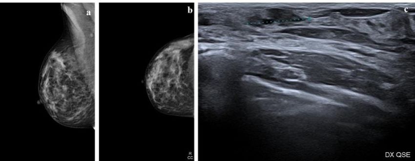
Figure 2. A 57 year-old woman with high-grade DCIS. (a,b) Mammograms and (c) Tomosyinthesis
imaging of the left, almost entirely fatty,breast shows a cluster of fine-linear branching calcifications
Cancers(circle) in609
2020, 12, the upper inner quadrant (UIQ) classified using breast imaging-reporting and data system4 of 11
(BI-RADS) as Category IVc.
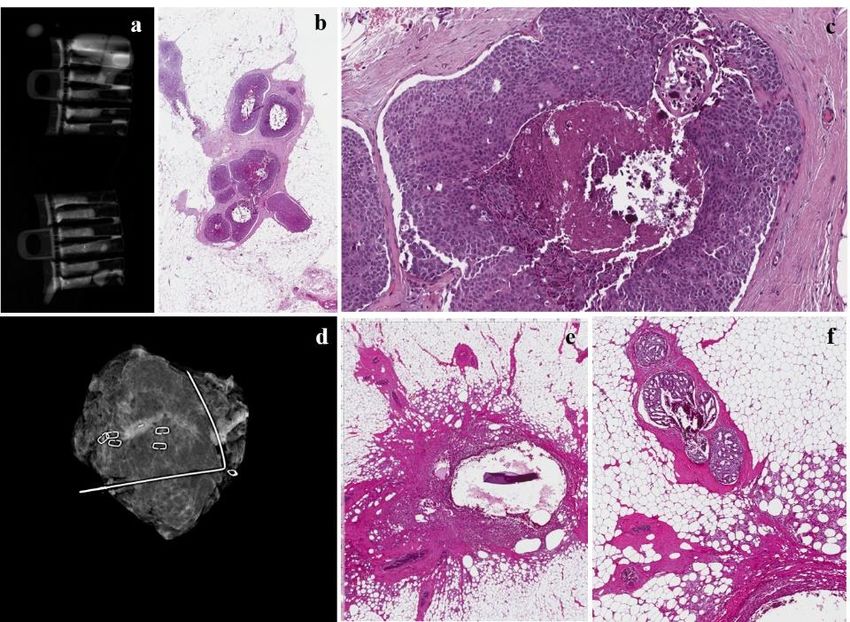
Figure 3. The same case as Figure 3. (a) Radiograph during localization of the microcalcification in
the samples collected in touch-free collection chambers using the Mammotome Revolve 10 gauge
biopsy system that reveals numerous microcalcifications in the cores; (b,c) mammotome biopsy shows
a high-grade DCIS with central comedonecrosis at low- and high-magnification; (d) mammogram
revealing successful retrieval of a cluster of pleomorphic calcifications with prior localization and
subsequent surgery; (e) inflammatory reaction around the previously placed clips; (f) a residual focus
of cribriform carcinoma in situwith central microcalcifications.
5. Magnetic Resonance Imaging
Magnetic resonance imaging (MRI) has been considered more sensitive than mammography for
detecting DCIS without calcifications and multifocal lesions [11]. However, several authors report
that MRI shows higher sensitivity for invasive carcinoma (up to 98%) than for DCIS (60–80%): DCIS
is typically not mass-forming, with adelayed peak enhancement profile; MRI can miss low-grade
DCIS, while it is more sensitive for high-grade DCIS, showing higher vascularity. A comparison of
morphological and immunohistochemical studies with MRI features of DCIS suggests that tumor
angiogenesis could contribute to MRI enhancement [12,13].
Contrast-enhanced MRI has been considered an effective method for the detection of a concurrent
contralateral carcinoma in situin women with a previous diagnosis of ipsilateral DCIS, an occult
primary tumor, an examination of dense breast tissue, a study of breasts in patients with BRCA
mutations, and neoplastic involvement of the chest wall.In the evaluation of DCIS, MRI could be useful
to assess disease extent, showing a diagnostic accuracy of more than 60%, compared tomammographic
accuracy of 55%; however, this methodology has a high rate of false negatives.
6. Morphological Features of Carcinoma In Situ
DCIS is a unifocal disease originally restricted to a single duct system but with the capability
to involve different lobules. The basal membrane and the layer of myoepithelial cells are—by
definition—preserved. The latter cells can be easily highlighted by means of immunohistochemistry
using different immunomarkers such as p63, high molecular-weight keratins 5/6, 14, calponin, S100,
and smooth muscle actin. DCIS is a heterogeneous disease not only biologically and genetically
but also morphologically. The pathology report should include several features according to the
College of American Protocol of Pathologists [14]: nuclear grade, architectural growth pattern, necrosis,
stromal reaction, tumor-infiltrating lymphocytes, immunohistochemical profile (estrogen receptor-ER,
Progesteron receptor-PgR, HER-2), size and extension, and surgical margins. Based on nuclear gradeCancers 2020, 12, 609 5 of 11
atypia, DCIS can be categorized as low, intermediate, and high nuclear grade lesions. It is not
unusual to detect two different nuclear grades in the same lesion as the result of genetic heterogeneity.
Low-grade DCIS is a proliferation of small and monomorphic neoplastic cells, with inconspicuous
nucleoli and few mitoses (Figure 4). Conversely, high-grade DCIS shows large-sized, pleomorphic
neoplastic cells with large and irregular nuclei, multiple and prominent nucleoli, high mitotic index,
and often necrosis (Figure 3). Intermediate-grade DCIS is a neoplasia with overlapping features
between low- and high-grade DCIS. Numerous meta-analysis studies suggest that high-grade DCIS
shows an increased risk of ipsilateral recurrence compared to low-grade DCIS. However, Zhang et al.
in his meta-analysis shows that high- and intermediate-grade DCIS are not significantly associated
with the risk of local invasive recurrence [15]. DCIS may exhibit different growth patterns, including
(i) cribriform, in which neoplastic cells form round and rigid arches giving a holing aspect to the
luminal space (Figure 4); (ii) papillary, in which neoplastic cells show a papillary architecture with
a fibrovascular core; (iii) micropapillary, in which tufts of monomorphic neoplastic cells without a
fibrovascular core, protrude into the glandular lumen; and (iv) solid, in which neoplastic cells form
sheets filling the glandular lumen. Several studies correlate solid, papillary, and micropapillary
growth patterns with a more consistent and strong adverse outcome. High-grade DCIS often shows
comedonecrosis, when the necrotic core extends into the ductal lumen that canundergo calcifications.
Cancers 2020, 12, x 5 of 11
Necrosis is also constantly and strongly associated with ipsilateral recurrence, with the hazard ratio
(HR), in general,
demonstrated above
that 2.0 of
the risk [16]. Some studies
ipsilateral demonstrated
recurrence is greater that the with
if DCIS risk of ipsilateral recurrence
comedonecrosis is only
is greater if DCIS with comedonecrosis is only treated with breast
treated with breast conserving surgery, compared to DCIS managed with mastectomy, conserving surgery, compared
skin -sparing
to DCIS managed
mastectomy, withconserving
or breast mastectomy, skin-sparing
surgery mastectomy,
plus radiotherapy or Zhang
[16]. breast conserving surgery plus
et al. also disagreeon the
radiotherapy
role of comedonecrosis, suggesting that there is no correlation between n ecrosis and the risk no
[16]. Zhang et al. also disagreeon the role of comedonecrosis, suggesting that there is of
correlation
invasive localbetween necrosis
recurrence and the risk of invasive local recurrence [15].
[15].
Figure4.4.Features
Figure Featuresof aoflow-grade
a low-grade
DCIS.DCIS. (a) Low-grade
(a) Low-grade DCIS in aDCIS in a core
core needle needle
biopsy; biopsy; (b,c)
(b,c) intermediate
intermediate and high-magnification of the lesion showing a cribriform growth pattern;
and high-magnification of the lesion showing a cribriform growth pattern; (d) DCIS with negative (d) DCIS
with negative
margins, but themargins, but the neoplasia
distance between distance between
and inkedneoplasia andmm;
margin isCancers 2020, 12, 609 6 of 11
younger age, larger size, comedonecrosis and ER negative, and HER2 overexpression. However, no
significant association was identified for longer follow-ups [21]. It should be noted that the evaluation
of TILs has a prognostic value and not a predictive value of response to therapies, therefore, this
criterion is not used to decide whether or not to administer chemotherapy or other systemic therapies.
DCIS tumor size could be difficult to measure on multiple slides; DCIS with a ≤20 mm diameter
is usually considered “small”. Several meta-analyses showed that among tumor characteristics,
tumor size was certainly positively correlated with a higher rate of recurrence of ipsilateral breast
cancer, although many of the estimates were not statistically significant [22]. Conversely, in another
meta-analysis, Zhang et al. disprove that tumor size is significantly associated with the risk of local
invasive recurrence [15]. DCIS multifocality is defined as multifocal foci with at least 5 mm of
intervening healthy tissue confined to a single breast quadrant [23]. Several studies of meta-analyses
correlate multifocal DCIS with increased risk of recurrence with an estimated risk of 1.95 [22].
Conversely, Zhang et al. show a multifocal DCIS lesion is also associated with an increased risk of
recurrence for invasive ductal carcinoma [15].
Several predictive score systems have been developed for DCIS, mainly based on pathological
features. A recent clinical risk score, proposed by Punglia et al., seems to be useful and easy to
use [24]. This score system includes a large number of patients (n. 2762) treated with breast-conserving
surgery (BCS) alone or plus radiation and/or hormonal therapy. The following factors were found to
be associated with ipsilateral recurrence within 5 years: (i) age ≤50 years; (ii) comedonecrosis; and
(iii) ER-negative status. It was calculated that the 5-year risk of ipsilateral recurrence after BCS alone
was 9% for the low-risk group, 23% for the intermediate-risk group, and 51% for the high-risk group.
Based on these findings, clinicians, after establishing the patient’s risk-group, can guide subsequent
adjuvant treatment (radiation with or without hormonal therapy) for patients with negative margins
after BCS.
7. Immunohistochemical Profile
Although several immunohistochemical studies have been performed on DCIS, with the aim
of correlating a specific immunohistochemical profile with the risk of local recurrence (in terms
of in situor invasive lesions), the results are conflicting [16]. This is true not only for each single
immunomarker tested but also after classifying DCIS into molecular subtypes, similarly to what has
been applied to invasive breast carcinoma [25]. Although ER-status has been incorporated as one of
the major pathologic factors, along with comedonecrosis, in a clinical score system for predicting DCIS
recurrence [24], the results of two clinical studies, UKANZ and NSABP B-24, suggest that ER status is a
weakly prognostic biomarker for local recurrence, but it is a strong predictor of response to endocrine
therapy to reduce local recurrence. Currently, ER is the only useful marker for planning a potential
endocrine therapy, and it should be performed on excision rather than in core biopsy.
8. Molecular Features
Breast cancer does not yet have a molecular signature that can predict the risk of recurrence or
that may indicate treatment. To date, the only validated and available molecular marker is Oncotype
DX Breast DCIS Score, which evaluates 12 genes derived from the 21 genes used for calculating the
Oncotype DX Recurrence Score for early invasive breast cancer. The DCIS score can be used to quantify
the risk within 10 years of developing a relapse, even invasive, following the diagnosis of DCIS without
radiotherapy. The histopathological aspects that correlate with the score are unclear, but several studies
explain that the predictive value of this score assumes greater value if correlated with age at diagnosis,
tumor size, and multifocality [16].
9. Surgical Margins
The standard treatment of DCIS is primarily surgical, including BCS for localized lesions, and
mastectomy for extensive or multicentric disease. Local recurrence rates (LRs) after BCS alone are high,Cancers 2020, 12, 609 7 of 11
ranging from 25% to 35% at 13–17 years of follow-up, and approximately half of all recurrences are
invasive [26]. As most patients with negative margins after BCS are at lower risk of LR, the optimal
margin width has been a matter of debate for many decades. Although multiple studies have shown
that positive margins or close to margins are both associated with a higher risk of LRs, there is no
consensus about the optimal adequate margin width. This is also complicated by the fact that many
patients treated by surgery, usually undergo subsequent whole breast radiation therapy (WBRT).
The Society of Surgical Oncology (SSO), the American Society for Radiation Oncology (ASTRO), and
the American Society of Clinical Oncology (ASCO) concluded that a positive margin, defined as ink on
tumor (DCIS), is associated with an increased risk in LR and that such risk is not nullified by the use
of WBRT. They also concluded that the free margin for BCS with WBRT is 2 mm [27]. At the same
time the LORIS trial metanalysis [28] of patients with DCIS, undergoing BCS plus WBRT, concluded
that margin distances above 2 mm are not significantly associated with a further reduction in odds
of LR. Even if histologic grade and architectural patterns, comedonecrosis, tumor size, and gene
expression profiles are associated with the risk of LR, they did not influence the recommended margin
width. The European Society for Medical Oncology (ESMO), the National Institute for Health and
Clinical Excellence (NICE), and the New Zealand Guidelines Group established that an appropriate
margin width should be 2 mm [29–31]. The National Comprehensive Cancer Network guidelines
now states that margins of at least 2 mm are associated with a reduced risk of ipsilateral breast tumor
recurrence [32]. A different perspective comes from the American Society of Breast Surgeons that
considers exclusively “no ink on the tumor” as a negative margin [33]. Notably the MD Anderson
experience [34], corroborated by the Memorial Sloan Kettering Cancer Center [35], reported that the
difference in Local recurrence rate (LRR) for patients with margins 5 cm),
or in the presence of multicentric disease or tumor mass diagnosed by an imaging study suspicious for
invasive cancer. The reason to perform SLNB for patients undergoing mastectomy is the impossibility
to perform this diagnostic procedure in the event that an invasive cancer is detected. Similarly, SLNB
should be performed if the lumpectomy will compromise drainage enough to prevent a future sentinel
lymph node procedure. This is most often considered for DCIS lesions located in the high axillary tail.
11. Natural History
It is extremely difficult to trace the natural history of DCIS. As in the last decades the rate of
invasive recurrence of DCIS has not changed significatively, despite an increased rate of diagnosesCancers 2020, 12, 609 8 of 11
basedon mammographic screening, it is likely that the majority of patients with DCIS does notdevelop
invasive carcinoma. This is why DCIS should be regarded as a “non-obligate” precursor of invasive
carcinoma. This concept is in line with what we know from carcinomas in situ occurring in other
organs. We can speculate about the natural history of DCIS by evaluating the clinco-pathological
studies based on the recurrence rate after a histologicallyproven diagnosis on core biopsy. A long-term
follow-up study, based on a systemic review, meta-analysis, and meta-regression analysis, revealed a
local recurrence rate of 40% after 15years [37]. Notably only 28.1% of the recurrence was in the form
of invasive carcinoma associated with a mortality rate of 18%. The most updated view about DCIS
considers this disease as a wide morphological and biological spectrum, ranging from small-sized,
usually low-grade, lesions that can be treated by surgical excision alone, to extensive, often high-grade
lesions, for which the best treatment seems to be mastectomy. The largest studies on the natural history
of DCIS suggest that more than 50% of patients with high-grade DCIS have the potential to progress
to an invasive carcinoma in less than 5 years if left untreated, while low-grade DCIS has a similar
progression but in a small percentage of patients (35–50%) and in a more prolongated time course,
up to 40 years [38–41].
12. Treatment
Based on the notions from the natural history of DCIS, patients with a histologically proven
diagnosis should be treated to prevent the possibility of local recurrence both in terms of non-invasive or
invasive carcinoma. DCIS treatment is still controversial, with a wide possibility of options, including
surveillance, breast-conserving surgery (BCS), BCS in association with radiotherapy and/or hormonal
therapy, and mastectomy with or without radiotherapy. Over time, there have been many efforts
to identify patients with low- or high-risk DCIS lesions, in order to avoid, respectively, over- or
under-treatment. The option of an active surveillance could be offered not only to older patients, but
also to all patients with mortality risks due to other diseases [42]. As far as surgery is concerned,
mastectomy, with immediate or subsequent breast reconstruction, is strongly recommended for those
patients with large-sized tumors, multifocal tumors, small-sized breasts (cosmetic problems), family
history, or documented BRCA mutations. Conversely, the proposal of BCS alone or in combination with
radiotherapy is still a matter of debate. Although BCS alone seems to be an effective treatment, it is not
enough for DCIS. This is supported by the evidence that patients both with low/intermediate-grade and
high-grade DCIS (60 years) [49].
Author Contributions: Conceptualization, L.S. and G.M.; methodology, G.M., L.P., V.V.; software, R.C.; validation,
G.M., L.P. and L.S.; formal analysis, G.M.V.; investigation, G.M.V.; resources, G.M.V.; data curation, L.S. and
L.P.; writing—original draft preparation, L.S. and R.C.; writing—review and editing, G.M.; visualization, L.P.;
supervision, G.M.; project administration, L.S. All authors have read and agreed to the published version of
the manuscript.
Funding: This research received no external funding.
Acknowledgments: We wish to thank the Scientific Bureau of the University of Catania for language support.
Conflicts of Interest: The authors declare no conflict of interest.Cancers 2020, 12, 609 9 of 11
References
1. WHO. Classification of Breast Tumours, 5th ed.; WHO Classification of Tumours Editorial Board: Geneva,
Switzerland, 2019; Volume 2.
2. Weaver, D.L.; Rosenberg, R.D.; Barlow, W.E.; Ichikawa, L.; Carney, P.A.; Kerlikowske, K.; Buist, D.; Geller, B.M.;
Key, C.R.; Maygarden, S.J.; et al. Pathologic findings from the Breast Cancer SurvellainceConsortium:
Vpopulation-based outcomes in women undergoing biopsy after screening mammography. Cancer 2006, 106,
732–742. [CrossRef]
3. Siegel, R.L.; Miller, K.D.; Jemal, A. Cancer statistics, 2017. CA Cancer J. Clin. 2017, 67, 7–30. [CrossRef]
4. Levinsohn, E.; Altman, M.; Chagpar, A.B. Controversies regarding the diagnosis and management of ductal
carcinoma in situ. Am. Surg. 2018, 84, 1–6. [PubMed]
5. Virnig, B.A.; Tuttle, T.M.; Shamliyan, T.; Shamliyan, T.; Kane, R.L. Ductal carcinoma in situ of the breast:
A systematic review of incidence, treatment, and outcomes. J. Natl. Cancer Inst. 2010, 170–178. [CrossRef]
6. Lakhani, S.R.; Ellis, I.O.; Schnitt, S.J. WHO/IARC Classification of Tumours of the Breast, 4th ed.; WHO/IARC:
Lyon, France, 2012; Volume 4.
7. Bijker, N.; Donker, M.; Wesseling, J.; Th Rutgers, E.J. Is DCIS breast cancer, and how do I treat it? Curr. Treat.
Options Oncol. 2013, 14, 75–87. [CrossRef] [PubMed]
8. Ernster, V.L.; Ballard-Barbash, R.; Barlow, W.E.; Zheng, Y.; Weaver, D.L.; Cutter, G.; Yankaskas, B.C.;
Rosenberg, R.; Carney, P.A.; Kerlikowske, K.; et al. Detection of ductal carcinoma in situin women
undergoing screening mammography. J. Natl. Cancer Inst. 2002, 94, 1546–1554. [CrossRef] [PubMed]
9. Stomper, P.C.; Connolly, J.L.; Meyer, J.E.; Harris, J.R. Clinically occult ductal carcinoma in situdetected with
mammography: Analysis of 100 cases with radiologic-pathologic correlation. Radiology 1989, 172, 235–241.
[CrossRef] [PubMed]
10. Bernardi, D.; Macaskill, P.; Pellegrini, M.; Valentini, M.; Fantò, C.; Ostillio, L.; Tuttobene, P.; Luparia, A.;
Houssami, N. Breast cancer screening with tomosynthesis (3D mammography) with acquired or synthetic
2D mammography compared with 2D mammography alone (STORM-2): A population-based prospective
study. Lancet Oncol. 2016, 17, 1105–1113. [CrossRef]
11. Mcneil, J.H.; Morris, E.A.; Dershaw, D.D.; Abramson, A.F.; Brogi, E.; Liberman, L. Determination of the
presence and extent of pure ductal carcinoma in situby mammography and magnetic resonance imaging.
Breast J. 2005, 11, 382–390. [CrossRef]
12. Pediconi, F.; Catalano, C.; Roselli, A.; Padula, S.; Altomari, F.; Moriconi, E.; Pronio, A.M.; Kirchin, M.A.;
Passariello, R. Contrast-enhanced MR mammography for evaluation of the contralateral breast in patients
with diagnosed unilateral breast cancer or high-risk lesions. Radiology 2007, 243, 670–680. [CrossRef]
13. Londero, V.; Zuiani, C.; Linda, A.; Girometti, R.; Bazzocchi, M.; Sardanelli, F. High-risk breast lesions at
imaging- guided needle biopsy: Usefulness of MRI fur treatment decision. AJR Am. J. Roentgenol. 2012, 199,
W240–W250. [CrossRef] [PubMed]
14. College of American Pathologists (CAP). Protocol for the Examination of Specimens from Patients with Ductal
Carcinoma in Situ(DCIS) of the Breast; Version: Breast DCIS 4.1.0.0.; CAP: Northfield, IL, USA, 2018.
15. Zhang, X.; Dai, H.; Liu, B.; Song, F.; Chen, K. Predictors for local invasive recurrence of ductal carcinoma in
situ of the breast: A meta-analysis. Eur. J. Cancer Prev. 2016, 25, 19–28. [CrossRef] [PubMed]
16. Hanna, W.M.; Parra-Herran, C.; Lu, F.I.; Slodkowska, E.; Rakovitch, E.; Nofech-Mozes, S. Ductal carcinoma
in situ of the breast: An update for the pathologist in the era of individualized risk assessment and tailored
therapies. Mod. Pathol. 2019, 32, 896–915. [CrossRef]
17. Sneige, N.; McNeese, M.D.; Atkinson, E.N.; Ames, F.C.; Kemp, B.; Sahin, A.; Ayala, A.G. Ductal carcinoma in
situtreated with lumpectomy and irradiation: Histopathological analysis of 49 specimens with emphasis on
risk factors and long term results. Hum. Pathol. 1995, 26, 642–649. [CrossRef]
18. Loi, S.; Sirtaine, N.; Piette, F.; Salgado, R.; Viale, G.; Van Eenoo, F.; Rouas, G.; Francis, P.; Crown, J.P.A.;
Hitre, E.; et al. Prognostic and predictive value of tumor-infiltrating lymphocytes in a phase III randomized
adjuvant breast cancer trial in node-positive breast cancer comparing the addition of docetaxel to doxorubicin
with doxorubicin-based chemotherapy: BIG 02-98. J. Clin. Oncol. 2013, 31, 860–867. [CrossRef]
19. Dieci, M.V.; Mathieu, M.C.; Guarneri, V.; Conte, P.; Delaloge, S.; Andre, F.; Goubar, A. Prognostic and
predictive value of tumor-infiltrating lymphocytes in two phase III randomized adjuvant breast cancer trials.
Ann. Oncol. 2015, 26, 1698–1704. [CrossRef] [PubMed]Cancers 2020, 12, 609 10 of 11
20. Stanton, S.E.; Adams, S.; Disis, M.L. Variation in the Incidence and Magnitude of Tumor-Infiltrating
Lymphocytes in Breast Cancer Subtypes: A Systematic Review. JAMA Oncol. 2016, 2, 1354–1360. [CrossRef]
[PubMed]
21. Pruneri, G.; Lazzeroni, M.; Bagnardi, V.; Tiburzio, V.; Rotmensz, N.; DeCensi, A.; Guerrieri-Gonzaga, A.;
Vingiani, A.; Curigliano, G.; Zurrida, S.; et al. The prevalence and clinical relevance of tumor-infiltrating
lymphocytes (TILs) in ductal carcinoma in situ of the breast. Ann. Oncol. 2017, 28, 321–328. [CrossRef]
[PubMed]
22. Shamliyan, T.; Wang, S.Y.; Virnig, B.A.; Tuttle, T.M.; Kane, R.M. Association between patient and tumor
characteristics with clinical outcomes in women with ductal carcinoma in situ. J. Natl. Cancer Inst. Monogr.
2010, 41, 121–129. [CrossRef]
23. Sikand, K.; Lee, A.H.; Pinder, S.E.; Elston, C.W.; Ellis, I.O. Sections of the nipple and quadrants in mastectomy
specimens for carcinoma are of limited value. J. Clin. Pathol. 2005, 58, 543–545. [CrossRef]
24. Punglia, R.S.; Jiang, W.; Lipsitz, S.R.; Hughes, M.E.; Schnitt, S.J.; Hassett, M.J.; Nekhlyudov, L.; Achacoso, N.;
Edge, S.; Javid, S.H.; et al. Clinical risk score to predict likelihood of recurrence after ductal carcinoma in
situtreated with breast-conserving surgery. Breast Cancer Res. Treat. 2018, 167, 751–759. [CrossRef] [PubMed]
25. Williams, K.E.; Barnes, N.L.; Cramer, A.; Johnson, R.; Cheema, K.; Morris, J.; Howe, M.; Bundred, N.J.
Molecular phenotypes of DCIS predict overall and invasive recurrence. Ann. Oncol. 2015, 26, 1019–1025.
[CrossRef] [PubMed]
26. Wapnir, I.L.; Dignam, J.J.; Fisher, B.; Mamounas, E.P.; Anderson, S.J.; Julian, T.B.; Land, A.R.; Margolese, R.G.;
Swain, S.M.; Costantino, J.P.; et al. Long-Term Outcomes of Invasive Ipsilateral Breast Tumor Recurrences
After Lumpectomy in NSABP B-17 and B-24 Randomized Clinical Trials for DCIS. J. Natl. Cancer Inst. 2011,
103, 478–488. [CrossRef] [PubMed]
27. Morrow, M.; Van Zee, K.J.; Solin, L.J.; Houssami, N.; Chavez-MacGregor, M.; Harris, J.R.; Horton, J.;
Hwang, S.; Johnson, P.L.; Marinovich, M.L.; et al. Society of Surgical Oncology-American Society for Radiation
Oncology-American Society of Clinical Oncology Consensus Guideline on Margins for Breast-Conserving
Surgery with Whole-Breast Irradiation in Ductal Carcinoma In Situ. Ann. Surg. Oncol. 2016, 23, 3801–3810.
[CrossRef]
28. Francis, A.; Thomas, J.; Fallowfield, L.; Wallis, M.; Bartlett, J.M.; Brookes, C.; Roberts, T.; Pirrie, S.; Gaunt, C.;
Young, J.; et al. Addressing overtreatment of screen detected DCIS; the LORIS trial. Eur. J. Cancer 2015, 51,
2296–2303. [CrossRef]
29. Senkus, E.; Kyriakides, S.; Ohno, S.; Penault-Llorca, F.; Poortmans, P.; Rutgers, E. Primary breast cancer:
ESMO Clinical Practice Guidelines for diagnosis, treatment and follow-up. Ann. Oncol. 2015, 26, V8–V30.
[CrossRef]
30. National Institute of Health and Clinical Excellence. Early and locally advanced breast cancer: Diagnosis
and treatment. NICE clinical guideline 80. In Surgery to the Breast (1.31); National Institute of Health and
Clinical Excellence: London, UK, 2009.
31. New Zealand Guidelines Group (NZGG). Ductal carcinoma in situ. In Management of Early Breast Cancer;
New Zealand Guidelines Group (NZGG): Wellington, New Zealand, 2009; pp. 133–141.
32. National Comprehensive Cancer Network (NCCN). NCCN Clinical Practice Guidelines; Breast Cancer Version
1; New Zealand Guidelines Group (NZGG): Wellington, New Zealand, 2017.
33. American Society of Breast Surgeons. The American Society of Breast Surgeons Position Statement on Breast
Cancer Lumpectomy Margins; American Society of Breast Surgeons: Colombia, MD, USA, 2015.
34. Tadros, A.B.; Smith, B.D.; Shen, Y.; Lin, H.; Krishnamurthy, S.; Lucci, A. Contemporary Breast Conservation
Patient Outcomes for Ductal Carcinoma in situand MarginsCancers 2020, 12, 609 11 of 11
37. Stuart, K.E.; Houssami, N.; Taylor, R.; Hayen, A.; Boyages, J. Long-term outcomes of ductal carcinoma in situ
of the breast: A systematic review, meta-analysis and meta-regression analysis. BMC Cancer 2015, 15, 890.
[CrossRef]
38. Collins, L.C.; Tamimi, R.; Baer, H.; Connolly, J.; Colditz, G.; Schnitt, S. Risk of invasive breast cancer in
patients with ductal carcinoma in situ [DCIS] treated by diagnostic biopsy alone:Results from the Nurses’
Health Study. Breast Cancer Res. Treat. 1994, 88, 1083. [CrossRef]
39. Page, D.L.; Dupont, W.D.; Rogers, L.W.; Jensen, R.A.; Schuyler, P.A. Continued local recurrence of carcinoma
15–25 years after a diagnosis of low grade ductal carcinoma in situ of the breast treated only by biopsy.
Cancer 1995, 76, 197–2000. [CrossRef]
40. Sanders, M.E.; Schuyler, P.A.; Dupont, W.D.; Page, D.L. The natural history of low-grade ductal carcinoma in
situ of the breast in women treated by biopsy only revealed over 30 years of long-term follow-up. Cancer
2005, 103, 2481–2484. [CrossRef] [PubMed]
41. Sanders, M.E.; Schuyler, P.A.; Simpson, J.F.; Page, D.L.; Dupont, W.D. Continued observation of the natural
history of low-grade ductal carcinoma in situreaffirms proclivity for local recurrence even after more than
30 years of follow-up. Mod. Pathol. 2015, 28, 662–669. [CrossRef] [PubMed]
42. Ryser, M.D.; Worni, M.; Turner, E.L.; Marks, J.R.; Durrett, R.; Hwang, E.S. Outcomes of active surveillance for
ductal carcinoma in situ: A computational risk analysis. J. Natl. Cancer Inst. 2015, 108. [CrossRef] [PubMed]
43. Solin, L.J.; Gray, R.; Hughes, L.L.; Wood, W.C.; Lowen, M.A.; Badve, S.S.; Baehner, F.L.; Ingle, J.N.; Perez, E.A.;
Recht, A.; et al. Surgical excision without radiation for ductal carcinoma in situ of the breast: 12-year results
from the ECOG-ACRIN E5194 Study. J. Clin. Oncol. 2015, 33, 3938–3944. [CrossRef]
44. Holmes, P.; Lloyd, J.; Chervoneva, I.; Pequinot, E.; Cornfield, D.B.; Schwartz, G.F.; Allen, K.G.; Palazzo, J.P.
Prognostic markers and long-term outcomes in ductal carcinoma in situ of the breast treated with excision
alone. Cancer 2011, 117, 3650–3657. [CrossRef]
45. Rakovitch, E.; Nofech-Mozes, S.; Hanna, W.; Narod, S.; Thiruchelvam, D.; Saskin, R.; Spayne, J.; Taylor, C.;
Paszat, L. HER2/neu and Ki-67 expression predict non-invasive recurrence following breast-conserving
therapy for ductal carcinoma in situ. Br. J. Cancer 2012, 106, 1160–1165. [CrossRef]
46. Donker, M.; Litière, S.; Werutsky, G.; Julien, J.P.; Fentiman, I.S.; Agresti, R.; Rouanet, P.; de Lara, C.T.;
Bartelink, H.; Duez, N.; et al. Breast-conserving treatment with or without radiotherapy in ductal carcinoma
in situ: 15-year recurrence rates and outcome after a recurrence, from the EORTC 10853 randomized phase
III trial. J. Clin. Oncol. 2013, 31, 4054–4059. [CrossRef]
47. Wärnberg, F.; Garmo, H.; Emdin, S.; Hedberg, V.; Adwall, L.; Sandelin, K.; Ringberg, A.; Karlsson, P.;
Arnesson, L.G.; Anderson, H.; et al. Effect of radiotherapy after breast-conserving surgery for ductal
carcinoma in situ: 20 years follow-up in the randomized SweDCIS Trial. J. Clin. Oncol. 2014, 32, 3613–3618.
[CrossRef]
48. Cuzick, J.; Sestak, I.; Pinder, S.E.; Ellis, I.O.; Forsyth, S.; Bundred, N.J.; Forbes, J.F.; Bishop, H.; Fentiman, I.S.;
George, W.D. Effect of tamoxifen and radiotherapy in women with locally excised ductal carcinoma in situ:
Long-term results from the UK/ANZ DCIS trial. Lancet Oncol. 2011, 12, 21–29. [CrossRef]
49. Guerrieri-Gonzaga, A.; Sestak, I.; Lazzeroni, M.; Serrano, D.; Rotmensz, N.; Cazzaniga, M.; Varricchio, C.;
Pruneri, G.; Leonardi, M.C.; Orecchia, R.; et al. Benefit of low-dose tamoxifen in a large observational cohort
of high risk ER positive breast DCIS. Int. J. Cancer 2016, 139, 2127–2134. [CrossRef] [PubMed]
© 2020 by the authors. Licensee MDPI, Basel, Switzerland. This article is an open access
article distributed under the terms and conditions of the Creative Commons Attribution
(CC BY) license (http://creativecommons.org/licenses/by/4.0/).You can also read