Action plan for Pressure ulcers
←
→
Page content transcription
If your browser does not render page correctly, please read the page content below
Action plan for
Pressure ulcers

                   1
Care of Sweden – Medical technology since 1992.

    At Care of Sweden we market and develop medical technology
    products and services for the health sector. We are specialised in
    mattresses with qualitative characteristics and functions. Caring
    accessories are also a part of our professional product range.

    Care of Sweden’s Action plan for Pressure ulcers is produced in
    order to increase awareness of pressure ulcer, underlying causes
    and risks. It can also be used as a guideline in the daily work on
    prevention and treatment of pressure ulcer.

2
Introduction
Pressure ulcers are a complication that causes considerable problems
not only for those who are affected, but also for others who are close to
them and for staff. Caring for people with pressure ulcers represents a
challenge for the health service in the form of increased costs.

Most pressure ulcers can be prevented by early identification of people who are in the risk zone
and by taking selective medical and nursing measures ( 1,2,3).

It is not completely understood how pressure ulcers form, but there are a few theories. One of
them argues that the injury begins at the surface of the skin and eventually reaches the deeper
tissues (”top to bottom”). Another one claims that a pressure ulcer occurs deep in the tissue and
then move towards the skin surface (”bottom to top”) when the muscle tissue is considered to be
more sensitive to reduced or turned off blood flow than the skin (3).

                                                                                                    3
Definition of pressure ulcers
    A pressure ulcer is a localised injury to the skin and/or underlying tissue, usually
    over a bone prominience, as a result of pressure alone or in combination with
    shearing. There are also a number of contributory factors that may be related
    to pressure ulcers, though the significance of these factors has not as yet been
    investigated (4).

    A large number of risk factors have been described for the occurrence of pressure ulcers.
    Whether or not a pressure ulcer occurs is determined by a combination of various risk factors
    together with external pressure. The pressure required for an ulcer to occur depends in part on
    the force of the pressure, and also the length of time the tissue is exposed to pressure. Sensitivity
    to pressure varies from one individual to another and for different types of tissue (2).
    A number of documented patient-related risk factors are: advanced age, reduced mobility,
    reduced general health, incontinence, acute illness, neurological conditions, cardiovascular
    disease, terminal stage illness and previous pressure ulcers.
    External causes, known as environment-related risk factors, are the effect of pressure, shearing
    (which occurs when different layers of tissue are shifted relative to each other), friction, temper-
    ature and moisture. Long periods of time spent lying down or sitting, especially on parts of the
    body that are exposed to pressure, incorrectly used transfer technique and transfer aids (5,6).

    Risk assessment
    The risk of developing pressure ulcers must be assessed as soon as possible after arrival. Pre-
    ventive measures should be taken and an individual care plan prepared for all patients at risk. The

4
care plan describes goals, planned measures are carried out and the results are described and
evaluated (2).
The risk assessment must then be repeated regularly and whenever there is a change in the
patient’s health, following major surgical intervention as well as prior to discharge, in order to
ensure that information is provided to everyone in the care chain. Risk assessment and a clear
policy for preventing pressure ulcers allow at-risk patients to be identified and selective preven-
tive measures to be taken.
All measures must be documented and followed up in order to ensure communication between
professional categories and permitted exchanges of information within the care team, so that the
planning of care is adequate and allows long-term monitoring of an individual’s condition. Risk
factors that are identified by risk assessment should result in an individualised care plan (4,8,11).
Risk assessment, skin inspection and preventive measures must be documented and entered
into the case notes in accordance with the Health and Medical Services Act. This is extremely
important for patient safety and for ensuring that the measures can be quality-assured and that
the communication provides additional information concerning the patient/user about what is
being planned so that everyone is working towards the same goal (2,12,13).
Risk assessment can be based on various risk assessment tools, and it can be used as a supple-
ment to clinical assessment (6).
The Modified Norton scale assesses eight areas: mental state, physical activity, ability to move,
food intake, fluid intake, incontinence and general health. The maximum score is 28, with individ-
uals scoring 20 or less being at risk of developing pressure ulcers (7).
RAPS/RBT assesses physical activity, ability to move, the degree to which the skin is exposed to
moisture, food intake, fluid intake, sensation, friction and shearing, body temperature and serum
albumin. The maximum score is 39, with individuals scoring 31 or less being at risk of developing
pressure ulcers (8).
Braden assesses five different areas: sensory perception, moisture, activity, mobility and friction
and shearing. A score of 18 or less indicates that the person is at risk of developing pressure
ulcers (9).
Waterlow assesses nine areas: BMI, incontinence, skin type, medication, length of operation,
neurological problems, mobility, screening for malnutrition, gender and age. A score of 10 or
higher indicates a risk of developing pressure ulcers (10).

  App to Care, the pressure ulcer app can be downloaded in App Store
  (iPhone, iPod Touch and iPad).

                                                                                                        5
Location
& classification
Pressure ulcers can occur on all parts of the body. Points that are particularly prone to pressure
ulcers are points of buttock, iliac crest, sacrum, shoulder blades, back of the head, heels and
ankles (14). Other parts of the body can also be affected, so be particularly aware that medical
equipment can exert pressure, e.g.: catheters, plaster casts, nasal tubes, or if the person is
intubated (4,6,12).
Skin inspection Ears
Back of the head
Areas that are exposed to pressure are
inspected as soon as possible after arrival.
Pressure ulcers are categorised on a scale of Shoulder
1-4. Inspection must be repeated daily for all
Shoulder blade
patients at risk, anyone who is bed-ridden or
who uses a wheelchair, or who spends much
of the day sitting (2). Trochanter Elbow
Sacrum
We differentiate between pressure/moisture
damage, although it can often be regarded as Gluteal
a combination.
Moisture damage is always caused by mois-
Points of buttock
ture. The edges of the ulcer are often diffuse
Knees
or irregular. Moisture damage never forms
necrosis. Often occurs in skin folds as super-
ficial damage.
Pressure damage often occurs over bone
prominence. The edges of wounds are Ankles
Heels
usually well defined, and necrosis may
occur(6).
© Care of Sweden
6Classification, pressure ulcers
Category I
Redness that does not reduce on pressure.
Intact skin with redness in a clearly defined
area, usually above a bone prominence, that
does not reduce on pressure. Dark, pigment-
ed skin might not show this sign, though the
colour is different from the surrounding areas
of skin. The area may be painful, solid or soft,
© Care of Sweden
and warmer or colder than other areas of
skin. Category I pressure ulcers may be diffi- Category I
Redness that does not reduce on pressure.
cult to detect in people with dark skin tones.
Category I pressure ulcers may be a sign that
the patient is in the risk zone for developing
deeper pressure ulcers (4).
Category II
Partial skin damage.
Partial skin damage that appears as a superfi-
cial open ulcer with a pinkish-red wound bed
without fibrin slough. It may also be an intact
or open/ruptured serum-filled or blood-filled
blister. Appears as a shiny or dry superficial
ulcer without fibrin slough or superficial hae- © Care of Sweden
matoma. This category should not be used Category II
to describe skin tears, tape burns, inconti- Partial skin damage.
nence-associated dermatitis or maceration (4).
7Category III
Full skin damage.
Subcutaneous fat is visible, though not bone,
tendon or muscle. Fibrin slough may be vis-
ible, though without obscuring the depth of
the injury. May include undermining and tun-
nelling. The depth of a Category III ulcer may
vary depending on its anatomical location. © Care of Sweden
The bridge of the nose, ears, back of the head
Category III
and ankles do not have any subcutaneous Full skin damage.
fatty tissue and Category III pressure ulcers
may be superficial at these locations. In con-
trast, areas of significant subcutaneous fatty
tissue can develop extremely deep Category
III pressure ulcers. Bone/tendon is not visible
or palpable (4).
Category IV
Deep full-tissue damage involving bone,
tendon or muscle. There may be visible fibrin
or necrosis. There is often undermining and
tunnelling. The depth of Category IV pressure
ulcers varies depending on anatomical
location. For example, there is no subcuta-
neous fatty tissue at the bridge of the nose,
ears, back of the head or ankles, and ulcers at © Care of Sweden
these locations may be superficial. Category Category IV
Deep full-tissue damage.
IV pressure ulcers may involve muscles and
supporting structures (e.g. fascia, tendons or
joint capsules), which means that osteomy-
elitis and otitis may occur. Exposed bone and
muscle is visible or directly palpable (4).
Severe necrosis is assessed as category 4 even if the skin is intact, as may be the case with the
heels, for example (2).
It is important that nursing staff who inspect the skin know how to assess and categorise pressure
ulcers. A web-based training programme, PUCLAS (Pressure Ulcer Classification) has been
developed within EUPAP in order to improve knowledge about pressure ulcer assessment (6,22).
89
Preventive measures
It is a team work to prevent pressure ulcers and following measurements
should be taken into consideration.
Skin assessment/skin care
Inspection of the skin must include assessment of whether the skin is free from moisture and the
presence of cracked skin, oedemas, increased heat, hardened or loosened skin, or the presence
of eczema or rash.
Regular inspections of the skin are necessary in order to detect early signs of pressure ulcers.
The skin must be kept dry and clean, soft and supple with moisturising cream. Skin care products
must be unperformed and free of allergenic substances. Loosened skin (maceration) must be
protected, using a barrier cream for example.
Moisture and increased temperature makes the skin more sensitive to pressure and shearing.
Never use massage as a preventive measure, since this compresses the capillaries and increases
the risk of damage. The skin must not be scrubbed when cleaned as scrubbing can cause tissue
damage, especially in delicate elderly patients (2,4,5,6).
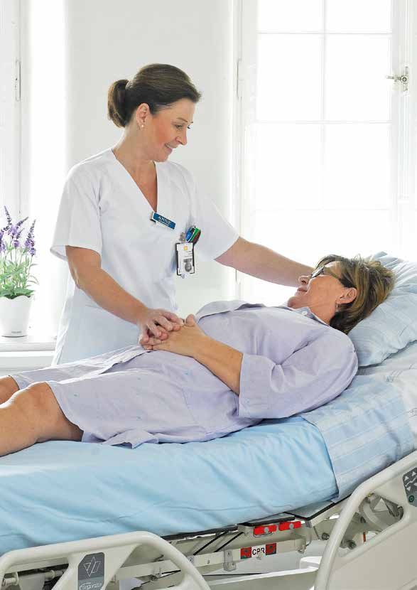
Pressure relief/pressure equalising
A first preventive measure is to look at what type of mattress or chair cushion is being used by a
person at risk of developing pressure ulcers. All persons in the risk zone should be prescribed a
pressure-equalising or pressure-relieving base.
Pressure equalisation reduces pressure on the skin; materials used are foam, fibre, gel and static
products. Pressure relieving eliminates pressure on the skin; alternating pressure is used (5).
The choice of base should be determined by the person’s individual needs for pressure relief and
in order to facilitate mobilisation. Attention must be paid to weight, occurrence of ulcers, ulcer
category, time spent lying/sitting and mobility. The type of bed also affects the choice. The bed
height must not be too high if the person is able to get into and out of bed themselves. Needs can
change over time, so the skin must always be checked regularly.
Regardless of which base is chosen, work with changes in position must always be in accordance
with the individual’s needs and wishes.
Bases that are used for 24 hours per day, 365 days per year, are exposed to extreme wear and
tear and must be inspected regularly and replaced if necessary. Both mattresses and chair
cushions are perishable products (2,5,6).
10Redistribution
of pressure
Increased contact
surface to reduce Pressure relief
pressure
Change of position to
Passive pressure Change of position to Active pressure relief Lift of pressure
increase contact surface
reduction bedding, remove pressure from bedding, exposed body parts,
between the body and
such as foam, a specific for example with for example heel
bedding, such as 30-
gel or air aids body part alternating aids reliefing aids
degree lateral position
Fig. 3. Methods for pressure distribution (2).
Reference to SKL pressure distribution methods, page 7(2).
Activity
Movement is the body’s defence against pressure ulcers and other complications from being
bedridden. People with reduced sensitivity, paralysis, contractions or unconsciousness do not
respond to the body’s warning signals, due to reduced or failed reaction or inability to feel pain
or discomfort. Therefore, help is needed regularly to change position. Small, frequent changes in
position are often sufficient for the circulation to work (15).
Changing position
This should be based on the person’s individual needs and status. Lying and sitting positions
must be adapted so as to keep the pressure between skin, bone prominience and the base to a
minimum. The time the person can sit or lie without changing position should not exceed two
hours, but must always be adapted to the individual. The pressure required for a pressure ulcer
to develop depends on the force of the pressure and the duration of pressure on the tissue. This
can vary from one person to another, and the location of the pressure and general health are of
decisive importance for any one individual.
Wherever possible, use aids to move patients or change position; this helps both staff and
user and reduces the risk of shearing or friction damage. Never place a user directly on bone
prominience or on a skin surface that is already reddened. Redness indicates that the body has
not yet recovered from earlier pressure and that the skin requires more time before any pressure
can be put on it.
In order to reduce the risk of someone developing pressure ulcers, it is important to reduce the
duration and force of the pressure to which the person is exposed (4,5,15 ).
11Modified flowchart over risk assessment by Eila Sterner, 2012
A need of Risk assessment related
to the patient’s situation
YES Modified Norton NO
< 20 > 20
NO Is the person ambulatory? YES
Needs of special
mattress/seat cushion?
Needs of repositioning?
NO Is the nutrition satisfying? YES
Nutritional supple-
ments + Care plan
12Lying
30° angle position, alternating between the left and right side, relieves the sacrum, iliac crest,
hips and heels. If necessary, use a cushion between the patient’s knees to relieve strain. Make
sure that there is no pressure on the inside and outside of the toes, ankles and feet; changes in
position always put greater pressure on some other part of the body.
A half-sitting position in bed increases pressure on the sacrum; the risk of shearing increases if
the patient slides down.
Different cushions can be used to relieve pressure and to position a patient based on need and
diagnosis (15,18,19,20).
Heels
Heels are an area at high risk of developing pressure ulcers. Micro circulation is low and the heel
bone is close to the skin. Total pressure relief is extremely important.
For example, a cushion can be placed along the calf. NB: make sure the pressure does not
increase on the Achilles tendon and that the heel is uncovered. The foot end of the bed can be
raised through 10° if necessary in order to increase venous return and reduce the risk of throm-
bosis in the calf (5,6).
Sitting
In a sitting position, the greatest part of your body weight is carried by the buttocks and thighs.
Ulcers across the sitting bones (tuber ischii) are caused by insufficient pressure relief in a sitting
position.
Time spent sitting is a decisive factor in the occurrence of pressure ulcers. The length of time a
person can sit depends on the risk or pressure ulcer category, and it must always be adapted to
the individual.
The correct sitting position is when the force of gravity falls in front of the back and down through
the sitting bones. The correct sitting height is when the knees rest at the same height as the hips.
The feet must always be supported by the floor or a footplate, otherwise sitting stability is lost and
the person risks sliding forwards.
An incorrect sitting position increases pressure on the sitting bones. Prevent shearing and fric-
tion by making sure the patient does not slide down the chair. The most important objective for
anyone sitting down for a long time is to sit comfortably, as comfort is an absolute requirement.
The only person who can decide whether a sitting position is good or bad is the person who is
sitting down.
Lying and sitting positions must be adapted so as to keep the pressure between the base and
bone prominience to a minimum (5,16).
13Micro climate and moist skin
The micro climate, in other words the temperature and humidity between the base and the per-
son’s skin, is very important in the development of pressure ulcers.
The skin’s mechanical properties change in response to moisture and temperature variations;
the skin becomes more sensitive and the risk of pressure, shearing and friction increases.
Diapers and other plastic materials can lead to a deterioration in both sitting and lying surfaces
and reduce the possibility of preventing pressure ulcers. All “unnecessary” material should
therefore be removed. The least possible material between the person and the base helps to
increase pressure relief (4,5,6).
Nutrition
People who are underweight or overweight, dehydrated or with reduced nutritional intake are at
a greater risk of developing pressure ulcers.
In order to meet their energy and food requirements, breakfast, lunch, dinner and three snacks
should be served. Food should be adapted to the individual’s needs, for example adapted food
consistency or energy and protein-rich food. Measures to help the patient eat may also need to
be reviewed. Nutritional status must be assessed in all patients and care recipients (17).
High-protein nutritional drinks can be offered as a supplement to ordinary food. These nutritional
drinks should be given between meals in order to avoid these having a negative effect on normal
food and fluid intake (2). Food and water intake should be recorded so that food can be adapted
according to the calculated energy intake (17).
14Example on nutritional assessment (26)
Mini Nutritional Assessment
MNA®
Last name: First name:
Sex: Age: Weight, kg: Height, cm: Date:
Complete the screen by filling in the boxes with the appropriate numbers. Add the numbers for the screen. If score is 11 or less, continue with the
assessment to gain a Malnutrition Indicator Score.
Screening J How many full meals does the patient eat daily?
0 = 1 meal
A Has food intake declined over the past 3 months due to 1 = 2 meals
loss of appetite, digestive problems, chewing or 2 = 3 meals
swallowing difficulties? K Selected consumption markers for protein intake
0 = severe decrease in food intake • At least one serving of dairy products
1 = moderate decrease in food intake (milk, cheese, yoghurt) per day yes no
2 = no decrease in food intake
• Two or more servings of legumes
B Weight loss during the last 3 months
or eggs per week yes no
0 = weight loss greater than 3kg (6.6lbs)
• Meat, fish or poultry every day yes no
1 = does not know
0.0 = if 0 or 1 yes
2 = weight loss between 1 and 3kg (2.2 and 6.6 lbs)
0.5 = if 2 yes
3 = no weight loss
1.0 = if 3 yes .
C Mobility
L Consumes two or more servings of fruit or vegetables per day?
0 = bed or chair bound
0 = no 1 = yes
1 = able to get out of bed / chair but does not go out
M How much fluid (water, juice, coffee, tea, milk...) is consumed per
2 = goes out
day?
D Has suffered psychological stress or acute disease in the
0.0 = less than 3 cups
past 3 months?
0.5 = 3 to 5 cups
0 = yes 2 = no
1.0 = more than 5 cups .
E Neuropsychological problems
N Mode of feeding
0 = severe dementia or depression
0 = unable to eat without assistance
1 = mild dementia
1 = self-fed with some difficulty
2 = no psychological problems
2 2 = self-fed without any problem
F Body Mass Index (BMI) (weight in kg) / (height in m )
O Self view of nutritional status
0 = BMI less than 19
0 = views self as being malnourished
1 = BMI 19 to less than 21
1 = is uncertain of nutritional state
2 = BMI 21 to less than 23
2 = views self as having no nutritional problem
3 = BMI 23 or greater
P In comparison with other people of the same age, how does the
Screening score patient consider his / her health status?
(subtotal max. 14 points) 0.0 = not as good
0.5 = does not know
12-14 points: Normal nutritional status 1.0 = as good
8-11 points: At risk of malnutrition 2.0 = better .
0-7 points: Malnourished Q Mid-arm circumference (MAC) in cm
0.0 = MAC less than 21
For a more in-depth assessment, continue with questions G-R 0.5 = MAC 21 to 22
1.0 = MAC 22 or greater .
Assessment R Calf circumference (CC) in cm
0 = CC less than 31
G Lives independently (not in nursing home or hospital) 1 = CC 31 or greater
1 = yes 0 = no
H Takes more than 3 prescription drugs per day Assessment (max. 16 points) .
0 = yes 1 = no
I Pressure sores or skin ulcers Screening score .
0 = yes 1 = no
Total Assessment (max. 30 points) .
Ref.
®
Vellas B, Villars H, Abellan G, et al. Overview of MNA - Its History and Challenges. Malnutrition Indicator Score
J Nut Health Aging 2006; 10: 456-465.
Rubenstein LZ, Harker JO, Salva A, Guigoz Y, Vellas B. Screening for
Undernutrition in Geriatric Practice: Developing the Short-Form Mini Nutritional 24 to 30 points normal nutritional status
Assessment (MNA-SF). J. Geront 2001; 56A: M366-377.
®
Guigoz Y. The Mini-Nutritional Assessment (MNA ) Review of the Literature – What 17 to 23.5 points at risk of malnutrition
does it tell us? J Nutr Health Aging 2006; 10: 466-487.
® Société des Produits Nestlé, S.A., Vevey, Switzerland, Trademark Owners
© Nestlé, 1994, Revision 2006. N67200 12/99 10M Less than 17 points malnourished
For more information: www.mna-elderly.com
© Nestlé, 1994, Revision 2009. N67200 12/99 10M.
15Treating pressure ulcers
Pressure ulcers heal slowly and healing times of up to one year are not uncom-
mon (6). Those treating pressure ulcers must be very familiar with how pressure
ulcers are to be treated, and also with assessment, documentation, care plans
and choice of dressing. It is best if treatment is provided by the fewest possible
people (6,15).
Clean the ulcer carefully before the assessment. An assessment must be made based on catego-
ry, location and size of the ulcer, signs of infection, pain, appearance of the edges of the wound
and the wound bed, amount of exudate, presence of dead tissue, growth of new epithelial cells
and granulation tissue (5,6,15). Supplement the documentation with a photo or a drawing of the ul-
cer. Lukewarm tap water is beneficial (depending on the quality of the water). Sterile procedures
must be used around joints and orifices.
If several ulcers are to be dressed at the same time, the cleanest ulcer must be dressed first and
the dirtiest-looking one last (6).
The aim of local treatment is to alleviate pain, reduce or eliminate any odour problem, reduce
ulcer secretion, reduce dressing changes and provide pain-free dressing changes (5).
Only someone who is very familiar with the task is allowed to clean up the ulcer with scissors,
tweezers or a curette (15).
Pain must be avoided by applying Emla cream or Xylocaine gel to the ulcerated area 0.5-1 hours
before any planned intervention. Peroral pain relief may also be required.
Different pressure ulcer categories require different dressing strategies and different dressings.
Choose dressings according to their properties and the type of ulcer. In general, there should
be as few dressings as possible, so that the ulcer has the chance to heal without unnecessary
dressing changes.
No pressure ulcers in categories II-IV should be aired, as air cools and prevents the healing of the
ulcers (6).
Dry ulcers must be protected, moist ulcers must be kept moist and the surrounding skin protect-
ed from loosening (maceration); necrotic tissue must be removed. Patients with diabetes and
reduced peripheral circulation with dry black necrosis on the feet must be treated with care and
the necrosis must be left intact (5,6,15).
16Equipment for ulcer treatment
If an increased amount of ulcer fluid is saturating the dressing and making it leak, it should be
changed more frequently; possibly another type of dressing should be used.
For foul-smelling ulcers, a combination dressing of charcoal and silver can be used as an alterna-
tive in order to prevent bad smells (5.6).
Factors that promote healing are moisture, slightly acidic pH, ulcer temperature approx. 32°,
oxygen saturation, pain relief and consistent treatment.
Reverse categorisation can never be used to describe the healing process of an ulcer; lost muscle
tissue, subcutaneous fat and dermis can never be replaced.
To prevent the spread of infection, basic hygiene procedures should be followed by all staff;
gloves and plastic aprons must be worn and hand sanitiser used before and after treatment.
The prevention and treatment of pressure ulcers requires structured tuition and training. This
should be directed at patients, personnel and those close to patients.
At each unit that cares for and treats patients there must be clear guidelines on how the work is to
be done (2,5,6,13).
Pain
A lot of people with pressure ulcers experience pain. People with deep ulcers in category III and
IV report more pain than those with superficial ulcers (6,20,21).
Pain should be assessed initially and continuously thereafter. Precise documentation is required
17in order to ensure that all patients with painful pressure ulcers get the right treatment (2). Pain is
assessed based on location, intensity, type of pain relief and its effect. Is pain caused by anything
in particular? When does the pain set in? When is the pain at its worst? Does anything help with
the pain? Can the pain be explained by an infected ulcer? Osteomyelitis (5)?
Quality of life
Pressure ulcers can affect a person both physi-
cally and in terms of their psychosocial situation.
The pain can severely limit day-to-day life and
result in reduced activity. Many people also
Smärtskala, VAS (Visuell Analog skala)
become anxious with the worry that the pressure
ulcer may deteriorate and lead to cancer, for example.
Reliance on the help of others increases, odour and dressing changes can disrupt patients’ social
relations, well-being and sleep.
For both those close to patients and personnel, the occurrence of pressure ulcers is often
charged and associated with guilt (5,6).
Information
It is extremely important that patients and those close to them are provided with information
about the mechanics of how pressure ulcers develop, how they are treated and what effect they
may have. All information material must be readily comprehensible and adapted for the target
group. Both patients and those close to them should take part in the provision of care (2).
Pressure ulcers in terminal stage illness
Pressure ulcers often develop in thin and emaciated patients in terminal stages of disease. The
primary objective in this case is to alleviate pain and discomfort. For these patients, comfort is
always the decisive factor in measures and treatment (5).
KTU (Kennedy terminal ulcer) is a commonly-occurring though little known pressure ulcer that
occurs towards the end of life. The skin change has an irregular pattern like a butterfly, a pear or
a horseshoe and it takes on a dark red/yellow/black colour. The skin change occurs suddenly
despite preventive measures and deteriorates quickly, sometimes developing into a deep ulcer
within twenty-four hours. They are due to the skin no longer functioning normally in conjunc-
tion with the rest of the body’s organs and circulation starting to fail. Combination with reduced
nutritional status, increased body temperature and reduced physical condition increases the risk
of developing a KTU (23,24,27).
18References
1. Socialstyrelsen, 1997. 12. SFS 1985:562. Patientjournallagen. 23. Bjerke J. Att dö med trycksår.
Boken om trycksår. En kunskaps- http://www.riksdagen.se Uppsats i Vårdvetenskap. Upp-
sammanställning. Ädel 50, SOS- sala Universitet Institutionen för
rapport, 1997:7. Socialstyrelsen 13. SOSFS 1993:17. Omvårdnad inom folkhälso- och vårdvetenskap. HT
Stockholm. hälso-och sjukvård. Socialstyrel- 2011.
sens Allmänna råd.
2. Sveriges Kommuner och Landsting. http://www.sos.se 24. Langemo D. When the goal is pal-
Nationell Satsning För Ökad liative care. Adv Skin Wound Care
patientsäkerhet. 2011. 14. Vanderwee K, Clark M, Dealey 2006: 19(3):148-153.
C, Gunningberg L, Defloor T.
3. Handbok för hälso- och sjukvård. Pressure ulcer Prevalence in Eu- 25. International review. Pressure ulcer
Sveriges kommuner och landsting/ rope: a pilot study. J Eval Clin Pract prevention: pressure, shear, fric-
Regioner, Kapitel Trycksår, 2009. 2007;13(2):227-35. tion and microclimate in context.
A consensus document. London
4. European Pressure Ulcer Advisory 15. Vårdhanboken, kapitel Trycksår. Wounds Internatioanl, 2010.
Panel and Nationel Pressure Ulcer Inera AB, 2011-11.
Advisory Panel. Prevention and 26. Vellas B, Villars H, Abellan G, et
treatment of pressure ulcers: quick 16. HJAELPMIDDEL INSTITUTET. al. Overview of the MNA® - Its
reference guide. Trycksårsförebyggelse i den sid- History and Challenges. J Nutr
Washington DC: National Pressure dende stilling. 2003. Health Aging 2006;10:456-465.
Advisory Panel;2009. Översättning Rubenstein LZ, Harker JO, Salva
till svenska har gjorts av Proffessor 17. Nutritionsbehandling i sjukvård A, Guigoz Y, Vellas B. Screening for
Christina Lindholm, Röda Korsets och omsorg. SWESPEN (Swedish Undernutrition in Geriatric
Högskola/Karolinska Universi- Society for Clinical Nutrition and Practice: Developing the Short-
tetssjukhuset och Docent Lena Metabolism. 2006, 2:a utgåvan. Form Mini Nutritional Assessment
Gunningberg, Uppsala universitet/ http://www.swespen.se/docu- (MNA-SF). J Geront 2001;56A:
Akademiska sjukhuset. ments/Nutritionshandboken.pdf M366-377.
Guigoz Y. The Mini-Nutritional
5. Lindholm C. SÅR. Studentlittera- 18. Ek A-C, Lindgren M, Boken om
Assessment (MNA®) Review of the
tur, 3:e upplagan, 2012, kapitel trycksår.Socialstyrelsen. SOS rap-
Literature - What does it tell us? J
trycksår. port, 4:e Tryckningen 1997:7.
Nutr Health Aging 2006; 10:466-
19. Colin D, Abraham P, Preault L.et al. 487.
6. Regionalt vårdprogram. Trycksår Kaiser MJ, Bauer JM, Ramsch C,
Prevention och behandling. Stock- Comparison of 90 degrees and 30
degrees laterally inclined positions et al. Validation of the Mini Nutri
holms läns landsting. 2010. tional Assessment Short-Form
in prevention of pressure ulcers,
transcutaneous Oxygen and CO? (MNA®-SF): A practical tool for
7. Ek AC, Unosson M, Bjurulf P. The
Pressures. Advanced Wound Care identification of nutritional status.
Modified Norton Scale and the
1996;9(39:35-8) J Nutr Health Aging 2009; 13:782-
nutrional intake. Scand J Caring Sci,
788. (for the MNA®-SF)
1989 3:4;183-187.
20. Using the 30° tilt to reduce
pressure ulcers. Nursing Times 27. Sibbald RG, Krasner DL, Lutz JB,
8. RBT skalan (Riskbedömning
24.01.12/Vol 108 No 4/ et al. The SCALE Expert Panel: Skin
Trycksår) The RAPS scale. Lindgren
www.nursingtimes.net Changes At Life’s End. Final
M, Ek A-C, Unosson M. 2002.
Consensus Document. October 1,
9. Braden Scale For Predicting 21. Lindholm C, Bergsten A & Berglund 2009.
Pressure Sore Risk. Braden B. E. Chronic wounds-prevalence,
Bergström N. 1988. demography and nursing care in
694 patients- a survey study of
10. Waterlow Assessment Score. Uppsala County, Sweden. J Wound
Waterlow J. 2005. Care 1999;8(1):5-10.
11. Hearley F. Root cause analysis 22. PUCLAS 2-Klassifikation av
for tissue viability incidents. trycksår. European Pressure Ulcer
Tissue Viability Society 2006 Advisory Panel, 2005. Utbildnings-
Feb;16(1):12-5. program - trycksår.http://www.
puclas.ugent.be/puclas/s.
197 331345 028316
Art No 95-001246
Supplier
Care of Sweden AB, Box 146, SE-514 23 Tranemo, SWEDEN
+46 (0)771 106 600 | info@careofsweden.se | www.careofsweden.comYou can also read