Acne Treatment With a 1,450 nm Wavelength Laser and Cryogen Spray Cooling
←
→
Page content transcription
If your browser does not render page correctly, please read the page content below
Lasers in Surgery and Medicine 31:106–114 (2002)

Acne Treatment With a 1,450 nm Wavelength Laser and
Cryogen Spray Cooling
Dilip Y. Paithankar, PhD,1* E. Victor Ross, MD,2 Bilal A. Saleh, MEng,1 Mark A. Blair, MD,2
and Bradley S. Graham, MD2
1
  Candela Corporation, 530 Boston Post Road, Wayland, Massachusetts
2
  Naval Medical Center, San Diego, 34520 Bob Wilson Drive, San Diego, California

Background and Objectives: A laser with a wavelength             can have profound and long-lasting psychological effects.
in the mid-IR range targeting the depth in skin where            Pustules and scarring occur at an age when the potential
sebaceous glands are located in combination with cryogen         impact on the patient is greatest. Acne appears to have the
spray cooling was evaluated for treatment of acne. In this       potential to damage, perhaps even in the long term, the
non-ablative treatment, the laser energy heats the dermal        emotional well-being of patients [2].
volume encompassing sebaceous glands whereas the cold               Acne is a disease of the pilosebaceous unit of the skin
cryogen spray preserves the epidermis from thermal               wherein there is an inflammatory reaction in the oil-
damage.                                                          producing follicle [3]. The basic lesion of acne is the comedo,
Study Design/Materials and Methods: Monte Carlo                  an enlargement of the sebaceous follicle. The formation of
simulations and heat transfer calculations were performed        the comedo begins with defective keratinization of the
to optimize the heating and cooling parameters. A variety        follicular duct, resulting in abnormally adherent epithelial
of heating and cooling parameters were tested in an in vivo      cells and plugging of the duct with sebum and keratin-
rabbit ear study to evaluate the histological effect of the      ous debris. When the lipid egress is blocked and the plug
device on sebaceous glands and skin. Similar experiments         pushes up to the surface, it causes a blackhead (or open
were performed on ex vivo human skin. A clinical study for       comedo). When the opening is very tightly closed, the
the treatment of acne on backs of human males was also           material behind it causes a whitehead (or closed comedo).
conducted.                                                       Some comedones evolve into inflammatory papules, pus-
Results: Monte Carlo simulations and heat transfer               tules, nodules, or chronic granulomatous lesions. Prolifera-
calculations resulted in a thermal damage profile that           tion of Propionibacterium acnes (P. acnes) results in the
showed epidermal preservation and peak damage in the             production of inflammatory compounds resulting in neu-
upper dermis where sebaceous glands are located. Ex vivo         trophil chemotaxis [2].
human skin histology confirmed the damage profile quali-            Acne patients routinely receive years of topical or syste-
tatively. In vivo rabbit ear histology studies indicated         mic therapies. Current treatment options include topical
short-term thermal alteration of sebaceous glands with           anti-inflammatory, topical peeling agents, topical and oral
epidermal preservation. In the human clinical study on the       antibiotics, topical and oral retinoids, and hormonal ago-
back, a statistically significant reduction in lesion count on   nists and antagonists. These treatments must be used over
the treated side compared to the control side was seen           long periods of time and are associated with several poten-
( p < 0.001). Side effects were transient and few.               tial side effects. Pervasive use of antibiotics can lead to the
Conclusions: The studies reported here demonstrate the           emergence of resistance in P. acnes [4]. Systemic isotreti-
feasibility of treating acne using a photothermal approach       noin has been successfully used to treat acne. However, it
with a mid-IR laser and cryogen cooling. Lasers Surg.            has extraordinary teratogenicity and is linked with side
Med. 31:106–114, 2002. ß 2002 Wiley-Liss, Inc.                   effects that include dry mouth and skin, itching, derma-
                                                                 titis, eye irritation, and hepatotoxicity [5]. Most of these
Key words: cryogen cooling; laser treatment of acne;             therapies are expensive and associated with at least mild
Monte Carlo light transport modeling; non-ablative;              systemic or localized side effects [1]. With the exception of
sebaceous glands                                                 systemic isotretinoin, traditional acne remedies do not alter
                                                                 the sebaceous glands from which acne lesions originate
INTRODUCTION
  Acne vulgaris is the most common skin disease in the             Grant sponsor: National Institutes of Health; Grant number:
United States, and accounts for 25% of all visits to derma-      1R43AR46938-01.
tologists [1]. While the highest incidence of acne occurs          *Correspondence to: Dilip Y. Paithankar, Candela Corporation,
                                                                 530 Boston Post Road, Wayland, MA 01778.
between the ages of 15 and 18 years in both males and            E-mail: dilip.paithankar@c1zr1.com
females, acne can begin at virtually any age and occasi-           Accepted 13 May 2002
                                                                   Published online in Wiley InterScience
onally persist into adulthood. Because it most commonly            (www.interscience.wiley.com).
affects the face and can lead to permanent scarring, acne          DOI 10.1002/lsm.10086

ß 2002 Wiley-Liss, Inc.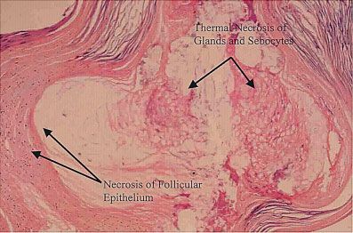
ACNE TREATMENT WITH A 1,450 NM WAVELENGTH LASER                                          107

and the remedies remain non-curative. For many patients,        total laser duration in the range of 160–200 milliseconds
however, acne tends to spontaneously involute after ado-        that was divided into four pulses of equal durations, inter-
lescence. This phenomenon is not well understood [6]. A         spersed with three cryogen sprays. The scheme of inter-
successful treatment, systemic isotretinoin is associated       spersing four laser sub-pulses with cryogen pulses was
with shrinking of sebaceous glands and a remarkable re-         implemented so as to avoid thermal damage to the epi-
duction in sebum output during treatment [6]. However,          dermis. The irradiance ranged from 87 to 110 W/cm2. In
1 year after cessation of treatment, sebum output is re-        addition, there was a pre-laser spray and a post-laser
stored to a level seen before treatment. Despite this return    spray. All sprays were adjustable for precise durations.
to the pre-treatment sebum level, many patients remain          The timing diagram is shown in Figure 1 which shows a
clear of acne. Thus, a temporary effect on sebaceous glands     pre-spray duration of 15 milliseconds, three intermediate
may be sufficient to cause a long-term or even permanent        sprays of 15 milliseconds duration each, and a post-laser
acne clearance. Whether such an effect can be brought           spray of 20 milliseconds duration. The laser light from the
about by a photothermal laser treatment targeting the           device was coupled into an optical fiber. Optics at the end
sebaceous glands in the upper dermis is examined in this        of the fiber produced a homogeneous collimated 4-mm
work. Here, a laser treatment is presented which is shown       diameter circular beam on skin.
to thermally alter the sebaceous glands while preserving
the epidermis. Results of modeling calculations, histology      Monte Carlo Simulations of Light Transport
studies, and human clinical studies are presented.              and Heat Transfer Calculations
                                                                  Light transport and heat transfer calculations were
MATERIALS AND METHODS                                           performed to serve as a guide in optimizing the treatment
                                                                parameters. The results that were obtained were not
Choice of Wavelength
                                                                expected to be exact but were useful in understanding the
  Skin can be divided into three layers: epidermis (up to       temperature distribution and thermal injury for various
a depth of 60–100 mm), the dermis (up to a depth of about       treatment parameters and for optimization of the same.
2–5 mm), and subcutaneous fat, just below the dermis.             In skin, the primary absorber at this wavelength is water
Within skin, sebaceous glands are located at depths from        and it is assumed that the water content does not vary as
about 200–1,000 mm [7] below the stratum corneum. Since         a function of depth. A single layer model with constant
the goal of the sub-surface treatment is to spare the epider-   absorption and scattering properties is used in skin. The
mis and thermally injure the dermis where the sebaceous         absorption coefficient of water is dependent on tempera-
gland structure including the infundibulum resides, the         ture. In an extreme case, the temperature of the skin upon
desired penetration depth of light in skin is about 400 mm.     treatment can increase from 308C to a maximum of 908C.
From Monte Carlo simulations discussed later, at the wave-      The change in ma of water with 18C temperature increase
length of 1,450 nm, the penetration depth is 435 mm with        has been reported to be 0.01475 cm1/8C [9]. A tempera-
water being the principal absorber in skin. Thus, this          ture change of 608C corresponds to a change in absorption
wavelength of 1,450 nm was chosen to produce an injury          coefficient of 0.885 cm1. If skin is 70% water, the change
zone in the dermal region where sebaceous glands are            in absorption coefficient of skin would be 0.6195 cm1. A
located.                                                        change of 0.6195 cm1 in the skin absorption coefficient of
                                                                20 cm1 is small and hence the dependence of absorption
Choice of Cooling                                               coefficient on temperature is neglected. The absorption
  Cutaneous laser treatments have been combined with            properties [9] and scattering properties [10] at 1,450 nm
various cooling methods that can be classified broadly into     wavelength as given in Table 1 were used as input for the
cryogen spray cooling, cold air-cooling, and contact cooling.   simulations. A circular homogeneous collimated 4-mm
Cryogen spray cooling, with its precise control of spray        diameter beam was incident on the tissue surface.
durations, can selectively cool the epidermis while leaving       The tissue volume was discretized into a three-dimen-
the temperature of the dermis unchanged [8]. Hence, cryo-       sional grid with 41, 41, and 1,001 grid points in the x, y,
gen spray cooling was used in this application. In this         and z directions, respectively, where z-direction is perpen-
method, cryogen spurts were applied to the skin surface         dicular to skin surface. The separation between grid points
for a period on the order of 10 milliseconds. The cryogen       was 0.025, 0.025, and 0.0025 cm in x, y, and z directions,
used was tetrafluoroethane, an EPA approved refrigerant
and FDA approved propellant with a boiling point of
268C at atmospheric pressure.

Treatment Device
  The laser device (Candela Corporation, Wayland, MA)
employed a combination of diode laser light at 1,450 nm
and an integrated dynamic cooling device (DCDTM) that
provides cryogen spray cooling. The cryogen cooling allowed
preservation of the epidermis, thus minimizing side effects.    Fig. 1. A timing diagram showing alternate cryogen spray
The radiant exposure range was from 14 to 22 J/cm2 with         and laser pulses used per treatment shot.
108                                                             PAITHANKAR ET AL.

                     TABLE 1. Optical Properties Used in the Monte Carlo Model for Calculation of
                     Light Fluence Rate Distribution

                                           Refractive             Absorption           Scattering              Anisotropy
                     Component              Index, n             coefficient, ma      coefficient, ms           factor, g
                     Air                         1                    0                     0                       0
                     Skin                      1.37                20 cm1              120 cm1                   0.9

respectively. Monte Carlo simulations were performed to                     x, y, and z directions, respectively. The time increment was
calculate light fluence rate at all grid points within the                  chosen as 3 milliseconds.
tissue using the MCML software, given the optical                              The kinetic thermal damage model relates the tempera-
absorption and scattering properties of skin [11,12].                       ture-time history of tissue to the thermal damage. The
   In a second step, heat transfer calculations were per-                   thermal damage measure, O, is traditionally defined as the
formed by solving the heat conduction equation, as given                    logarithm of the ratio of the original concentration of
in Eq. (1), numerically by a finite-difference method.                      native tissue, C(0), to the remaining native state tissue,
                                                                            C(t), and by using an Arrhenius-type kinetic model, it is
      qTðx; y; z; tÞ    k                    m fðx; y; z; tÞ
                     ¼     r2 Tðx; y; z; tÞ þ a              ;       ð1Þ    given at a time t by Eq. (3).
          qT           rCp                       rCp
                                                                                                        ðt
T(x, y, z, t) is the temperature at location (x, y, z) and time                OðtÞ ¼ lnfCð0Þ=CðtÞg ¼ fA expðEa =RTðtÞÞgdt              ð3Þ
t; k, r, and Cp are the thermal conductivity, density,
                                                                                                        0
and specific heat of skin, respectively. The last term on the
right represents heat generation within tissue due to                       where A is a pre-exponential factor, Ea is the activation
absorption of light in which f(x, y, z, t) is the fluence rate.             energy, R is the universal gas constant, and T(t) is the
The boundary condition at the top surface (perpendicular                    thermal history as a function of time t [16]. The parameters
to the z-axis) is described by the convective boundary                      A and Ea are typically determined by fitting experimental
condition as described by Eq. (2).                                          measurements of damaged and undamaged tissue concen-
                 qT                                                         trations as a function of time and temperature. This be-
            k      ¼ hcoolant ðTtissuesurface  Tcoolant Þ:        ð2Þ    havior is to be expected from the exponential nature of the
                 qz
                                                                            function. A set of parameters, Ea ¼ 6.28  105 J/mole and
In the above equation, h is the convective heat transfer                    A ¼ 3.1  1098 sec1 has been reported [16]. The damage
coefficient for either air-skin or cryogen-skin interface.                  was calculated as a function of depth in skin through the
Tcoolant is the temperature of either cryogen or air that is                center of the treated spot by numerically evaluating
in contact with the tissue. The air-skin heat transfer co-                  the integral given in Eq. (3) with the above parameters
efficient and air temperature are used for the top surface                  and the calculated temperature evolution with time.
except on the treatment spot where the respective values
for cryogen-skin are used during the time period when                       Ex Vivo Human Skin Histology
cryogen spray is incident on skin. The value of the cryogen-                  A human skin sample was obtained from an elective
skin heat transfer coefficient has been reported as high as                 breast reduction at the University of Massachusetts
40,000 W/m2K [13] and as low as 2,400 W/m2K [14]. An                        Memorial Hospital, Worcester, MA. The sample was trans-
intermediate value of 4,000 W/m2K has been reported by                      ported at 48C and used in the experiments within 8 hours.
Pikkula and we used this value [15]. Torres et al. [14] re-                 During the experiment, the sample was placed on a warm
ported the cryogen temperature to be 448C and we used                      metal plate, the temperature of which was maintained at
this value. The values of air-skin heat transfer coefficient                328C by immersing part of it in a temperature-controlled
and air temperature chosen were 50 W/m2K and 308C,                          water bath. Treatments were performed on different spots
respectively. The parameters used in the heat transfer cal-                 with a 4-mm diameter spot with various combinations of
culations are provided in Table 2. For the finite difference                spray and radiant exposures. Biopsies with a 3-mm punch
heat transfer calculations, the tissue volume was discretiz-                were taken and fixed in a 10% buffered formalin solution
ed into a three-dimensional grid with 21, 21, and 101 grid                  within 20 minutes after the treatment. The samples were
points in the x, y, and z directions, respectively. The sep-                processed and stained by hematoxylin and eosin (H&E)
aration between grid points was 0.05, 0.05, and 0.005 cm in                 stain and examined microscopically.

TABLE 2. Values of Parameters Used in the Heat Transfer Calculations

Laser                  Laser                               Intermediate                                          Thermal       Cryogen-skin
radiant       Spot     pulse     Cryogen      Pre-laser   spray duration   Post-laser                         diffusivity of   heat transfer
exposure      size    duration temperature spray duration   (split in 3) spray duration                       tissue, k/rCp     coefficient
16 J/cm2     4 mm      210 ms          448C             15 ms              45 ms            20 ms           8  104 cm2/sec 4,000 W/m2K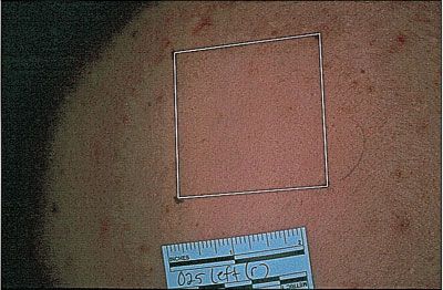
ACNE TREATMENT WITH A 1,450 NM WAVELENGTH LASER                                           109

Rabbit Ear Histology Study                                      on both sides of the back. Institutional Review Board
   The rabbit ear model has been developed by Kligman           approval was obtained prior to initiation of the study and
and Mills [17]. The rabbit ear histological study was           informed consent was obtained from each of the patients
intended to test the following hypothesis: Thermal injury       prior to enrollment.
to sebaceous glands residing within the dermis is caused           Bilateral areas of the treated and control sites were
by the laser whereas the thermal injury to the epidermis is     mapped on a transparent paper to track the location of
prevented by the DCD cryogen spray. The thermal injury          lesions and ensure the accuracy of site selection and lesion
to the epidermis, the dermis, sebaceous glands and asso-        counts at all time points. The selection of treated and con-
ciated structures in the dermal region was evaluated after      trol sides was randomized. The treatment area received
treatment with the test laser device in a rabbit ear model      laser and cryogen, while the control area received only
with a wide range of treatment parameters.                      cryogen spray. The areas of treated and control sites on the
   The study was conducted at Primedica Corporation,            back were up to approximately 36 cm2 each. Four treat-
Worcester, MA. Institutional Animal Care and Use Com-           ments separated by a period of 3 weeks were administered
mittees (IACUC) approval was obtained prior to the animal       to the same treated area. The treatment was performed on
study. The procedures and animal husbandry was per-             the entire selected area and not necessarily on lesions
formed as described in the ‘‘Guide for the Care and Use of      only.
Laboratory Animals,’’ National Research Council, revised           After the first treatment, patients were seen for a 1-day
1996 and/or in accordance with the standard operating pro-      and a 1-week follow-up. For subsequent treatments, they
cedures of Primedica. The 19 New Zealand white rabbits in       were seen every 3 weeks for follow-ups and treatments
this study, aged 6–9 months, underwent procedures as            until each completed a total of four treatments. After the
indicated in Table 3. Six treatment sites per ear were          fourth treatment, patients were seen for follow-up visits at
marked by tattoo ink on the ventral aspects of each ear.        6, 12, and 24 weeks. Photographs of the treated and control
Hair was clipped from the ears prior to tattooing and           sides were taken before the initial treatment at every
treatment. Rabbits were tranquilized with a subcutaneous        treatment or follow-up visit. The radiant exposure was
injection of medetomidine prior to tattooing, laser treat-      chosen by the clinical investigator so as to be lower than
ment, punch biopsy collection and as necessary to facilitate    the radiant exposure that caused epidermal whitening.
handling. A single treatment parameter set was used on          Radiant exposure values ranged between 14 and 22 J/cm2;
each ear on six treatment spots. On day 1, laser treatment      the cooling parameters for each subject were kept the
was applied within the boundaries of each treatment site        same or varied slightly. The average radiant exposure was
with a 4-mm diameter spot. Within an hour after treat-          18 J/cm2.
ment, tissue samples from two adjacent sites per ear for           During all treatments and follow-up visits, the physi-
one treatment parameter set and a single untreated control      cians and staff recorded and maintained records of all
site were obtained by a 3-mm punch biopsy. Two more             patients describing clinical observations associated with
punch biopsies of treated sites of each ear were also col-      the treatments, including lesion counts, acne severity, as
lected on day 3. The final treatment sites and control tissue   well as before and after photographs. Lesion counts in-
were collected at necropsy on day 7. Prior to necropsy,         cluded all non-inflammatory and inflammatory lesions.
euthanasia was performed by deep anesthesia with IV             Biopsies of treatment sites were obtained immediately
sodium pentobarbital, followed by exsanguination. Tissue        after treatment in four subjects. Additional biopsies were
samples from the treated and control sites were evaluated       obtained at 6, 12, or 24-week follow-ups. The biopsy sam-
histopathologically. Several radiant exposures and DCD          ples taken immediately after treatment were fixed in for-
settings were evaluated.                                        malin and examined microscopically after H&E staining.
                                                                Histological analyses of the treatment effects on both the
                                                                skin and the sebaceous glands were performed.
Human Clinical Study                                               Clinical observations of the treated and control sides
  The objective of this study was to evaluate the effective-    were graded and recorded. These observations included new
ness of the 1,450 nm laser for the treatment of acne.           or recurrent lesion counts, acne severity, erythema, edema,
Twenty seven subjects were enrolled in the study con-           blistering, abnormal pigmentation (hyper- or hypo-), and
ducted at the Naval Medical Center in San Diego, CA.            scarring. The assessment of the above observations was
Volunteers with acne on bilateral areas of the upper back       performed at all time points on a scale of 0–3 (0, absent; 1,
were enrolled. At baseline, the acne severity was similar       mild; 2, moderate; and 3, severe).

                TABLE 3. Rabbit Ear Histology Study Design Table

                Number of         Right             Left             Laser            Punch
                animals            ear              ear            treatment          biopsy      Necropsy
                19            6 treatment      6 treatment      Day 1, each        Day 1 and 3      Day 7
                                sites/animal     sites/animal    treatment site
                Day 1 punch biopsy ¼ just after treatment (within 1 hour).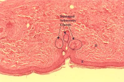
110                                                PAITHANKAR ET AL.

  In the data analysis, the mean lesion count and the
standard deviation at baseline and at different follow-
up time points were calculated. Student t-test (paired
samples) was performed comparing the lesion counts at
the baseline with that at different follow-up time points for
both the treated and control sides.

RESULTS
Monte Carlo Simulations of Light Transport
and Heat Transfer Calculations
  The results of the Monte Carlo simulations yielded the
penetration depth, defined as the depth at which the flu-
ence rate reaches (1/e), i.e., 36.8% of the fluence rate at
the surface, as 439 mm. If scattering effects were absent,
the penetration depth, according to Beer law, is 1/ma or
1/20 cm1 or 500 mm. Thus, scattering effects reduce the
penetration depth to 439 mm. The results of one repre-             Fig. 3. A plot of thermal damage versus depth in skin.
sentative calculation are discussed. Laser energy of 2.01 J
with a 4-mm circular spot that corresponds to a radiant
exposure of 16 J/cm2 was used. A cooling scheme that pro-
vides a pre-laser spray of 15 milliseconds, three inter-
                                                                the damage can be increased or decreased by adjusting the
mediate sprays of 15 milliseconds each, and a final post-
                                                                laser radiant exposure.
laser spray of 20 milliseconds was employed. This scheme
was expected to lead to epidermal preservation. The total
laser time of 210 milliseconds was divided into four pulses     Ex Vivo Human Skin Histology
of equal durations and equal energies. The evolution of           Figure 4 shows a histological section of skin after
spatial temperature profiles with time was calculated.          treatment with radiant exposure of 20.6 J/cm2, pre-laser
  Figure 2 shows a color plot of calculated temperature         spray of 10 milliseconds, intermediate spray consisting of
versus time and depth. Different colors represent different     three sprays of 10 milliseconds each, and a post-laser
levels of temperature. The repeated cryogen sprays cooled       spray of 20 milliseconds. It shows that the epidermis is
the epidermis whereas laser caused heating of the upper         preserved whereas the upper dermis is darker and coagu-
dermis. The peak temperature was calculated to be 88.88C        lated indicating thermal damage. The typical laser radiant
at the end of the last laser sub-pulse at a depth of 150 mm.    exposure values used in non-ablative treatment of subjects
Figure 3 shows the damage profile predicted by the kinetic      as discussed later are lower than the value used in
thermal damage model on a log scale as a function of depth      demonstrating the histology and hence the treatments
along the center of the treatment spot. The magnitude of        created a milder thermal injury.

                                                                Fig. 4. Histological section through the treatment spot of
                                                                ex vivo human skin immediately after treatment processed
Fig. 2. A color plot of calculated temperature versus time      with H&E staining. The arrow indicates the zone of thermal
and depth. Five cryogen pulses result in epidermal cooling.     damage. Note epidermal preservation and thermal damage
Thermal heating of the upper dermis is achieved.                within the upper dermis.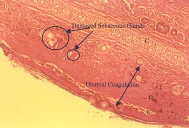
ACNE TREATMENT WITH A 1,450 NM WAVELENGTH LASER 111
Rabbit Ear Histology
Sebaceous necrosis with minimal epidermal damage
was seen for several parameter sets on the treated side. No
changes were seen on the control side. Figure 5 shows a
photograph of the biopsy section at day 1, after a radiant
exposure of 15 J/cm2 and no DCD. Full thickness necrosis
is seen. The dermis and the associated sebaceous glands
are completely destroyed. Also, the epidermis is also des-
troyed since no cooling was used. Figure 6 shows a photo-
graph of the slide at day 1 after treatment with 24 J/cm2
and a DCD consisting of a pre-spray of 10 milliseconds,
three intermediate sprays of 13.3 milliseconds each, and a
post-spray of 20 milliseconds; see Figure 1 for the timing
diagram of sprays. The epidermis was mostly preserved
whereas the dermis was damaged; some epidermal separa-
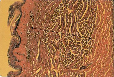
tion was noted however. Three lobes of sebaceous glands Fig. 6. Histology of rabbit ear with 24 J/cm2 and DCD con-
were thermally damaged. Both tinctorial changes and py- sisting of a pre-spray of 10 milliseconds, three interme-
knotic nuclei were seen in the damaged sebaceous glands. diate sprays of 13.3 milliseconds each, and a post-spray of
A close up of the damaged sebaceous gland is shown in 20 milliseconds at day 1.
Figure 7. Similar epidermal preservation and damage to
the dermis and sebaceous glands was seen at day 3 for
the parameter sets of 24 J/cm2 and DCD consisting of a
pre-spray of 10 milliseconds, three intermediate sprays of the treated side and a small change on the control side. A
10 milliseconds each, and post-spray of 20 milliseconds as statistically and clinically significant reduction ( p < 0.01)
shown in Figure 8. A close-up photograph of damaged in lesion counts was seen on the treated side. While the
sebaceous glands is shown in Figure 9. At day 7, several number of subjects at different follow-up times varied, for
sebaceous glands were found to be present and undamaged all 15 subjects completing their 24-week follow-up, no acne
under the histological examination for most of the para- lesions were seen on the treated areas of 14 subjects.
meter sets. Figure 10 is a photograph of the treated side on a
subject’s back 3 weeks after the second treatment. Four
Human Clinical Study marks delineate the treatment area within which clear-
Clinical efficacy: acne improvement. The mean ance of lesions is seen while lesions are seen outside the
lesion count and the standard deviation at baseline and treated area. Figure 11 shows a photograph of the control
at different follow-up time points are given in Table 4. The side of the same subject’s back at the same time point.
p-values from a Student t-test (paired samples) comparing Several lesions are seen on the control side.
the lesion counts at baseline with those at different follow- Safety and side effects. There were no unusual side
up time points for both the treated and control sides are effects or adverse reactions. In brief, the most common
also given. For the 6, 12, and 24-week follow-up time clinical effect seen in subjects as a response to the treat-
points, there is a mean reduction of five or more lesions on ments was erythema, which was expected. Erythema and
Fig. 5. Histology of rabbit ear after treatment with 15 J/cm2
and no DCD at day 1. ‘E’, epidermis; ‘D’, dermis; and ‘C’, Fig. 7. A close-up of the damaged sebaceous glands showed in
cartilage in this and all following figures. Figure 6.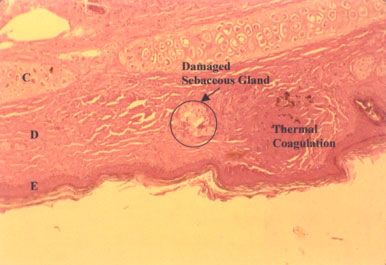
112 PAITHANKAR ET AL.
TABLE 4. Results of Student t-Test (Paired Samples) Comparing the Lesion Count Between Follow-Up and Baseline for the Treated and Control Sides
p-value
0.75
0.55
0.78
0.05
0.24
0.68
(standard deviation)
Mean lesion count
at follow-up
5.93 (3.05)
5.48 (2.04)
5.68 (2.23)
4.95 (1.88)
5.06 (2.16)
4.73 (1.98)
Control
(standard deviation)
Mean lesion count
Fig. 8. Histology of rabbit ear after treatment with 24 J/cm2
at baseline
5.81 (2.37)
5.70 (2.40)
5.77 (2.43)
5.65 (2.43)
5.53 (2.58)
5.00 (2.14)
and DCD consisting of a pre-spray of 10 milliseconds, three
intermediate sprays of 10 milliseconds each, and post-spray of
20 milliseconds at day 3.
edema had resolved by the next follow-up observation.
Three out of twenty-seven subjects showed hyperpigmen-
The mean difference, standard deviation, and the p-value are presented at each follow-up time point.
tation at some time during the treatment regimen. Hyper-
10
2.19 1010
8.22 1011
1.64 107
1.56 108
2.34 109
p-value
pigmentation was graded ‘‘severe’’ for one subject at one
1.09 10
time point; the other two were classified as ‘‘mild.’’ Hyper-
pigmentation uniformly resolved and was absent for all
subjects at the 6, 12, and 24-week follow-ups after the
fourth treatment. No signs of purpura or scarring were
evident in any subject at any time point. (standard deviation)
Mean lesion count
Histological changes after treatment. The density at follow-up
of sebaceous glands on the back skin is not high and only
2.67 (2.24)
1.04 (1.66)
0.68 (1.86)
0.15 (0.67)
0.12 (0.33)
0.13 (0.52)
one of the biopsy samples taken immediately after treat-
ment yielded sebaceous glands. In this biopsy sample
obtained after treatment with 17.2 J/cm2 and DCD with
Treatment
pre-spray of 10 milliseconds, three intermediate sprays of
10 milliseconds each, and a post-spray of 20 milliseconds, a
rupture of the pilosebaceous unit with thermal coagulation
of the sebaceous lobule and follicle was noted as seen in
(standard deviation)
Mean lesion count
Figure 12. The overlying epidermis was unaltered. Long
term biopsies taken at 2 and 6 months after the treatment
at baseline
7.22 (3.59)
7.13 (3.49)
7.18 (3.57)
6.80 (3.12)
6.94 (3.36)
5.67 (1.29)
27
23
22
20
17
15
N
3 weeks spost Tx no. 1
12 weeks post Tx no. 4
24 weeks post Tx no. 4
3 weeks post Tx no. 2
3 weeks post Tx no. 3
6 weeks post Tx no. 4
Follow-up time-point
Fig. 9. A close-up of the damaged sebaceous glands shown in
Figure 8.
ACNE TREATMENT WITH A 1,450 NM WAVELENGTH LASER                                           113

Fig. 10. Photograph of the treated area at 3 weeks after the    Fig. 12. Histology of treated human back skin after treat-
second treatment. Lesions clearance is seen.                    ment; a close-up of a sebaceous gland. A complete rupture of
                                                                the pilosebaceous unit with thermal coagulation of the entire
                                                                sebaceous lobule and follicle is seen.
on the back and face with similar treatment parameters
showed sebaceous glands and associated ductal structures
that were unaltered from their pre-treatment (control)          (ALA)-photodynamic therapy (PDT). ALA was applied topi-
counterparts. Therefore, on routine microscopy, there           cally and the skin was irradiated with light in the 550–
appeared to be no long-term alteration in adnexal struc-        700 nm wavelength range. Clinically and statistically
ture architecture. The epidermis and the follicular             significant clearance of inflammatory acne for 10 weeks
structures also appeared normal.                                after a single treatment and at least 20 weeks after
                                                                multiple treatments was observed. However, transient
DISCUSSION AND CONCLUSIONS                                      hyperpigmentation, superficial exfoliation, and crusting
  Various light-based therapies are under development           were observed. They concluded that topical ALA plus red
for treatment of acne. Reduction in acne lesion count upon      light is an effective treatment for acne vulgaris, although
exposure to blue, red, violet, and ultra-violet light have      associated with significant side effects. In another study
been reported [18–22]. The mechanism of treatment with          on ALA-PDT [24], all patients had apparent improvement
blue light is believed to be absorption by endogenous           of facial appearance and reduction of new acne lesions at 1,
porphyrins produced by P. acnes and subsequent photo-           3, and 6 months following PDT treatment. The adverse
toxic effect on P. acnes to cause a beneficial effect in acne   effects were discomfort, burning, and stinging during
symptoms [18]. These therapies do not target sebaceous          irradiation, edematous erythema for 3 days after PDT,
glands as such and long-term remission is not proven and        epidermal exfoliation from the 4th to the 10th day,
may not be possible since repopulation by P. acnes is likely    irritation and hypersensitivity to physical stimulation for
to occur after cessation of treatment. Hongcharu et al. [23]    10 days after PDT, and pigmentation or erythema after
have reported treatment of acne with aminolevulinic acid        epidermal exfoliation. In summary, ALA-PDT may be an
                                                                effective treatment but is associated with side effects.
                                                                   In this article, a device combining a diode laser at
                                                                1,450 nm wavelength and cryogen cooling has been studied
                                                                for non-ablative treatment of acne vulgaris. Monte Carlo
                                                                modeling and heat transfer calculations predict that the
                                                                combination of DCD cooling and 1,450 nm laser can be used
                                                                to achieve thermal damage that is peaked around a depth
                                                                of 150–200 mm while minimizing the damage in the epi-
                                                                dermis and in the deeper dermis. Only qualitative and not
                                                                quantitative comparisons can be made between the model-
                                                                ing calculations and the experiments due to the many
                                                                approximations implicit in the model calculations. Histo-
                                                                logical examination of ex vivo human skin showed that the
                                                                concept of achieving thermal injury to the upper dermis
                                                                while preserving the epidermis is feasible. Rabbit ear was
                                                                used as a model to study the histological effects of the
                                                                irradiation with laser and cooling with DCD. Various treat-
Fig. 11. Photograph of the control area at 3 weeks after the    ment parameters were used. Histological analysis at day 1
second treatment. Lesions are still present.                    and 3 with certain treatment parameters showed damage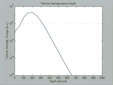
114                                                    PAITHANKAR ET AL.

to the dermis and thermal alteration of sebaceous glands             3. Sykes NL, Webster GJ. Acne: A review of optimum treat-
located within the dermis while the epidermis was pre-                  ment. Drugs 1994;48:59–70.
                                                                     4. Eady EA. Bacterial resistance in acne. Dermatology 1998;
served. Histological examination of biopsies taken at day 7             196:59–66.
indicated that sebaceous glands were intact or had recover-          5. Turkington CA, Dover JS. Skin Deep: An A–Z of Skin
ed from initial injury. Biopsies and histology of ex vivo               Disorders, Treatment and Health. New York: Facts on File,
                                                                        Inc; 1996. p 8.
human skin showed that it is possible to achieve heat-               6. Plewig G, Kligman AM. Acne and Rosacea, 2nd edn. Berlin:
ing of the dermal region containing the sebaceous glands                Springer-Verlag; 1993. p 711.
while preserving the epidermis. Immediate as well as 3-              7. Montagna W, Kligman AM, Carlisle KS. Estimation from
and 7-day histological response to treatment was studied                histology photographs in Atlas of Normal Human Skin. New
                                                                        York: Springer-Verlag; 1992.
on the rabbit ears. This showed thermal alteration of the            8. Anvari B, Tanenbaum BS, Milner TE, Kimel S, Svaasand LO,
sebaceous glands immediately and at 3 days; the sebac-                  Nelson JS. A theoretical study of the thermal response of skin
eous glands do not show pronounced histological effect at               to cryogen spray cooling and pulsed laser irradiation: Impli-
                                                                        cations for treatment of port wine stain birthmarks. Phys
the 7-day time point.                                                   Med Biol 1995;40:1451–1465. [published erratum appears in
   A human clinical study for the treatment of acne vul-                Phys Med Biol 1996; 41:1245.]
garis was conducted on backs of males with a 1,450 nm                9. Gardner CM. Absorption coefficient of water. Personal
laser combined with cryogen spray cooling. A reduction in               Communication, Brookline, MA, 5/6/1998.
                                                                    10. Lask GP, Lee PK, Seyfzadeh M, Nelson JS, Milner TE,
lesion count was seen immediately after first treatment. A              Anvari B, Dave D, Geronemus RG, Bernstein LJ, Mittelman
statistically and clinically significant reduction in lesion            H, Ridener LA, Coulson WF, Sand B, Baumgarder J,
counts was seen on the treated side compared to the control             Hennings DR, Menefee RF, Berry M. Nonablative laser
                                                                        treatment of facial rhytides. Proc SPIE 1997;2970:338–349.
side at the 6, 12, and 24-week follow-ups after the fourth          11. Jacques SL, Wang LH. Monte Carlo modeling of light trans-
treatment. Longer-term follow-ups will be performed to                  port in tissue. Optical thermal response of laser irradiated
see if these results persist. Side effects included transitory          tissue. Chapter 4. New York: Plenum Press; 1995. 73–100.
                                                                    12. Wang LH, Jacques SL, Zheng LQ. MCML—Monte Carlo
erythema and edema. Although four treatments were used
                                                                        modeling of photon transport in multi-layered tissues.
in this study, reduction in acne lesions was seen after a               Comput Methods Programs Biomed 1995;47:131.
single treatment. Thus, lower number of treatments may              13. Anvari B, Milner TE, Tanenbaum BS, Nelson JS. A
suffice. Determination of optimum number of treatments                  comparative study of human skin thermal response to sap-
                                                                        phire contact and cryogen spray cooling. IEEE Trans Biomed
and interval between treatments is being done. Histo-                   Eng 1998;45:934–941.
logical analysis of biopsies obtained immediately after             14. Torres JH, Nelson JS, Tanenbaum BS, Milner T, Goodman
treatment indicated an alteration in sebaceous glands                   DM, Anvari B. Estimation of internal skin temperatures in
                                                                        response to cryogen spray cooling: Implications for laser
structure. Long term biopsies after the treatment on the                Therapy of port wine stains. IEEE J Selected Top Quantum
back showed sebaceous glands and associated ductal struc-               Electron 1999;5:1058–1066.
tures that were unaltered from their pre-treatment (control)        15. Pikkula B. Heat transfer coefficient at the skin-cryogen
counterparts. The reduction in acne lesion count is likely              interface. Personal Communication, Houston, TX, 12/13/
                                                                        2001.
due to a slight functional impairment of the glands secon-          16. Pearce J, Thomsen S. Rate process analysis of thermal
dary to mild thermal damage created at the time of irra-                damage. Chapter 17. In: Welch AJ, van Gemert MJC, editors.
diation. Accutane, an effective treatment of acne, also has             Optical-thermal response of laser-irradiated tissue. New
                                                                        York: Plenum Press; 1995. pp 160–162.
a temporary effect on sebaceous glands. Further studies on          17. Kligman AM, Mills OH. Acne cosmetica. Arch Deramotol
histological and clinical effects of this treatment on the              1972;106:843.
facial acne in humans are in progress. This treatment may           18. Shalita AA, Harth Y, Elman M, Slatkine M, Talpalariu G,
                                                                        Rosenberg Y, Korman A, Klein A. Acne phototherapy using
have a secondary effect on sebum production rate and P.
                                                                        u.v. free high intensity narrow band blue light—3 center
acnes population, both of which are associated with acne.               clinical study. Proc SPIE 2001;4244:61–73.
The effect of the laser treatment on these two is being             19. Cunliffe WJ, Goulden V. Phototherapy and acne vulgaris. Br
evaluated in ongoing studies.                                           J Dermatol 2000;142:855–856.
                                                                    20. Papageorgiou P, Katsambas A, Chu A. Phototherapy with
                                                                        blue (415 nm) and red (660 nm) light in the treatment of acne
ACKNOWLEDGMENTS                                                         vulgaris. Br J Dermatol 2000;142:973–978.
                                                                    21. Konig K, Ruck A, Schneckenburger H. Fluorescence detec-
  The rabbit ear histology study was supported by the                   tion and photodynamic activity of endogeneous protopor-
National Institutes of Health Grant Number 1R43AR4-                     phyrin in human skin. Opt Eng 1992;31:1470–1474.
6938-01. The authors thank Dr. Elliot Lach for providing            22. Meffert H, Scherf HP, Sonnichsen N. Treatment of acne vul-
                                                                        garis with visible light. Dermatol Monatsschr 1987;173:678–
the skin samples used in the ex vivo human skin study.                  679.
                                                                    23. Hongcharu W, Taylor CR, Chang Y, Aghassi D, Suthamjariya
                                                                        K, Anderson RR. Topical ALA-photodynamic therapy for the
REFERENCES                                                              treatment of acne vulgaris. J Invest Dermatol 2000;115:183–
 1. Bergfeld WF, Odom RB. New perspectives on acne. Clinician           192.
    1994;12:3–29.                                                   24. Itoh Y, Ninomiya Y, Tajima S, Ishibashi A. Photodynamic
 2. Kellett SC, Gawkrodger DJ. The psychological and emotional          therapy of acne vulgaris with topical delta-aminolaevulinic
    impact of acne and the effect of treatment with isotretinoin.       acid and incoherent light in Japanese patients. Br J Dermatol
    Br J Dermatol 1999;140:273–282.                                     2001;144:575–579.
You can also read