The impact of testosterone levels on J-wave patterns observed in healthy Turkish males - DergiPark
←
→
Page content transcription
If your browser does not render page correctly, please read the page content below
Early Online

The European Research Journal 2019;0(0):0-0                                                                                ORIGINAL A RTICLE

The impact of testosterone levels on J-wave patterns observed
in healthy Turkish males
Burak Hünük

Department of Cardiology, Yeditepe University School of Medicine, İstanbul, Turkey

                                                                                                                                DOI: 10.18621/eurj.519192

ABSTRACT
Objectives: Early-repolarization (ER) and Brugada-type-ECG-patterns (BTEP) have recently been grouped
under a common terminology called “J-wave patterns” (JWP) and have been associated with an increased risk
of sudden-cardiac-death. Scarce data is present about the male dominance in JWP and the probable effects of
gonadal hormones on cardiac ion-channel functions. We sought to evaluate the relationship of testosterone-
levels and the presence of JWP in healthy Turkish-males.
Methods: One hundred eighty-five healthy male volunteers between ≥18 to ≤50 years old without any cardiac
disorders were evaluated. ECG, blood biochemistry and total testosterone levels were obtained together with
thorough physical examination. Subjects with complete-bundle-branch-block, non-sinus-rhythms and any
abnormality on cardiac examination were excluded from the study. BTEP was searched according to the
EHRA/HRS 2016 Consensus Conference on V1-V3. ER on ECG was defined as J-point elevation of ≥ 0.1 mV
in ≥ 2 leads in the inferior (II, III, aVF) (Inferior ER), lateral (DI, aVL, V4-6) (Lateral ER) or both (Inferolateral
ER).
Results: A total of 179 subjects (mean age 34.9 ± 7.9 years) were included in our analyses. Three BTEP (1.7%)
and 45 ER (26%) were detected. 22 were lateral (49%), 13 inferior (29%) and 10 were (22%) inferolateral ER.
JWP (+) subjects (n = 48, 27%) were demonstrating significantly lower basal heart rates (73.9 ± 11bpm vs
68.4 ± 10.3 bpm, p = 0.001) and longer PR intervals (153.9 ± 20.3 ms vs 163.3 ± 21.6 ms, p = 0.01). JWP (+)
subjects had significantly higher testosterone levels compared with the ones without (485.5 ± 128.3 ng/dl vs
559.3 ± 167.7, p < 0.001). In the subgroup analyses, BTEP and inferior/inferolateral ER patterns were
significantly associated with higher testosterone levels compared with the JWP (-) population, while
testosterone levels of subjects with lateral ER was not significantly higher. Electrolytes and blood chemistry
values were non-significant between JWP + and - subjects. In the ROC analysis, the cut-off value for predicting
the presence of a JWP on ECG was 629 ng/dl with a sensitivity of 44% and specificity of 86% [AUC = 0.66
(95% CI: 0.56-0.75) p = 0.001]. In multivariate analysis, total testosterone level > 629 ng/dl was significantly
predicting a JWP on ECG, even outperforming age and hs-CRP levels with an OR of 4.57 (95% CI 1.910-
10.9, p = 0.001).
Conclusions: Testosterone might be associated with the male predominance observed in the JWP. More
malignant inferior/inferolateral ER seems to be mainly associated with the high testosterone levels in Turkish
male population. This finding might be attributed to the previously demonstrated effects of testosterone on
cardiac ion-channel functions, especially outward-K channels.

Keywords: Electrocardiogram, j-wave pattern, male, testosterone

                              Received: February 3, 2019; Accepted: March 4, 2019; Published Online: March 11, 2019
                     Address for correspondence: Burak Hünük, MD., Yeditepe University School of Medicine, Department of Cardiology, İstanbul, Turkey
                     E-mail: burakhunuk@hotmail.com

                                                           Copyright © 2019 by The Association of Health Research & Strategy
 e-ISSN: 2149-3189
                                                                        Available at http://dergipark.gov.tr/eurj

The European Research Journal Volume                          Issue             2019                                                                    1
Eur Res J 2019;0(0):0-0                                                             Testosterone and J-waves patterns in males

E     lectrocardiographically the point where the QRS
      complex ends and a prominent ST segment starts
is called the J point [1]. In 1953, Osborn et al. [2] de-
                                                              Ito channels in the male right ventricular outflow tract
                                                              and right ventricular epicardium is shown to be one of
                                                              the reasons, but some aspects of the BS clinic cannot
fined J points in the form of a positive deflection from      be fully explained in this way, such as its clinical pre-
the isoelectric line in experimental hypothermia which        ponderance after the puberty and attenuation of the
were clinically referred to as “Osborn Waves” and then        malignant clinical features after the 6th decade in
as “J waves”. The fact that such changes are frequently       males [10, 18, 19]. In research studies, testosterone
observed in the young male population also led to the         and dihydrotestosterone levels effected the Na, Ca and
association of J waves with ST-segment elevation as           K (especially Ito) channels forming the action poten-
a benign electrocardiographic finding called “Early           tial dome leading to a repolarization dispersion simi-
Repolarization Variant/ Pattern (ER) [3–5]. In recent         larly observed in J-wave syndromes [20]. It was also
years, several population-based studies in which J            shown that the Brugada pattern was lost in men who
waves such as inferolateral ER and Brugada type ECG           underwent orchiectomy due to prostate cancer [21].
patterns (BTEP) were determined to be associated              Regarding the ER patterns, young athletic male pre-
with an increased sudden cardiac death (SCD) and              dominance might give a clue about the probable con-
lethal ventricular arrhythmias (VA) have been pub-            tributing effects of higher testosterone levels to the ER
lished worldwide [6-11]. Although they have different         phenomenon which share similar ionic and clinical
diagnostic criteria, clinical phenotypes such as Bru-         characteristics with BS [12].
gada Syndrome (BS) and Early Repolarization Syn-                   Hence, we aimed to evaluate the relationship of
drome (ERS) which are associated with specific J              testosterone-levels and the presence/distribution and
wave findings / J wave patterns with different disper-        clinical features of JWP in healthy Turkish-males and
sion of repolarization mechanisms in different regions        compared the effects with other clinical and laboratory
of the heart and causing life-threatening arrhythmias,        parameters which might contribute to the frequency
have been grouped under a new terminology and con-            of JWP such as age and inflammation parameters.
cept as “J Wave Syndromes” in recent years [12, 13].
National and international research on the etiology,
mechanisms and frequency of J wave syndromes are              METHODS
increasing and new studies are designed to recognize
this assumingly rare but fatal syndrome earlier and re-           One hundred eighty-five healthy male volunteers
veal its physiopathology [13-16]. In these studies, the       between ≥ 18 to ≤ 50 years old without any diagnosed
frequencies of observed J wave patterns (JWP) were            cardiac/systemic history and no family history of SCD
highly heterogenous between 3-5% to 0.1% in the gen-          were prospectively and consecutively evaluated for
eral population according to the race, age and comor-         inclusion to the study. The study complies with the
bidities of the study population, however constantly          Declaration of Helsinki, patients provided signed
and significantly found to be highest in the male sam-        informed consent and the local ethical committee
ple groups irrespective of the other features of the          approval obtained. Supine 12-lead surface ECG,
study population [10, 12, 13]. Even though, the repo-         complete blood count, high sensitivity (hs)-CRP,
larization theory is the most popular explanation about       renal/thyroid function tests and total testosterone
the observed transmural epi-endocardial voltage gra-          levels obtained together with thorough physical
dient in JWPs, contemporary findings in elec-                 examination. Subjects with complete bundle branch
troanatomic mapping/ablation studies state that JWPs          blocks on ECG, non-sinus rhythms and any abnormal
might also occur because of delayed depolarization on         findings on cardiac examination were excluded from
the related segments [13, 17].                                the study.
    Although J wave syndromes such as autosomal
dominant BS genetically demonstrate an equal transi-          Electrocardiographic Assessment
tion in males and females, it is extremely rare to have           The 12-lead ECGs were recorded at 25 mm/s with
a phenotypic appearance in women compared with                a calibration of 10 mm/mV (Nihon Kohden, Tokyo,
men (1:10 ratio) [10, 12, 18]. The presence of more           Japan) and uploaded on the hospital ECG database at

2                                                           The European Research Journal Volume       Issue            2019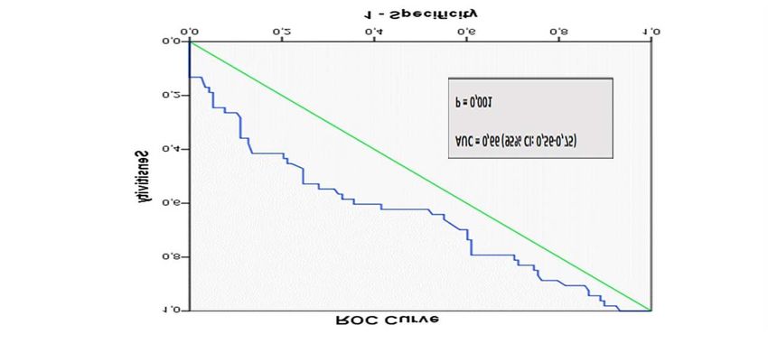
Eur Res J 2019;0(0):0-0                                                                                              Hünük

300 DPI. These images were amplified x10 and then               venous stasis through a 21-gauge cannula inserted into
baseline heart rates, PR, QRS, QTc (Bazett) intervals           an antecubital vein using ethylenediamine tetraacetic
were manually measured by electronic calipers. The              acid containing monovettes (Sarstedt, Nuembrecht,
presence of a lateral (I, aVL, V5-V6), inferior (II, III,       Germany), and transferred immediately to the
aVF) or inferolateral (II, III, aVF, I, aVL, V5-V6) ER          laboratory to be centrifuged. Hs-CRP level was
was defined as an evident J-point elevation of at least         measured on Cobas Integra 400 Plus using a latex
1 mm (0.1 mV) above the isoelectric line in at least            particle-enhanced     immunoturbidimetric       assay
two consecutive leads with either QRS slurring (i.e. a          following the manufacturer's instructions (Roche
smooth transition from the end QRS to the beginning             Diagnostics, Indianapolis, IN). Testosterone levels
of ST-segment) or notching [positive deflection (J-             were determined using electrochemiluminescence
wave) occurring immediately after a positive QRS                immunoassay method and an auto-analyser (Cobas
complex at the onset of the ST-segment] according to            6000, E 601 Roche Diagnostics, GmbH, Mannheim,
the most recently proposed terminology from the latest          Germany). The complete blood counts were evaluated
international consensus documents for the ECG                   using an auto-analyzer Sysmex XT-1800i
definition of ER [13, 18], and “J peak” was accepted            Haematology Analyzer (Sysmex Corporation, Kobe,
as “J point” denoting the peak of a notch or onset of a         Japan). The remaining routine biochemistry
slur. BTEPs were searched according to the                      parameters have been determined by the core
EHRA/HRS 2016 Consensus Conference on V1-V3                     laboratory.
leads [10, 18]. All the ECGs were analysed by the
                                                    Statistical Analysis
author in a blinded fashion for the laboratory findings
of the patients with borderline results being reassessed Continuous variables are expressed as mean ± SD
by another experienced cardiologist.                (if the parameter is normally distributed) or standard
                                                    error of mean (SEM ± SD) whichever is suitable. If
Blood Tests and Analysis                            appropriate, they were compared using the Student’s
    Venous blood samples were drawn with patients t-test. Categorical variables are expressed as numbers
after they rest supine for about 15 min prior to and percentages and, if appropriate, were compared
sampling. Samples were drawn atraumatically without with the Chi-square analysis. Univariate and later

Figure 1. Prevalence of the J-wave patterns in study population. ER = Early repolarization pattern, BTEP = Brugada type ECG
pattern.

The European Research Journal Volume      Issue          2019                                                            3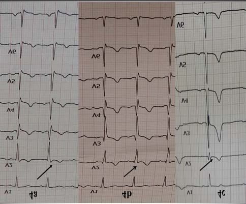
Eur Res J 2019;0(0):0-0 Testosterone and J-waves patterns in males
!
Figure 2. (a) Lateral early repolarization pattern example. Arrows indicate QRS slurring with around 2 mm J point elevation followed
by upsloping benign ST segment changes. (b) Inferior early repolarization pattern example. Arrows indicate QRS slurring with
around 1 mm J point elevation and a malignant type horizontal ST segment elevation is followed.
multivariate analysis was performed to determine the RESULTS
predictive value of significant and predetermined
confounders on the JWP observations using the Of 185 volunteers, 179 male subjects (mean age
logistic regression model. A Receiver Operating 34.9 ± 7.9 years) were included in our analyses after
Characteristic (ROC) curve was plotted in order to the application of inclusion criteria. In total 48 JWPs
determine the diagnostic accuracy of a certain (26.8%) were detected on surface ECG; consisting of
laboratory value. A p value < 0.05 was accepted 3 BTEPs (1 Type-2, 2 type-3) and 45 ER patterns (22
statistically significant. Statistical analysis was lateral, 13 inferior and 10 of them were inferolateral
performed using SPSS 16.0 (IBM Inc., Armonk, New ER) (Figures 1 to 4). JWP (+) subjects were
York, USA). significantly younger, with a lower basal heart rate,
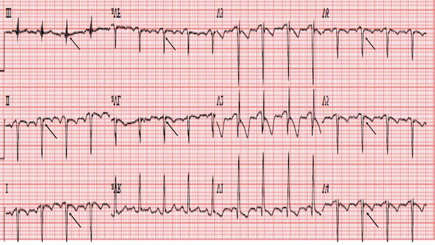
Figure 3. Inferolateral early repolarization pattern. Arrows indicate QRS slurring with around 1 mm J point elevation and a benign
!
type upsloping ST segment elevation is followed in inferior leads and QRS notching on lateral leads followed by upslope ST elevations
most evident on DI, DII, aVF, aVL, V4 to V5.
4 The European Research Journal Volume Issue 2019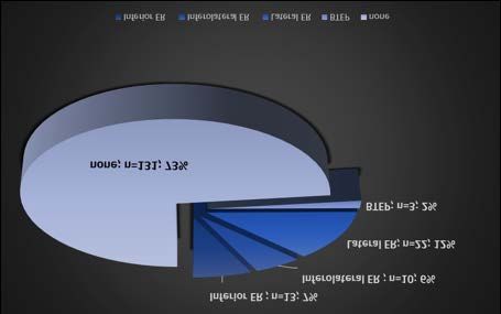
Eur Res J 2019;0(0):0-0                                                                                                         Hünük

Figure 4. Brugada-type-ECG-patterns revealed in our study. 4a: Type-2 Brugada pattern, 4b and c: Type-3 Brugada pattern. Arrow-
heads denote the diagnostic j-waves. No type-1 pattern was found.

longer PR interval and had significantly higher hs-                   testosterone levels, we divided the testosterone levels
CRP and testosterone levels compared with the ones                    into 25th, 50th, 75th and 90th percentiles and showed that
without (Table 1). However, no significant association                significantly more JWPs were demonstrated after the
could be demonstrated with ESR, white blood cell                      50th percentile and significance continues as the
count, neutrophil to lymphocyte ratio and uric acid                   testosterone percentiles rises, when compared with
levels. For the subgroup analyses and to efficiently                  each other (Figure 5). Among the JWPs, BTEPs have
define a probable dose-response effect with the                       the highest mean testosterone value and it was

Figure 5. The frequency of J wave patterns according to the testosterone percentiles Asterix denote a statistically significant and ev-
                                                                                                                !

idently higher J wave patterns in the groups higher than 50th percentile when compared with the low testosterone levels

The European Research Journal Volume          Issue            2019                                                                  5
Eur Res J 2019;0(0):0-0 Testosterone and J-waves patterns in males
!
Table 1. Baseline characteristics of the subjects with and without J-wave patterns
JWP (-) JWP (+) p value
(n = 131) (n = 48)
Age (years) 35.5 ± 8.1 33 ± 5.8 0.048
Weight (kg) 83.1 ± 11.6 81.5 ± 10.6 0.42
Height (cm) 176.2 ± 5.6 176.3 ± 4.8 0.82
Body mass index (kg/m2) 26.7 ± 3.3 26.2 ± 3.5 0.40
ECG parameters
Heart rate (bpm) 74 ± 11.5 68 ± 10 < 0.001
QTc duration (ms) 404.1 ± 18 404 ± 14.3 0.89
PR duration (ms) 153 ± 20.3 163.5 ± 22 0.01
QRS duration (ms) 90.2 ± 11 92 ± 10 0.30
Laboratory results
Glucose (mg/dL) 87.1 ± 11.1 85.9 ± 10.5 0.51
BUN (mg/dL) 29.2 ± 7.2 29.5 ± 6.7 0.75
Creatinine (mg/dL) 0.91 ± 0.12 0.89 ± 0.10 0.28
Uric acid (mg/dL) 5.3 ± 1.01 5.4 ± 1.1 0.53
Sodium (mmol/L) 143.1 ± 2.5 143.1± 2.4 0.94
Potassium (mmol/L) 4.5 ± 0.3 4.5 ± 0.3 0.96
Hemoglobin (g/dL) 14.5 ± 0.9 14.4 ± 0.9 0.57
Platelet count (103/µL) 232.9 ± 57.9 235.1 ± 50.2 0.81
WBC (103/µL) 5.97 ± 1.4 5.63 ± 1.1 0.13
Neutrophil (103/µL) 3,15 ± 1.03 2.86 ± 0.88 0.08
Lymphocyte (103/µL) 2.02 ± 0.46 1.891 ± 0.52 0.17
NLR (% ) 1.56 ± 0.47 1.63 ± 0.68 0.50
hsCRP (mg/L) 1.6 ± 1.7 3.0 ± 2.9 0.004
ESR (mm/h) 6.7 ± 4.6 8.4 ± 6.2 0.07
Total cholesterol (mg/dL) 182.5 ± 34.3 184.2 ± 33.8 0.78
HDL (mg/dL) 41.5 ± 8.7 42.1 ± 9.8 0.75
LDL (mg/dL) 112.2 ± 27.5 112.1 ± 26.3 0.99
Triglycerides (mg/dL) 140.9 ± 78.4 143.0 ± 78.6 0.87
TSH (mIU/L) 1.7 ± 0.9 1.8 ± 0.96 0.73
Vitamin D, 25-Hydroxy (ng/mL) 12.8 ± 6.03 13.2 ± 5.08 0.73
Vitamin B12 (pg/mL) 270.5 ± 118.9 305.9 ± 113.6 0.18
Total Testosterone (ng/dL) 481.6 ± 125.7 556.4 ± 165.3 0.002
LDH (U/L) 346.8 ± 49.9 341 ± 62.1 0.57
Iron (Fe) (µg/dL) 102.6 ± 35.8 89.2 ± 57.5 0.35
Ferritin (mg/L) 92.1 ± 70.8 13.2 ± 5.08 0.79
Folate (ng/mL) 7.58 ± 1.8 7.83 ± 2.05 0.45
Fibrinogen (mg/dL) 270.3 ± 89.4 274.6 ± 96.01 0.83
BMI = body mass index, BUN =blood urea nitrogen, ECG = electrocardiography, ESR = erythrocyte
sedimentation rate, HDL = high density lipoprotein, hsCRP = high-sensitive-C-reactive peptide, JWP = J-wave
patterns, LDH = lactate dehydrogenase, LDL = high density lipoprotein, NLR = neutrophil-to-lymphocyte ratio,
TSH = thyroid stimulating hormone, WBC = white blood cell count
6 The European Research Journal Volume Issue 2019Eur Res J 2019;0(0):0-0 Hünük
Table 2. Testosterone levels among the J-wave patterns
J-wave Pattern Testosterone levels p value*
Mean ± SD 95% Confidence
Interval
Inferolateral ER 600.2 ± 163.3 483.3 717.1 0.005
Inferior ER 593.4 ± 201.6 471.6 715.3
Lateral ER 520.6 ± 145.5 456.1 585.1
BTEP 627.6 ± 206.5 114.6 1140.7
ER = early repolarization pattern, BTEP = Brugada type ECG pattern, *P value is for the statistical significance
of the higher testosterone levels observed in inferior/inferolateral ER and BTEP compared with the lowest
testosterone levels observed in lateral ER group.
!
significant when compared with JWP (-) population testosterone level > 629 ng/dl was significantly
and it was followed by the inferior and inferolateral predicting a JWP on ECG, even outperforming age
ER patterns with significantly higher testosterone and hs-CRP levels with an OR of 4.57 (95% CI 1.910-
levels (Table 2). However, lateral ER patterns did not 10.9, p = 0.001) (Table 3).
demonstrate any significant difference regarding the
mean testosterone levels when compared with the JWP
(-) subjects. In the ROC curve, the Area Under the DISCUSSION
Curve was determined 0.66 (95% CI: 0.56-0.75) (p =
0.001) and a cut-off value for testosterone was In our study, a JWP prevalence of 27% and a
determined as 629 ng/dl with a sensitivity of 44% and BTEP percentage of 1.7% was consistent with the
a reasonable specificity of 86% to predict the presence findings of the previous similar age and gender
of JWP (Figure-6). In multivariate analysis, total matched population based studies [12]. No type-1
Figure 6. ROC analysis to define the predictive value of higher testosterone levels and the presence of J wave pattern on ECG. ROC
= Receiver Operating Characteristics, AUC = Area Under the Curve, Cutt off value for total testosterone level : 629 ng/dL (sensitivity
44%, specificity 86%)
The European Research Journal Volume Issue 2019 7
!Eur Res J 2019;0(0):0-0 Testosterone and J-waves patterns in males
Table 3. Multivariate regression model for the testosterone cut-off value determined in ROC
analysis predicting the odds that the subject might demonstrate a JWP on ECG.
Parameters Odds ratio 95% confidence interval p value
Age 0.95 0.90-1.02 0.950
hsCRP 1.26 1.08-1.47 0.004
BMI 0.97 0.85-1.11 0.662
Vitamin D 1.01 0.93-1.08 0.653
TSH 1.11 0.73-1.68 0.620
Testosterone value > 629 ng/dL 4.57 1.910-10.9 0.001
hsCRP = high-sensitive-C-reactive peptide, BMI = Body mass index, TSH = thyroid stimulating hormone
!
BTEP was found. The ones with JWP were a transmural voltage gradient between endocardium
significantly demonstrating a higher vagal tone and epicardium on partial regions of the heart leading
(relatively low basal heart rate, longer PR interval) to lethal ventricular arrhythmias [12].
compared with the ones without JWP, a finding also
compatible with the previous population based studies Clues about the possible role of the gonadal hormones
and proposed mechanisms[12, 13, 22]. Our healthy on electrocardiogram
male population gave out evidently higher prevalance It has formerly been demonstrated that, on
than the general population for these relatively rare average, adult men have shorter QT intervals than do
ECG findings however, it was the result of our design women, but gender differences become apparent after
to increase the expected number of cases in order to the onset of puberty [23]. This gender difference is
perform significant statistical calculations. absent at birth and in young children [24]. Throughout
Testosterone demonstrated a dose response effect on puberty, the QTc interval in males shortens by 20 ms,
the prevalence and type of JWPs. Significantly more whereas the QTc of females remains unchanged,
JWPs were being observed in the higher levels of resulting in a 6% shorter QTc in males compared to
testosterone and suggestively high-risk patterns such females [25] Pecori-Geraldi et al. [26] compared the
as BTEP and inferior/inferolateral ER patterns were QTc in 26 men with hypogonadism with the QTc in
related with the highest percentile of testosterone 26 age-matched controls. They reported a higher
distribution. In the ROC analysis a cut of testosterone prevalence of a prolonged QTc in hypogonadal men
value of 629 ng/dl were significantly found to be (15%) than in controls (0%) (p < 0.05) and
predictive of a JWP on ECG with a good specificity demonstrated normalization of the QTc after
and a reasonable sensitivity. In the multivariate testosterone therapy. Similarly, a case-control study
regression model, predetermined testosterone level of with 27 orchiectomized men with no exogenous
629 ng/dl were evidently and significantly predicting testosterone therapy and 53 non-orchiectomized
the presence of JWP on the surface ECG and even controls demonstrated that orchiectomized men had
outperforming previously known predictors of JWP significantly longer JTc intervals (from the start of the
and cardiac risk including young age and J wave to the end of the T wave) than non-
inflammation. orchiectomized men [27]. In fertile women it was
In J wave syndromes, main mechanism leading to demonstrated that menstrual cycle influences QT
ERs and Brugada pattern is explained by an outward interval and responses to drugs. During the follicular
shift in cardiac action potential repolarizing current phase, when oestradiol rises, there is a greater
due to a decrease in Na+ or Ca+2 channel currents or sensitivity to potassium channel blockers (Ibutilide)
an increase in outward currents (Ito, IK-ATP, IK-ACh, resulting in a greater QT prolongation. During luteal
or other) leading to a more negative intracellular ionic phase progesterone seems to shorten QT interval.
balance on the related myocardial sites giving rise to Furthermore, there are changes in autonomic tone
8 The European Research Journal Volume Issue 2019Eur Res J 2019;0(0):0-0 Hünük
resulting in higher level of noradrenaline (but not a letter by Juhani-Junttila et al. [36], authors
adrenaline or vagal tone) in luteal phase in respect demonstrated a higher prevalence of ER patterns on
with follicular phase [28]. Males manifest a greater the highest tertile of the testosterone levels consistent
transient outward potassium current (Ito)-mediated with our findings however they demonstrated a higher
phase 1 notch in the right ventricular epicardium than incidence of the lateral ER patterns with benign type
females [29], however after puberty, the J point upsloping ST elevations in their Finnish male
amplitude gets higher and the ST segment angle population. Oppositely, we demonstrated that in
becomes steeper suggesting additive role of the ever- Turkish male subjects, highest testosterone tertiles
changing hormonal status on the JWPs observed apart were associated with the inferior/inferolateral ER
from the microstructural differences observed in males patterns or BTEPs which theoretically have the highest
[30, 31]. It has also been demonstrated that diurnal risk characteristics according to the former population-
changes in the testosterone levels effect the based studies [13].
augmentation or disappearance of the BTEPS In our work, subclinical inflammatory parameter
observed in BS patients, even in the same day and (hs-CRP) was also associated with an increased
night. frequency of JWPs with a borderline significance as a
secondary finding of our work and it was also
Cellular effects of gonadal hormones on cardiac ion consistent with the previously published data on the
channels effects of inflammation and JWPs [37], yet
It has previously been demonstrated that sex testosterone levels outweighed this significance in the
steroid hormones exert their effects via transcriptional multivariate regression analysis revealing itself as a
regulation and in order to do that, they bind to sex significantly more dominant risk factor.
hormone receptors and then translocate into the
nucleus leading to regulation of gene expression [32]. Limitations
That means that, it was formerly thought that gonadal In this study we only considered the admittance
hormones need several hours or days to establish their surface ECG and the well-known dynamic character
effects. However, in the recent literature, new research of the JWPs [12] might be the leading limitation for a
challenged this idea and has shown that sex steroids probable underestimation. This aspect of the J-wave
might also acutely affect the cardiac ion channel phenomenon will always be there in the clinical
activity PI3K/Akt/eNOS pathway. It has been shown studies conducted on this concept because of its
that testosterone induced phosphorylation of the susceptibility to the ever changing vagal/hormonal
Ser/Thr kinase and endothelial nitric oxide (NO) tonus and environmental factors like temperature,
synthase leads to NO -synthase-3 activation and diurnal vagal and hormonal changes and even by the
production of NO [33]. NO leads to s-nitrosylation of food intake [12, 13]. We tried to overcome this
cysteine residues on the channel underlying the slow underestimation problem by trying to conduct our
delayed rectifier K+ current (IKs) [34]. L-type Ca2+ study on young and male volunteers and
current (ICa,L) is conversely suppressed by NO via a hypothetically increasing our expected JWP
cGMP dependent pathway. Regulation of IKs and prevalence. Because of the highly influential JWP
ICa,L by testosterone is dose-dependent and leads to changes in the menstrual cycle period of women [29],
shortening of action potential duration and QT we did not include female subjects in our study
intervals [32, 33]. In the contemporary literature however it would be good to compare the dose-
emerging evidence accumulates about the non- response effects of male and female gonadal hormones
genomic acute effects of sex steroids such as directly on the ECG manifestations by a more complicated
binding to IKs channel to modify its functions directly study design. It would also be better to look for the Na
to a special site on channel and effecting its gating and K channel mutations in the JWP group for causal
functions suggesting that even non-genomic actions and additive definitions in spite of the fact that recent
of testosterone and progesterone on cardiac ion data shows sex hormones failure to effect the functions
channels are likely to contribute to the gender of mutant Na channels [38]. Even though our study
differences in cardiac repolarization processes [35]. In design was cross-sectional, it might be good to follow
The European Research Journal Volume Issue 2019 9Eur Res J 2019;0(0):0-0 Testosterone and J-waves patterns in males
the patients with highest percentile testosterone and [6] Haïssaguerre M, Derval N, Sacher F, Jesel L, Deisenhofer I,
JWP in the long term for any arrhythmic event and de Roy L, et al. Sudden cardiac arrest associated with early
repolarization. N Engl J Med 2008;358:2016-23.
changes in JWP. [7] Nam G-B, Kim Y-H, AntC zelevitch. Augmentation of J
waves and electrical storms in patients with early repolarization.
N Engl J Med 2008;358:2078-9.
CONCLUSION [8] Tikkanen JT, Anttonen O, Junttila MJ, Aro AL, Kerola T,
Rissanen HA, et al. Long-term outcome associated with early
repolarization on electrocardiography. N Engl J Med
The prevalence of JWPs observed in young health 2009;361:2529-37.
Turkish males are consistent with the frequencies [9] Haruta D, Matsuo K, Tsuneto A, Ichimaru S, Hida A, Sera N,
observed in other Caucasian population studies et al. Incidence and prognostic value of early repolarization
revealing more lateral ER and similar BTEP pattern in the 12-lead electrocardiogram. Circulation
prevalence. Testosterone levels seemingly influence 2011;123:2931-7.
[10] Antzelevitch C, Brugada P, Borggrefe M, Brugada J,
the prevalence of JWP observed in young healthy Brugada R, Corrado D, et al. Brugada Syndrome: Report of the
males with a dose-response relationship demonstrating Second Consensus Conference. Circulation 2005;111:659-70.
the highest frequency and suggestively highest JWPs. [11] Kamakura S, Ohe T, Nakazawa K, Aizawa Y, Shimizu A,
This finding might be attributed to the experimentally Horie M, et al. Long-term prognosis of probands with Brugada-
demonstrated effects of sex-hormones on various pattern ST-elevation in leads V1-V3. Circ Arrhythm
Electrophysiol 2009;2:495-503.
cardiac ion channel functions taking part in the cardiac [12] Antzelevitch C. J wave syndromes: molecular and cellular
action potential and might be related to non-genomic mechanisms. J Electrocardiol 2013;46:510-8.
direct effects on different races and geographical areas [13] Macfarlane PW, Antzelevitch C, Haissaguerre M, Huikuri
[32, 35]. Larger, population-based studies with a long H V., Potse M, Rosso R, et al. The early repolarization pattern: a
term follow up might be designed to elucidate the consensus paper. J Am Coll Cardiol 2015;66:470-7.
[14] Bozkurt A, Yas D, Seydaoglu G, Acartürk E. Frequency of
mechanistic pathways between the gonadal hormones Brugada-type ECG pattern (Brugada sign) in southern Turkey.
and JWPs. Int Heart J 2006;47:541-7.
[15] Hünük B, Kepez A, Erdoğan O. The prevalence of early
Conflict of interest repolarization variant in Turkish male subjects: a clinical single
The author disclosed no conflict of interest during center study. Turk Kardiyol Dern Ars 2012;40:409-13.
[16] Rollin A, Maury P, Bongard V, Sacher F, Delay M, Duparc
the preparation or publication of this manuscript. A, et al. Prevalence, prognosis, and identification of the
malignant form of early repolarization pattern in a population-
Financing based study. Am J Cardiol 2012;110:1302-08.
The author disclosed that they did not receive any [17] Di Diego JM, Antzelevitch C. Inferolateral J-wave
grant during conduction or writing of this study. syndromes: A reflection of abnormal repolarization,
depolarization, or both? Heart Rhythm 2018.
doi:10.1016/j.hrthm.2018.11.020.
[18] Antzelevitch C, Yan G-X, Ackerman MJ, Borggrefe M,
REFERENCES Corrado D, Guo J, et al. J-Wave syndromes expert consensus
conference report: Emerging concepts and gaps in knowledge.
[1] Barnes AR, Katz LN, Levine SA, Pardee HEB, White PD, Europace 2017;19:665-94.
Wilson FN. The standardization of electrocardiographic [19] Benito B, Sarkozy A, Mont L, Henkens S, Berruezo A,
nomenclature. J Am Med Assoc 1943;121:1347-9. Tamborero D, et al. Gender differences in clinical manifestations
[2] Osborn JJ. Experimental hypothermia: respiratory and blood of Brugada syndrome. J Am Coll Cardiol 2008;52:1567-73.
pH changes in relation to cardiac function. Am J Physiol Content [20] James AF, Choisy SCM, Hancox JC. Recent advances in
1953;175:389-98. understanding sex differences in cardiac repolarization. Prog
[3] Myers GB, Klein HA. Normal variations in multiple Biophys Mol Biol 2007;94:265-319.
precordial leads. Am Heart J 1947;34:785-808. [21] Shimizu W, Matsuo K, Kokubo Y, Satomi K, Kurita T, Noda
[4] Goldman MJ. RS-T segment elevation in mid- and left T, et al. Sex hormone and gender difference--role of testosterone
precordial leads as a normal variant. Am Heart J 1953;46:817- on male predominance in Brugada syndrome. J Cardiovasc
20. Electrophysiol 2007;18:415-21.
[5] Klatsky AL, Oehm R, Cooper RA, Udaltsova N, Armstrong [22] Patton KK, Ellinor PT, Ezekowitz M, Kowey P, Lubitz SA,
MA. The early repolarization normal variant electrocardiogram: Perez M, et al. Electrocardiographic early repolarization.
correlates and consequences. Am J Med 2003;115:171-77. Circulation 2016;133:1520-9.
10 The European Research Journal Volume Issue 2019Eur Res J 2019;0(0):0-0 Hünük
[23] Nakagawa M, Ooie T, Ou B, Ichinose M, Takahashi N, Hara androgen-deprivation therapy and possible role of testosterone.
M, et al. Gender differences in autonomic modulation of Circ J 2010;74:2448-54.
ventricular repolarization in humans. J Cardiovasc Electrophysiol [31] Nakagawa M, Takahashi N, Watanabe M, Ichinose M, Nobe
2005;16:278-84. S, Yonemochi H, et al. Gender differences in ventricular
[24] Stramba-Badiale M, Spagnolo D, Bosi G, Schwartz PJ. Are repolarization: terminal T wave interval was shorter in women
gender differences in QTc present at birth? MISNES than in men. Pacing Clin Electrophysiol 2003;26:59-64.
Investigators. Multicenter Italian Study on neonatal [32] Yang P-C, Kurokawa J, Furukawa T, Clancy CE. Acute
electrocardiography and sudden infant death syndrome. Am J effects of sex steroid hormones on susceptibility to cardiac
Cardiol 1995;75:1277-8. arrhythmias: a simulation study. PLoS Comput Biol
[25] Rautaharju PM, Zhou SH, Wong S, Calhoun HP, Berenson 2010;6:e1000658.
GS, Prineas R, et al. Sex differences in the evolution of the [33] Bai C-X, Kurokawa J, Tamagawa M, Nakaya H, Furukawa
electrocardiographic QT interval with age. Can J Cardiol T. Nontranscriptional regulation of cardiac repolarization currents
1992;8:690-5. by testosterone. Circulation 2005;112:1701-10.
[26] Pecori-Giraldi F, Toja PM, Filippini B, Michailidis J, Scacchi [34] Asada K, Kurokawa J, Furukawa T. Redox- and calmodulin-
M, Stramba-Badiale M, et al. Increased prevalence of prolonged dependent S-nitrosylation of the KCNQ1 channel. J Biol Chem
QT interval in males with primary or secondary hypogonadism: 2009;284:6014-20.
a pilot study. Int J Androl 2010;33:e132-8. [35] Kurokawa J, Furukawa T. Non-genomic action of sex steroid
[27] Bidoggia H, Maciel JP, Capalozza N, Mosca S, Blaksley EJ, hormones and cardiac repolarization. Biol Pharm Bull 2013;36:8-
Valverde E, et al. Sex differences on the electrocardiographic 12.
pattern of cardiac repolarization: possible role of testosterone. [36] Juhani-Junttila M, Tikkanen JT, Porthan K, Oikarinen L, Jula
Am Heart J 2000;140:678-83. A, Kenttä T, et al. Relationship between testosterone level and
[28] Nakagawa M, Ooie T, Takahashi N, Taniguchi Y, Anan F, early repolarization on 12-lead electrocardiograms in men. JACC
Yonemochi H, et al. Influence of menstrual cycle on QT interval 2013;62:1633-4.
dynamics. Pacing Clin Electrophysiol 2006;29:607-13. [37] Hussein AA, Gottdiener JS, Bartz TM, Sotoodehnia N,
[29] Di Diego JM, Cordeiro JM, Goodrow RJ, Fish JM, Zygmunt DeFilippi C, See V, et al. Inflammation and sudden cardiac death
AC, Pérez GJ, et al. Ionic and cellular basis for the predominance in a community-based population of older adults: The
of the Brugada syndrome phenotype in males. Circulation Cardiovascular Health Study. Heart Rhythm 2013;10:1425-32.
2002;106:2004-11. [38] Yang G, Liu J, Wang Y, Du Y, Ma A, Wang T. Lack of
[30] Ezaki K, Nakagawa M, Taniguchi Y, Nagano Y, Teshima Y, influence of sex hormones on Brugada syndrome-associated
Yufu K, et al. Gender differences in the ST segment: effect of mutant Nav1.5 sodium channel. J Electrocardiol 2019;52:82-7.
This is an open access article distributed under the terms of Creative Common
Attribution-NonCommercial-NoDerivatives 4.0 International License.
The European Research Journal Volume Issue 2019 11You can also read