Larvae of the Clothing Moth Tineola bisselliella Maintain Gut Bacteria that Secrete Enzyme Cocktails to Facilitate the Digestion of Keratin - MDPI
←
→
Page content transcription
If your browser does not render page correctly, please read the page content below
microorganisms
Article
Larvae of the Clothing Moth Tineola bisselliella
Maintain Gut Bacteria that Secrete Enzyme Cocktails
to Facilitate the Digestion of Keratin
Andreas Vilcinskas 1,2 , Michael Schwabe 2 , Karina Brinkrolf 2 , Rudy Plarre 3 , Natalie Wielsch 4
and Heiko Vogel 5, *
 1   Institute for Insect Biotechnology, Justus-Liebig-University of Giessen, Heinrich-Buff-Ring 26-32,
     35392 Giessen, Germany; Andreas.Vilcinskas@ime.fraunhofer.de
 2   Department of Bioresources, Fraunhofer Institute for Molecular Biology and Applied Ecology,
     Ohlebergsweg 12, 35392 Giessen, Germany; michael.schwabe@ime.fraunhofer.de (M.S.);
     karina.brinkrolf@bio.uni-giessen.de (K.B.)
 3   Biologische Materialschädigung und Referenzorganismen, Bundesanstalt für Materialforschung und
     -prüfung (BAM), Unter den Eichen 87, 12205 Berlin, Germany; Ruediger.Plarre@bam.de
 4   Research Group Mass Spectrometry/Proteomics, Max-Planck Institute for Chemical Ecology,
     Hans-Knoell-Strasse 8, 07745 Jena, Germany; nwielsch@ice.mpg.de
 5   Entomology Department, Max-Planck Institute for Chemical Ecology, Hans-Knoell-Strasse 8,
     07745 Jena, Germany
 *   Correspondence: hvogel@ice.mpg.de

 Received: 24 July 2020; Accepted: 10 September 2020; Published: 14 September 2020

 Abstract: The evolutionary success of insects is promoted by their association with beneficial microbes
 that enable the utilization of unusual diets. The synanthropic clothing moth Tineola bisselliella provides
 an intriguing example of this phenomenon. The caterpillars of this species have adapted to feed on
 keratin-rich diets such as feathers and wool, which cannot be digested by most other animals and are
 resistant to common digestive enzymes. Inspired by the hypothesis that this ability may be conferred
 by symbiotic microbes, we utilized a simple assay to detect keratinase activity and a method to screen
 gut bacteria for candidate enzymes, which were isolated from feather-fed larvae. The isolation of
 DNA from keratin-degrading bacterial strains followed by de novo genome sequencing resulted in the
 identification of a novel bacterial strain related to Bacillus sp. FDAARGOS_235. Genome annotation
 identified 20 genes with keratinase domains. Proteomic analysis of the culture supernatant from this
 gut bacterium grown in non-nutrient buffer supplemented with feathers revealed several candidate
 enzymes potentially responsible for keratin degradation, including a thiol-disulfide oxidoreductase
 and multiple proteases. Our results suggest that the unusual diet of T. bisselliella larvae promotes
 their association with keratinolytic microorganisms and that the ability of larvae to feed on keratin
 can at least partially be attributed to bacteria that produce a cocktail of keratin-degrading enzymes.

 Keywords: insect biotechnology; beneficial microbes; symbiosis; keratin; dietary adaptation;
 Tineola bisselliella

1. Introduction
     In terms of species numbers, insects are the most diverse group of organisms on earth.
Their evolutionary success has in part been attributed to their ability to manage associated beneficial
microbes in order to utilize unusual diets [1]. Insect-derived microbes are therefore recognized as
a bioresource to be explored for human welfare, e.g., for the bioconversion of organic waste [2].
The clothing moth Tineola bisselliella is a global synanthropic pest because the caterpillars can feed on

Microorganisms 2020, 8, 1415; doi:10.3390/microorganisms8091415          www.mdpi.com/journal/microorganisms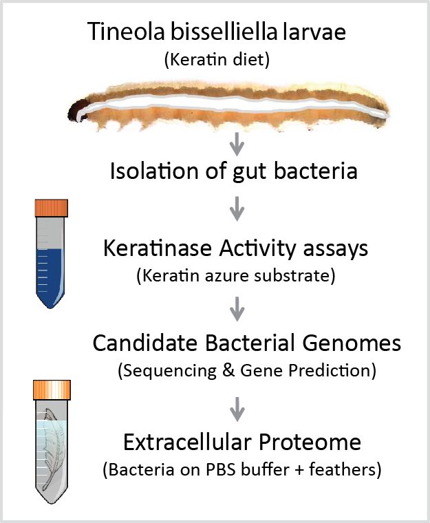
Microorganisms 2020, 8, 1415 2 of 11
clothes and carpets made of wool [3]. Autecological, behavioral, and historical data combined with
published faunistic records suggest that this species probably originated from Central or Southern
Africa and was introduced into Europe in the late 18th century. Its preference for dry environments
promoted its worldwide spread during the 20th century when indoor climates changed because of the
introduction of domestic central heating systems [3]. T. bisselliella has adapted to feed predominantly
on materials rich in keratin, such as feathers, woolen clothes, and carpets. Keratin is particularly
resistant to proteolytic degradation because of its abundant disulfide bonds (cysteine content = 7–13%),
which distinguishes it from other structural proteins such as collagen and elastin [4]. Only sophisticated
microbes can degrade keratin [5]. A subtractive library of transcripts from the gut of T. bisselliella
larvae identified serine-like and chymotrypsin-like proteases as candidate keratinases, but neither
cysteine-like proteases nor metalloproteinases were detected [6]. To date, there are no candidate
keratinase enzymes reported, which could mediate keratin digestion in the gut of T. bisselliella.
Given that many insects are associated with beneficial microbes providing enzymes which are
essential for the adaptation to unusual diets, we postulated that microbes could play a similar role
in T. bisselliella. To determine whether the ability of T. bisselliella larvae to utilize keratin is assisted
by its gut microbiome, we isolated bacteria from the guts of caterpillars fed on a diet of feathers as a
sole nutrient source. The culture supernatants were screened for keratinase activity using a simple
photometric assay and the most promising strains were analyzed in more detail. Here, we report the
identification of novel Bacillus strains found in the T. bisselliella larval gut with the apparent ability to
digest keratin. Cultivation of these strains on sterilized feathers followed by proteomic analysis of the
culture supernatant resulted in the identification of complex mixtures of enzymes and other proteins
that could contribute to the digestion of keratin by T. bisselliella caterpillars.
2. Materials and Methods
2.1. Insect Rearing and Bacterial Isolation
Specimens of T. bisselliella were obtained from the Federal Institute for Materials Research
and Testing (Berlin, Germany) and were reared on an exclusive diet of feathers at room temperature.
The feathers were surface sterilized with 80% ethanol to kill attached bacteria before feeding. Caterpillars
were surface sterilized before dissection and the extraction of gut bacteria, which was achieved by
macerating the entire gut in sterile phosphate-buffered saline (PBS) and preparing serial dilutions
of the homogenate. Samples were taken from different regions of the gut. Isolates were tested for
general proteolytic activity on casein plates, and positive isolates were cultivated with a keratin azure
substrate (blue-colored sheep wool) in PBS (37 ◦ C) to allow the photometric (A 595 nm) quantification
of keratinase activity, which releases a blue dye into the supernatant (Figures 1 and 2).
One unit of keratinase activity was defined as an increase in absorbance of 0.01 when comparing
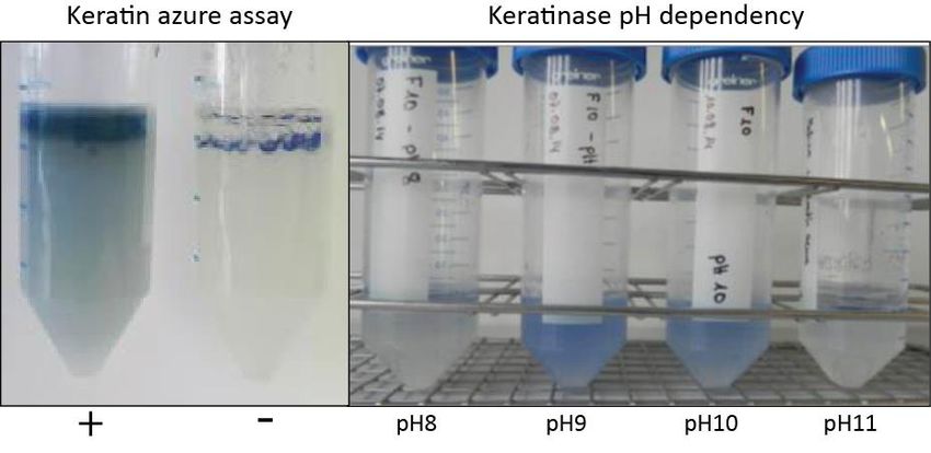
the sample to the PBS control. Keratinolysis is pH dependent, so we tested all isolates in buffers with
a pH range of 6–12 (Figure 2). The isolates with the highest keratinolytic activity were selected for
genomic analysis. The experimental keratinase activity quantification was repeated two times with
three falcon tubes per tested pH-value.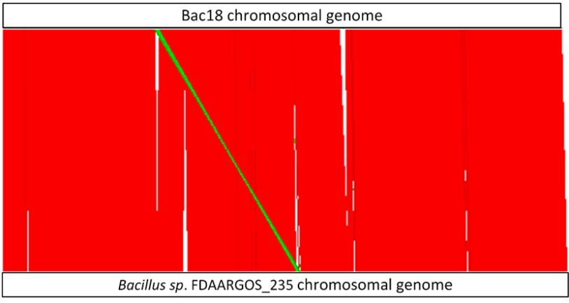
Microorganisms 2020, 8, 1415                                                                                          3 of 11
Microorganisms 2020, 8, x FOR PEER REVIEW                                                                                        3 of 11

           Figure 1. Experimental workflow for the identification of keratin-degrading proteins in bacteria
     Figure 1. Experimental workflow for the identification of keratin-degrading proteins in bacteria
          associated with the clothing moth Tineola bisselliella. Clothing moth larvae were reared on a keratin-based
     associated with the clothing moth Tineola bisselliella. Clothing moth larvae were reared on a keratin-
          diet for several generations before the isolation of gut bacteria. Individual bacterial colonies were
     based   diet
          then     for several
                tested            generations
                        for keratinase    activitybefore  the isolation
                                                   using keratin  azure asofa gut bacteria.
                                                                              substrate. DNA Individual    bacterial
                                                                                               was extracted     from colonies
                                                                                                                       two
     werebacterial
            then tested    forshowing
                     isolates   keratinase    activity using
                                         the strongest activitykeratin  azure azure
                                                                in the keratin as a substrate.  DNA
                                                                                     assay as well      was
                                                                                                   as the     extracted
                                                                                                           ability to growfrom
     two bacterial
          on whole isolates
                      feathers showing      the strongest
                                 as a sole nutrient  source.activity  in thesequence
                                                              The genome     keratin azure   assay asfor
                                                                                      was screened      well  as the ability to
                                                                                                          protein-coding
     growgenes
             on whole    feathers
                  allowing           as a sole nutrient
                             the identification            source.
                                                  of candidate       The genome
                                                                 keratinases.       sequence
                                                                               Extracellular    was screened
                                                                                             proteins              for protein-
                                                                                                       isolated from    the
     Microorganisms 2020, 8, x FOR PEER REVIEW                                                                             4 of 11
          supernatant
     coding              of bacteria
              genes allowing      thegrown     on feathers
                                       identification      as a sole nutrient
                                                       of candidate           source were
                                                                       keratinases.        analyzed proteins
                                                                                     Extracellular   by LC MS-MS.isolated from
     the supernatant of bacteria grown on feathers as a sole nutrient source were analyzed by LC MS-MS.

     One unit of keratinase activity was defined as an increase in absorbance of 0.01 when comparing
the sample to the PBS control. Keratinolysis is pH dependent, so we tested all isolates in buffers with
a pH range of 6–12 (Figure 2). The isolates with the highest keratinolytic activity were selected for
genomic analysis. The experimental keratinase activity quantification was repeated two times with
three falcon tubes per tested pH-value.

          Figure 2. Keratin azure assay for bacterial strain keratinase activity. Individual bacterial colonies
          Figure 2. Keratin azure assay for bacterial strain keratinase activity. Individual bacterial colonies were
          were inoculated in PBS buffer with the substrate keratin azure (blue-colored sheep wool) to allow the
          inoculated in PBS buffer with the substrate keratin azure (blue-colored sheep wool) to allow the
          photometric quantification of keratinase activity, which releases a blue dye into the supernatant (left
          photometric quantification of keratinase activity, which releases a blue dye into the supernatant (left
          panel). Addition of bacterial culture medium without bacteria was used as a negative control (−).
          panel). Addition of bacterial culture medium without bacteria was used as a negative control (−).
          Addition of bacterial culture medium with highest keratinolytic activity was used as a positive control
          Addition
          (+). Sinceof bacterial culture
                     keratinolysis  is pHmedium  with all
                                          dependent,   highest  keratinolytic
                                                           isolates           activity
                                                                    were tested        was used
                                                                                 in buffers with as a positive
                                                                                                 a pH  range ofcontrol
                                                                                                                  6–12
          (+). Since keratinolysis
          (right panel).           is pH  dependent,  all isolates  were tested in buffers  with a pH  range   of 6–12
          (right panel).

     2.2. Genome Sequencing and Characterization of Bac18 Isolated from T. bisselliella
          Sequencing was carried out at the Max Planck Genome Center (Cologne, Germany) using
     technology of a Pacific Biosciences Sequel instrument. A total of 90,570 PacBio raw reads were
Microorganisms 2020, 8, 1415 4 of 11
2.2. Genome Sequencing and Characterization of Bac18 Isolated from T. bisselliella
Sequencing was carried out at the Max Planck Genome Center (Cologne, Germany) using
technology of a Pacific Biosciences Sequel instrument. A total of 90,570 PacBio raw reads were
assembled with HGAP3 (Hierarchical Genome Assembly Process 3) [7] with default options and an
estimated genome size of 5 Mb. We checked for circular contigs using Circlator 1.5.1 [8] in “all” mode
using the corrected reads from the HGAP3 pipeline. Genome annotation was carried out using results
from Circlator for Prokka 1.11 [9] in default mode with “Bacteria” as kingdom. Nucleotide sequence
alignments of the 16S rRNA genes copies were aligned with Clustal X [10]. The closest reference
genomes (fully sequenced and assembled) were estimated using ReferenceSeeker 1.10 [11] in default
mode with the implemented database “bacteria” (August 2019). Bac18 was analyzed for its global
sequence identity to ReferenceSeeker’s best hit with the MUMmer3 package [12].
2.3. Target Gene Prediction
For the detection of genes involved in keratin degradation, UniProt (The UniProt Consortium, 2019,
date: 29 August 2019) was searched for the keyword “keratinase” and the resulting FASTA (canonical)
and associated text files were downloaded. Non-keratinase entries (A0A1S6Y3A5, A0A0G3BBA5)
were manually identified and removed before further analysis. Pfam [13] domains were extracted
from the corrected UniProt text file. In a second step, the coding sequences in Bac18 (predicted using
Prokka) were compared to the PfamA-31 database using the commands hmmscan –domtblout, –noali,
and –notextw in HMMER v3.1b2. The corresponding Pfam annotations were searched for keratinase
domains and classified using an in-house script. In the last step, we performed a local alignment of all
coding sequences and keratinase results from UniProt using BLASTp in BLAST v2.6.0+ [14].
2.4. Proteomic Analysis
Extracellular bacterial proteins contained in feather-supplemented media were concentrated
using the Vivaspin™ ultrafiltration spin columns (GE Healthcare Life Sciences) according to the
manufacturers’ recommendations. Protein mixtures were subsequently separated by sodium
dodecylsulfate polyacrylamide gel electrophoresis (SDS-PAGE) on 4–12% Criterion XT gradient
gels (BioRad, Hercules, CA, USA) with XT MES running buffer at 125 V for 1.5 h. The molecular
weights (kDa) of the separated proteins were estimated using pre-stained markers and unstained
high-mass-precision protein markers. For LC-MS/MS analysis, protein bands were excised from
the gel as 10 molecular weight blocks per lane, followed by tryptic digestion as described by [15].
LC-MS sample processing, data acquisition, and data processing were carried out as previously
described [16]. MS BLAST was then used to search a database derived from the in silico translation of
the Bac18 genome. Search parameters specifying mass measurement accuracy, minimum number of
product ion matches per peptide, minimum number of product ion matches per protein, minimum
number of peptide matches, and maximum number of missed tryptic cleavage sites are detailed in the
Supplementary Materials.
3. Results
3.1. Identification of Bacterial Candidates and Genome Sequencing
Keratin-degrading bacteria and candidate keratinases in the T. bisselliella larval gut were identified
by following the scheme shown in Figure 1. First, we dissected the guts of T. bisselliella larvae feeding
on an exclusive keratin-based diet and cultivated serially diluted gut homogenates on casein to identify
colonies with extracellular proteolytic activity. Individual colonies were then inoculated into the
substrate keratin azure, which produces an easily detected blue dye when digested (Figure 2). We also
tested for keratinolytic activity by incubating bacteria in a non-nutrient buffer containing feathers,
which began to fragment in the presence of keratinase activity. Based on these results, we selected
individual bacterial colonies with the highest overall keratinolytic activity for genome sequencing.Microorganisms 2020, 8, 1415 5 of 11
Alignment of the 16S rRNA genes indicated that the selected bacterial isolates represented the same
species and strain. We therefore focused on one of the two Bacillus candidates (Bac18) for further
genome analysis, proteomics, and keratinase assays.
The final draft genome of Bac18 is represented by one circular chromosomal genome with a
size of 5.2 Mb and additional five small contigs with a total length of 827 kb (Table 1). We identified
5946 protein coding sequences, 14 rRNA gene clusters, and 106 tRNA genes. A nucleotide sequence
alignment of the 16S rRNA gene copies led to the identification of eight different variants that differ
from each other by SNPs at six variable positions. The closest relative on the whole genome level
was calculated using all publicly available genomes that are fully sequenced and assembled (Table 2).
This calculation includes small contigs of Bac18 as well as potential plasmid sequences of the references.
The conserved DNA is the fraction of both genomes that are homologous while the ANI is the average
nucleotide identity of this homologous region.
Table 1. Assembly statistics for Bac18. Circularized contigs are marked with •.
Bac18
Contig Name Contig Length [bp] Sequencing Coverage
Bac18_0 • 5,236,241 131
Bac18_2 745,926 164
Bac18_3 • 16,484 479
Bac18_4 39,074 365
Bac18_5 12,019 167
Bac18_6 14,098 173
P
6,063,842
Table 2. Closest reference genome for Bac18 calculated with ReferenceSeeker.
ID Related Genome ANI Cons. DNA Taxonomy ID Organism Name
GCF_002073415.2 99.56% 83.93% 1839798 Bacillus sp. FDAARGOS_235
Bac18 GCF_000300475.1 98.54% 84.50% 1195464 Bacillus thuringiensis MC28
GCF_000496285.1 99.44% 78.04% 1415784 Bacillus toyonensis BCT-7112
We found that Bac18 and Bacillus sp. FDAARGOS_235 share 83.93% of their genomes (conserved
DNA) with an overall average nucleotide identity (ANI) of 99.56%. In this regard it has to be noted
that the small contigs of Bac18 make up ~14% of the length of the genome. A visualization of the
structural similarities of the Bac18 chromosomal sequence with Bacillus sp. FDAARGOS_235 is shown
in Figure 3, where homologous regions between both genomes are represented in red (homologous
sequences with same orientation) and green (reverse complement, but homologous region). No such
whole-contig similarities were detected for the respective small contigs of Bac18 with the plasmids of
the closest reference. Nevertheless, we were able to detect homologies of fragments of the small Bac18
contigs with publicly available plasmid sequences using BLASTn analysis. However, none of these
plasmids were covered completely.structural similarities of the Bac18 chromosomal sequence with Bacillus sp. FDAARGOS_235 is shown
in Figure 3, where homologous regions between both genomes are represented in red (homologous
sequences with same orientation) and green (reverse complement, but homologous region). No such
whole-contig similarities were detected for the respective small contigs of Bac18 with the plasmids of
the closest reference. Nevertheless, we were able to detect homologies of fragments of the small Bac18
Microorganisms 2020,
contigs 8, 1415
with publicly available plasmid sequences using BLASTn analysis. However, none of these 6 of 11
plasmids were covered completely.
Visualization
Figure 3.Figure of the
3. Visualization structural
of the structuralsimilarities ofBac18
similarities of Bac18 andand Bacillus
Bacillus sp. FDAARGOS_235
sp. FDAARGOS_235
chromosomal genomes calculated with Nucmer. Genomes are
chromosomal genomes calculated with Nucmer. Genomes are given as white boxes given as white boxes at the top
at (Bac18)
the top (Bac18)
and the bottom (Bacillus sp.) of the illustration. For comparison, both circular genomes are opened at
and the bottom (Bacillus sp.) of the illustration. For comparison, both circular genomes are opened at
the starting position of the dnaA gene. Red: homologous sequences with the same orientation; green:
the starting position
reverse complement, dnaA
of the but gene. Red:
homologous homologous
sequences, sequences
white: regions withoutwith the same orientation; green:
homologies.
reverse complement, but homologous sequences, white: regions without homologies.
3.2. Candidate Gene Prediction and Proteomic Analysis
3.2. Candidate Gene Prediction and Proteomic Analysis
We searched the Bac18 genome for potential keratinase-encoding genes and identified 20
sequencesthe
We searched withBac18
predicted keratinase
genome domains as
for potential found in Pfam. The amino
keratinase-encoding acid
genes sequences
and of the
identified 2020sequences
candidates were used as BLAST queries against keratinase entries in UniProt. Table 3 shows the best
with predicted keratinase domains as found in Pfam. The amino acid sequences of the 20 candidates
hits against UniProt filtered for sequence coverage (≥70%) and sequence identity (≥40%). With a
were usedsequence
as BLAST queries against keratinase entries in UniProt. Table 3 shows the best hits against
coverage of 100% and a sequence identity of 98% the gene PROKKA_03102 has the highest
UniProt filtered for sequence coverage
blast score against a keratinase (≥70%)
included and sequence
in UniProt. identityfrom
The supernatant (≥40%). With a sequence
the non-nutrient medium coverage
of 100% and a sequence
containing identity
fragmented of 98%
feathers the geneafter
was collected PROKKA_03102 has thefor
Bac18 had been growing highest blast score
72 h, allowing us to against a
keratinaseidentify
includedcandidate extracellular
in UniProt. proteins withfrom
The supernatant keratinase activity. The medium
the non-nutrient concentrated extracellular
containing fragmented
proteins were separated by SDS-PAGE and bands were observed with apparent molecular masses
feathers was collected after Bac18 had been growing for 72 h, allowing us to identify candidate
ranging from 100 kDa (Figure 4). The gel lanes were divided into ten blocks covering
extracellular proteins
different withfor
size ranges keratinase activity.
in-gel digestion withThe concentrated
trypsin. The resultingextracellular
peptides wereproteins werebyseparated
then analyzed
by SDS-PAGE
LC-MS/MSandusing
bands were
protein observed
sequences with in
predicted apparent
silico frommolecular masses
the Bac18 genome a database.from 100 kDa (Figure 4). The gel lanes were divided into ten blocks covering different size ranges for
in-gel digestion with trypsin. The resulting peptides were then analyzed by LC-MS/MS using protein
sequences predicted
Microorganisms in8,silico
2020, fromREVIEW
x FOR PEER the Bac18 genome as a database. 9 of 11
4. Analysis
Figure Figure of extracellular
4. Analysis proteins
of extracellular isolated
proteins from
isolated thethe
from supernatant
supernatantof ofBac18
Bac18 growing
growing ononfeathers
feathers
as nutrient
as a sole a sole nutrient source.
source. Left Left
panelpanel shows
shows SDS-PAGE
SDS-PAGE protein
protein geldivided
gel dividedinto
into 10
10 zones
zones with
withdifferent
different
molecular
molecular weightweight (PM = protein
ranges ranges (PM = protein size markers).
size markers). Rightshows
Right panel paneltheshows the properties
properties of the
of the identified
identified
proteins, includingproteins, including
predicted masses,predicted masses,
descriptions, descriptions,
and the number and
of the number
assigned of assigned
peptides. Thepeptides.
matching
Theproteins
predicted matchinginpredicted proteins
the bacterial genomein the
arebacterial
listed in genome areSupplementary
electronic listed in electronic supplementary
Materials, File S1.
material, File S1.
We identified 63 different proteins in the feather-supplemented culture supernatant that
matched sequences predicted in the Bac18 genome using our proteomics cut-off criteria
(Supplementary material, File S1). We subsequently focused on those candidates for which there was
evidence for direct or indirect involvement in keratin utilization. The largest group of these proteinsMicroorganisms 2020, 8, 1415 7 of 11
Table 3. Predicted genes of Bac18 that include Pfam domains related to keratinases.
Prokka ID Prokka Annotation Pfam Domains Keratinase Blast Hit Proteomics ID
PROKKA_00129 Aminopeptidase YwaD precursor PA, Peptidase_M28 n. h. 02318
PROKKA_00135 Thermolysin precursor FTP, Peptidase_M4_C, Peptidase_M4 n. h. 02311
PROKKA_00413 Neutral protease B precursor FTP, PepSY, Peptidase_M4, Peptidase_M4_C A0A0B4ZU77 02032
PROKKA_00751 Peptidase propeptide and YPEB domain protein PepSY n. h. 01721
PROKKA_01081 Minor extracellular protease vpr precursor Peptidase_S8, Inhibitor_I9, PA n. h. 01389
PROKKA_01290 Peptidase T Peptidase_M28 * n. h. 01180
PROKKA_01747 Microbial collagenase precursor Peptidase_S8 *, PPC n. h. 00731
PROKKA_02134 Bacillolysin precursor FTP, PepSY, Peptidase_M4, Peptidase_M4_C n. h. 00344
PROKKA_02729 Bacillolysin precursor FTP, PepSY, Peptidase_M4, Peptidase_M4_C n. h.
PROKKA_02930 Thermolysin precursor FTP, PepSY, Peptidase_M4, Peptidase_M4_C n. h.
PROKKA_03102 Thermitase Peptidase_S8 F8SVT0 **
PROKKA_03248 Bacillolysin precursor FTP *, PepSY n. h. 02311
PROKKA_03479 Intracellular serine protease Peptidase_S8 Q45GC8
PROKKA_04411 hypothetical protein PepSY n. h. 03334
PROKKA_04743 Minor extracellular protease vpr Peptidase_S8, Inhibitor_I9, PA n. h. 03020
PROKKA_04780 Bacillolysin precursor FTP, PepSY, Peptidase_M4, Peptidase_M4_C A0A0B4ZU77 02981
PROKKA_05541 Major intracellular serine protease Peptidase_S8 * n. h.
PROKKA_05686 Neutral protease B precursor FTP, PepSY, Peptidase_M4, Peptidase_M4_C A0A0B4ZU77 02032
PROKKA_05790 Thermophilic serine proteinase Peptidase_S8 * n. h.
PROKKA_05803 Chitinase A1 precursor Peptidase_S8 * n. h.
* Pfam domain is not complete (domain coverage < 90%); ** Best BLAST hit; n. h.: no hit.Microorganisms 2020, 8, 1415 8 of 11
We identified 63 different proteins in the feather-supplemented culture supernatant that matched
sequences predicted in the Bac18 genome using our proteomics cut-off criteria (Supplementary Materials,
File S1). We subsequently focused on those candidates for which there was evidence for direct or indirect
involvement in keratin utilization. The largest group of these proteins was annotated as proteases.
We identified 16 different proteases, including serine-type and metalloproteinases, with predicted
protein masses of 18–135 kDa (Figure 4). Of particular interest were candidate proteases related to
previously characterized bacterial keratinases, such as specific extracellular metalloproteases and the
subtilisin family of serine proteases. We also identified a thiol-disulfide oxidoreductase, which is likely
to facilitate the proteolytic degradation of keratin by promoting the hydrolysis of disulfide bonds.
4. Discussion
Caterpillars of the clothing moth T. bisselliella feed predominantly on keratin-based materials such
as feathers, hairs, and wool. The midgut of these insects is anaerobic with a negative redox potential,
which was proposed to facilitate the solubilization of keratin by reducing disulfide bonds, enabling
digestion by more widespread proteases [17]. However, even in a high redox potential environment,
only a subset of proteases can degrade keratin substrates [18]. Analysis of the crude gut preparations
resulted in identification of aminopeptidase activity and a l-Cysteine lyase, but neither cysteine
endopeptidase nor metalloprotease activities were found [19,20]. Although multiple serine proteases
with high expression levels were identified in the T. bisselliella gut transcriptome [6], their involvement
in keratin proteolysis is unclear.
Therefore, it remains to be determined whether serine proteases encoded by the host insect
genome can process keratin, or whether additional enzymatic or physiological functions are required
to initiate the degradation of this molecule by reducing the abundant disulfide bonds.
An alternative explanation for the adaptation of the clothing moth to keratin-based diets is that
keratinase activity is conferred by specific bacterial symbionts in the gut. Although previous studies
found no abundant microorganisms in T. bisselliella larvae [21,22], we were able to reproducibly isolate
bacteria from the midgut of larvae fed on a feather diet. Most of the isolates were identified as
Clostridiales, Lactobacillales, and the genus Bacillus, similar to the bacteria associated with the brown
house moth Hofmannophila pseudospretella, another keratin-feeding lepidopteran [23]. After screening
individual gut-isolated bacterial strains for their ability to degrade keratin, we selected two isolates
showing the highest levels of keratinase activity in both the keratin azure and whole-feather assays.
These were found to represent the same species, and genomic sequencing of isolate Bac18 identified
a new Bacillus strain with a close relationship to Bacillus sp. FDAARGOS_235 and B. thuringiensis.
Interestingly, Bacillus species are predominant among bacteria displaying keratinase activity, and are
known to encode for several keratin-degrading proteases.
On a keratin diet, larvae of the clothing moth develop very slowly, possibly owing to the limited
availability of an easily accessible nitrogen source. Likewise, in our assays using non-nutrient buffer
supplemented with whole feathers, the inoculated gut-associated bacteria showed slow growth. When
clothing moth larvae or their gut-associated bacteria were provided solely with keratin (whole feathers
in our experiments) as a nitrogen source, we hypothesized that nitrogen assimilation mechanisms are
induced, including the expression and secretion of keratin-active enzymes [24–26], which subsequently
requires polypeptide processing and active transport. Accordingly, proteomic analysis of culture
supernatants from Bac18 grown on non-nutrient medium supplemented with feathers revealed
the presence of enzymes with the potential to degrade keratin, including collagenases and other
serine proteases, metalloproteases, and a thiol-disulfide oxidoreductase. These findings support our
hypothesis that the unusual diet of T. bisselliella larvae promotes their association with keratinolytic
microorganisms and that keratin digestion is potentially supported by bacteria in the larval gut.
The thiol-disulfide oxidoreductase was particularly interesting because this enzyme can hydrolyze
disulfide bonds, thus enabling the complete proteolytic degradation of keratin and subsequent nitrogen
assimilation [25,26].Microorganisms 2020, 8, 1415 9 of 11
We detected a surprisingly complex mixture of numerous extracellular proteases as well as
enzymes that modify bacterial cell walls, peptide-binding proteins, transporters, and stress-response
factors. These functions are commensurate with the stress anticipated when bacteria are forced to grow
on keratin as a sole nutrient source. Previous studies of bacterial keratinases reviewed in [18,19] have
focused on a single enzyme while ignoring the complexity of the extracellular proteome. The presence
of such a rich cocktail of extracellular proteins suggests that the combinatorial action of multiple
enzymes, transporters, and stress-response proteins ensures the more efficient degradation of keratin
compared to a single, isolated protease. This hypothesis is supported by previous reports that certain
Bacillus species encode multiple proteases representing different functional classes and different protein
masses that nevertheless possess keratinase activity [27,28].
Based on the proteins detected in supernatants of Bac18 cultivated in nutrient-free medium
supplemented with feathers, we propose that keratin digestion is a multistep process beginning with the
cleavage of disulfide bonds by a thiol-disulfide oxidoreductase, enhancing the accessibility of the reduced
keratin polypeptide for a cocktail of exopeptidases (aminopeptidases and dipeptidyl peptidases) [22,28]
and endo-active proteases such as subtilisins, collagenases, and other metalloendoproteases,
and oligoendopeptidases. These digest keratin into shorter peptides and free amino acids, which are
sequestered by transport proteins and assimilated. Our results do not rule out the possibility
that T. bisselliella also produces its own enzymes that contribute to keratin digestion, but such
native keratinases have yet to be identified [6]. The isolated keratin-degrading bacteria help to
explain the ability of T. bisselliella to occupy a unique ecological niche. Strikingly, the tarantula
Chilobrachys guangxiensis, which requires powerful enzymes to digest the immobilized preys down
into a liquid taken up with straw-like mouthparts into the intestines, was also found to harbor
feather-degrading bacteria in the gut producing keratinases [29]. Such keratinase-producing bacteria
are of interest for biotechnological applications, particularly in the bioconversion of keratin-rich waste
such as feathers, hair and woolen textiles [2]. Further, we expanded the number of bacteria associated
with moth pest insects [30].
Supplementary Materials: The following are available online at http://www.mdpi.com/2076-2607/8/9/1415/s1,
File S1: MSE-based Identification of Proteins.
Author Contributions: Conceptualization, A.V. and H.V.; methodology, N.W., M.S., H.V., and K.B.; formal
analysis, M.S., K.B., N.W., H.V.; resources, R.P.; data curation, M.S., K.B., N.W., H.V.; writing—original draft
preparation, A.V.; writing—review and editing, A.V., H.V., K.B., M.S., and R.P.; visualization, K.B., H.V., and M.S.;
supervision, A.V., K.B., and H.V.; funding acquisition, A.V. and H.V. All authors have read and agreed to the
published version of the manuscript.
Funding: This research was funded by the Fraunhofer Society and Max Planck Society through the collaborative
project grant AIM-Biotech (Application of Insect-associated Microbes in industrial biotechnology). A.V. also
acknowledges funding received form the Hessen Ministry of Science and Art (HMWK) via the LOEWE Center for
Insect Biotechnology and Bioresources.
Acknowledgments: The authors wish to thank Rebecca Dietz and Henriette Ringys-Beckstein for her support in
general lab work and for obtaining high-molecular weight genomic DNA from bacterial cultures. We also thank
Richard M Twyman for editing the manuscript.
Conflicts of Interest: The authors declare no conflict of interest. The funders had no role in the design of the
study; in the collection, analyses, or interpretation of data; in the writing of the manuscript, or in the decision to
publish the results.
References
1. Douglas, A.E. Multiorganismal insects: Diversity and function of resident microorganisms. Annu. Rev. Entomol.
2015, 60, 17–34. [CrossRef] [PubMed]
2. Berasategui, A.; Shukla, S.; Salem, H.; Kaltenpoth, M. Potential applications of insect symbionts in
biotechnology. Appl. Microbiol. Biotechnol. 2016, 100, 1567–1577. [CrossRef] [PubMed]
3. Plarre, R.; Krüger-Carstensen, B. An attempt to reconstruct the natural and cultural history of the webbing
clothes moth Tineola bisselliella Hummel (Lepidoptera: Tineidae). J. Entomol. Acarol. Res. 2011, 43, 83–93.
[CrossRef]Microorganisms 2020, 8, 1415 10 of 11
4. Verma, A.; Singh, H.; Anwar, S.; Chattopadhyay, A.; Tiwari, K.K.; Kaur, S.; Dhilon, G. Keratin: Dissolution,
extraction and biomedical application. Crit. Rev. Biotechnol. 2017, 37, 476–491. [CrossRef]
5. Wan, W.-L.; Chen, M.-Y.; Tu, I.-F.; Lin, Y.-C.; EswarKumar, N.; Chen, M.-Y.; Ho, M.-C.; Wu, S.-H. The discovery
of novel heat-stable keratinases from Meiothermus taiwanensis WR-220 and other extremophiles. Sci. Rep.
2017, 7, 4658. [CrossRef]
6. Hughes, J.; Vogler, A.P. Gene expression in the gut of keratin-feeding clothes moths (Tineola) and keratin
beetles (Trox) revealed by subtracted cDNA libraries. Insect Biochem. Mol. Biol. 2006, 36, 584–592. [CrossRef]
7. Chin, C.S.; Alexander, D.H.; Marks, P.; Klammer, A.A.; Drake, J.; Heiner, C.; Clum, A.; Copeland, A.;
Huddleston, J.; Eichler, E.E.; et al. Nonhybrid, finished microbial genome assemblies from long-read SMRT
sequencing data. Nat. Methods 2013, 10, 563–569. [CrossRef]
8. Hunt, M.; Silva, N.D.; Otto, T.D.; Parkhill, J.; Keane, J.A.; Harris, S.R. Circlator: Automated circularization of
genome assemblies using long sequencing reads. Genome Biol. 2015, 16, 1–10. [CrossRef]
9. Seemann, T. Prokka: Rapid prokaryotic genome annotation. Bioinformatics 2014, 30, 2068–2069. [CrossRef]
10. Larkin, M.A.; Blackshields, G.; Brown, N.P.; Chenna, R.; McGettigan, P.A.; McWilliam, H.; Balentin, F.;
Vallace, I.M.; Wilm, A.; Lopez, R.; et al. Clustal W and Clustal X version 2.0. Bioinformatics 2007, 23, 2947–2948.
[CrossRef]
11. Schwengers, O.; Hain, T.; Chakraborty, T.; Goesmann, A. ReferenceSeeker: Rapid Determination of
Appropriate Reference Genomes. GitHub. 2019. Available online: https://github.com/oschwengers/
referenceseeker (accessed on 13 September 2020).
12. Kurtz, S.; Phillippy, A.; Delcher, A.L.; Smoot, M.; Shumway, M.; Antonescu, C.; Salzberg, S.L. Versatile and
open software for comparing large genomes. Genome Biol. 2004, 5, R12. [CrossRef] [PubMed]
13. El-Gebali, S.; Mistry, J.; Bateman, A.; Eddy, S.R.; Luciani, A.; Potter, S.C.; Qureshi, M.; Richardson, L.J.;
Salazar, G.A.; Smart, A.; et al. The Pfam protein families database in 2019. Nucleic Acids Res. 2019, 47,
D427–D432. [CrossRef]
14. Altschul, S.F.; Gish, W.; Miller, W.; Myers, E.W.; Lipman, D.J. Basic local alignment search tool. J. Mol. Biol.
1990, 215, 403–410. [CrossRef]
15. Shevchenko, A.; Tomas, H.; Havlis, J.; Olsen, J.V.; Mann, M. In-gel digestion for mass spectrometric
characterization of proteins and proteomes. Nat. Protoc. 2006, 1, 2856–2860. [CrossRef]
16. Skaljac, M.; Vogel, H.; Wielsch, N.; Mihajlovic, S.; Vilcinskas, A. Transmission of a protease-secreting bacterial
symbiont among pea aphids via host plants. Front. Physiol. 2019, 10, 438. [CrossRef] [PubMed]
17. Waterhouse, D.F. Wool digestion and mothproofing. In Advances in Pest Control Research, 2nd ed.; Metcalf, R.L.,
Ed.; Interscience: New York, NY, USA, 1958; pp. 207–262.
18. Ramnani, P.; Gupta, R. Keratinases vis-a-vis conventional proteases and feather degradation. World J.
Microbiol. Biotechnol. 2007, 23, 1537–1540. [CrossRef]
19. Christeller, J.T.; Markwick, N.P.; Burgess, E.P. Midgut proteinase activities of three keratinolytic larvae,
Hofmannophila pseudospretella, Tineola bisseliella, and Anthrenocerus australis, and the effect of proteinase
inhibitors on proteolysis. Arch. Insect Biochem. Physiol. 1994, 25, 159–173. [CrossRef]
20. Yoshimura, T.; Tabata, H.; Nishio, M.; Ide, E.; Yamaoka, R.; Hayashiya, K. L-Cystein lyase of the webbing
clothes moth, Tineola bisselliella. Insect Biochem. 1988, 18, 771–777. [CrossRef]
21. Crewther, W.G.; McQuade, A.B. The intestinal microflora of the clothes moth Tineola bisselliella in relation to
wool digestion. J. Gen. Microbiol. 1955, 12, 311–313. [CrossRef]
22. Kasper, C.S. Ultrastructure of the Digestive System of the Larval Clothes Moth Tineola bisselliella (Humm.).
Ph.D. Thesis, Boston University Graduate School, Boston, MA, USA, 1978; Unpublished work.
23. Shannon, A.L.; Attwood, G.; Hopcroft, D.H.; Christeller, J.T. Characterization of lactic acid bacteria in the
larval midgut of the keratinophagous lepidopteran, Hofmannophila pseudospretella. Lett. Appl. Microbiol. 2001,
32, 36–41. [CrossRef]
24. Daroit, D.J.; Brandelli, A. A current assessment on the production of bacterial keratinases. Crit. Rev. Biotechnol.
2014, 34, 372–384. [CrossRef] [PubMed]
25. Yamamura, S.; Morita, Y.; Hasan, Q.; Yokoyama, K.; Tamiya, E. Keratin degradation: A cooperative action of
two enzymes from Stenotrophomonas sp. Biochem. Biophys. Res. Commun. 2002, 294, 1138–1143. [CrossRef]
26. Lange, L.; Huang, Y.; Busk, P.K. Microbial decomposition of keratin in nature—A new hypothesis of industrial
relevance. Appl. Microbiol. Biotechnol. 2016, 100, 2083–2096. [CrossRef] [PubMed]Microorganisms 2020, 8, 1415 11 of 11
27. Brandelli, A.; Daroit, D.J.; Riffel, A. Biochemical features of microbial keratinases and their production and
applications. Appl. Microbiol. Biotechnol. 2010, 85, 1735–1750. [CrossRef]
28. Gupta, R.; Ramnani, P. Microbial keratinases and their prospective applications: An overview.
Appl. Microbiol. Biotechnol. 2006, 70, 21–33. [CrossRef]
29. Liu, Q.; Zhang, T.; Song, N.; Li, Q.; Wang, Z.; Zhang, X.; Lu, X.; Fang, J.; Chen, J. Purification and
characterization of four key enzymes from a feather-degrading Bacillus subtilis from the gut of tarantula
Chilobrachys guangxiensis. Int. Biodeterior. Biodegrad. 2014, 96, 26–32. [CrossRef]
30. Mereghetti, V.; Chouaia, B.; Montagna, M. New insights into the microbiota of moth pests. Int. J. Mol. Sci.
2017, 18, 2450. [CrossRef]
© 2020 by the authors. Licensee MDPI, Basel, Switzerland. This article is an open access
article distributed under the terms and conditions of the Creative Commons Attribution
(CC BY) license (http://creativecommons.org/licenses/by/4.0/).You can also read