The role of ASICs in cerebral ischemia - Zhi-Gang Xiong1 and Tian-Le Xu2
←
→
Page content transcription
If your browser does not render page correctly, please read the page content below
Focus Article

The role of ASICs in cerebral
ischemia
Zhi-Gang Xiong1∗ and Tian-Le Xu2

                 Cerebral ischemia is a leading cause of death and long-term disabilities worldwide.
                 Excessive intracellular Ca2+ accumulation in neurons has been considered essential
                 for neuronal injury associated with cerebral ischemia. Although the involvement
                 of glutamate receptors in neuronal Ca2+ accumulation and toxicity has been the
                 subject of intensive investigation, inhibitors for these receptors showed little effect
                 in clinical trials. Thus, additional Ca2+ toxicity pathway(s) must be involved.
                 Acidosis is a common feature in cerebral ischemia and was known to cause brain
                 injury. The mechanisms were, however, unclear. The finding that ASIC1a channels
                 are highly enriched in brain neurons, their activation by ischemic acidosis, and
                 their demonstrated Ca2+ permeability suggested a role for these channels in Ca2+
                 accumulation and neuronal injury associated with cerebral ischemia. Indeed, a
                 number of studies have now provided solid evidence supporting the involvement
                 of ASIC1a channel activation in ischemic brain injury. © 2012 WILEY-VCH Verlag GmbH
                 & Co. KGaA, Weinheim.

                                                         How to cite this article:
                              WIREs Membr Transp Signal 2012, 1:655–662. doi: 10.1002/wmts.57

INTRODUCTION                                                                 Although multiple pathways and biochemical
                                                                      changes contribute to ischemic brain injury, exces-
I  schemic stroke, or cerebral ischemia, is the third
   most common cause of death in most industrialized
countries. Although major advances have occurred
                                                                      sive intracellular Ca2+ accumulation and resultant
                                                                      toxicity has been considered essential in the pathol-
                                                                      ogy of cerebral ischemia.2 In the resting conditions,
in the prevention of stroke during the past several
                                                                      free intracellular Ca2+ concentration ([Ca2+ ]i ) in neu-
decades, no effective treatment is now available.
                                                                      rons is maintained at nanomolar range. Following
Current clinical practices for stroke patients utilize
                                                                      cerebral ischemia, however, [Ca2+ ]i can rise to as
thrombolytic agent tissue plasminogen activator (tPA)
                                                                      high as several micromolars. Excessive accumula-
to reopen the clotted vessels.1 This approach,
                                                                      tion of Ca2+ in neurons leads to uncontrolled acti-
however, has very limited success due to a short
                                                                      vation of various enzymes causing breakdown of
therapeutic time window of 3 h and side effect of
                                                                      proteins, lipids, and nucleic acids, and the destruc-
intracranial hemorrhage. On the other hand, cell
                                                                      tion of neurons.3–5 In addition, overloading Ca2+ in
death is prominent following stroke. Therefore, the
                                                                      mitochondria can cause opening of mitochondria per-
need for a continuous search of neuronal damage
                                                                      meability transition pore (PTP), promoting apoptosis
mechanisms and effective therapeutic strategies for
                                                                      through release of cytochrome c and activation of
neuroprotection remains high.
                                                                      caspases.6
                                                                             Ca2+ can enter neurons through various path-
Conflict of interest: Both authors have no conflict of interest to    ways, among which glutamate receptor-gated chan-
declare.                                                              nels have received the most attention. Unfortunately,
∗
    Correspondence to: zxiong@msm.edu                                 clinical trials targeting these channels have shown
1
 Neuroscience Institute, Morehouse School of Medicine, Atlanta,       little effect in improving the outcome of cerebral
GA 30310, USA                                                         ischemia.7 Multiple factors may have contributed
2
 Neuroscience Division, Department of Biochemistry and Molec-         to the failure of the trials. In particular, additional
ular Cell Biology, Institute of Medical Sciences, Shanghai
Jiao Tong University School of Medicine, Shanghai 200025,             glutamate-independent Ca2+ entry and toxicity path-
China                                                                 ways must be considered.

Volume 1, September/October 2012 © 2012 WILEY-VCH Verlag GmbH & Co. KGaA, Weinheim.                                        655
Focus Article wires.wiley.com/mts
BRAIN ACIDOSIS IN CEREBRAL To provide a link between ASIC1a activation
ISCHEMIA and ischemic brain injury, both in vitro neuronal
injury and in vivo cerebral ischemia models were
Acidosis, a condition characterized by too much acid employed. A brief (1 h) acid incubation, in the pres-
in the tissue or body fluid, is one of the most common ence of blockers of glutamate receptors and voltage-
pathophysiological changes in the brain associated gated Ca2+ channels, was able to induce substantial
with acute neurological conditions such as cerebral neuronal injury measured at 6 h or 24 h after acid
ischemia.8,9 In the ischemic core, for example, a rapid treatment. This acid-induced, glutamate-independent
drop of pH to 6.5 or lower is frequently observed.10,11 neuronal injury was inhibited by amiloride or PcTX1,
The lack of oxygen supply promotes anaerobic supporting the involvement of homomeric ASIC1a
glycolysis which leads to increased production of channels. Consistent with an essential role for ASIC1a
lactic acid.11 Accumulation of lactic acid, along with subunit in acid injury, neurons cultured from ASIC1
increased production of H+ from ATP hydrolysis, knockout mice were resistant to acid incubation.15,18
and release of H+ from presynaptic terminals,12 Reducing the concentration of extracellular Ca2+ ,
contributes to the acid buildup in the brain. Acidosis which decreases the driving force for Ca2+ entry, also
has long been recognized to aggravate brain injury ameliorated acid injury. To know whether ASIC1a-
associated with cerebral ischemia.8,9 However, the mediated injury can also take place in ischemic
detailed mechanism(s) remained elusive, although a condition, acid-mediated neuronal injury was stud-
number of possibilities have been suggested long ied in the condition of oxygen glucose deprivation
before the role of acid-sensing ion channels (ASICs) (OGD). OGD, in the presence of blockers of glu-
was recognized.8,13,14 tamate receptors and voltage-gated Ca2+ channels,
enhanced the acid-induced neuronal injury which
ASIC1a ACTIVATION IS INVOLVED was inhibited by amiloride and PcTX1. Thus, in
vitro studies support a role for ASIC1a activation
IN ACIDOSIS-MEDIATED ISCHEMIC in acidosis-mediated, Ca2+ -dependent, ischemic neu-
BRAIN INJURY ronal injury. Since a recent study showed that PcTX1
On the basis of the evidence that ASIC1a subunits also inhibits heteromeric ASIC1a/ASIC2b channels in
are highly expressed in brain neurons, their activa- addition to homomeric ASIC1a channels,19 the poten-
tion by pH drops to the level commonly seen in tial contribution of heteromeric ASIC1a/ASIC2b chan-
cerebral ischemia, and their permeability to Ca2+ nels to acidosis-mediated neuronal injury cannot be
and Na+ , Xiong and colleagues tested the hypoth- excluded.
esis that activation of ASIC1a channels is involved Does activation of ASIC1a channels also play a
in neuronal Ca2+ accumulation and injury associated role in ischemic brain injury in vivo? To answer this
with cerebral ischemia.15 Using patch-clamp recording question, two sets of experiments were performed.
and fast-perfusion technique, large inward currents The first experiment examined whether application
were recorded in cultured mouse cortical neurons in of ASIC inhibitors reduces ischemic brain injury, and
response to rapid perfusion of acidic solutions at pH the second tested whether knockout of the ASIC1
levels relevant to cerebral ischemia. The acid-activated gene renders the animal resistant to cerebral ischemia.
currents in cortical neurons were sensitive to non- Rodent model of focal ischemia, by middle cerebral
specific ASIC blocker amiloride and partially inhibited artery occlusion (MCAO), was employed for both
by ASIC1a-specific inhibitor PcTX1, suggesting that experiments. In rats and mice, intracerebroventricu-
the currents were mediated by ASIC1a-containing lar injection of ASIC1a inhibitor PcTX1 reduced the
channels. Consistent with the presence of func- infarct volume by up to 60%, measured at 24 h after
tional homomeric ASIC1a channels, which are Ca2+ - MCAO.15,20 Similar to the pharmacological blockade,
permeable,16 perfusion of acidic solution in these ASIC1 gene knockout provided a comparable degree
neurons increased intracellular Ca2+ concentration, of protection against ischemic brain injury.15 Remark-
even in the presence of blockers of voltage-gated Ca2+ ably, in contrast to most glutamate antagonists which
channels and glutamate receptors. As expected, the have only a short time window ofWIREs Membrane Transport and Signaling Role of ASICs in cerebral ischemia
of human brain neurons in culture. Thus, Ca2+ - ischemic conditions and whether the effects of
permeable ASIC1a channels represent a promising ASIC1a activation (e.g., membrane depolarization
therapeutic target for human cerebral ischemia. and intracellular Ca2+ accumulation) could be long-
lasting and detrimental are crucial in determining the
pathological functions of these channels.
ISCHEMIA-RELATED SIGNALS To this end, Xiong and colleagues first showed
ENHANCE THE ACTIVATION OF ASICs that OGD treatment can dramatically potentiate
the activity of neuronal ASICs—the amplitude of
Even though the results from both in vitro and the acid-activated current was enhanced while the
in vivo pharmacological and molecular biological decay of the current was reduced following 1 h
interventions clearly supported an important role for OGD.15 The overall outcome of these two effects
ASIC1a activation in neuronal injury associated with would be a dramatically increased ASIC-mediated
cerebral ischemia, several questions remained to be response, which might be translated into enlarged
addressed. and longer-lasting intracellular Ca2+ elevation. What
Under in vitro experimental conditions, currents could be the underlying mechanisms responsible for
of most ASIC subtypes, particularly the homomeric the changes of electrophysiological property of ASICs
ASIC1a channels, decay rapidly in the continuous observed after OGD or ischemia? This question was
presence of acidic pH,23 a phenomena of channel not directly answered by the studies of Xiong et al.
desensitization. In addition, pre-exposure of these However, later studies by different laboratories have
channels to small pH drops (e.g., from 7.4 to provided important missing links. It is now known
7.2) that are insufficient to activate the channel that various ischemia-related signals, for example,
also suppresses the channel activity in response to arrachidonic acid, dynorphine, lactate, and spermine,
subsequent, large pH drops, a process termed steady- can dramatically enhance the amplitude and/or reduce
state desensitization.24,25 Furthermore, it has been the desensitization of ASICs27 (Figure 1).
shown that the activities and/or expression of some
ion channels could be dramatically down-regulated
following the ischemia/hypoxia, as exemplified Arachidonic Acid
by N-methyl d-aspartate (NMDA) channels in Arachidonic acid (AA) is a polyunsaturated fatty acid
hypoxic turtle brain.26 Thus, whether a significant present in the phospholipids of all cell membranes and
amount of ASIC1a current can be activated in one of the most abundant fatty acids in the brain. In
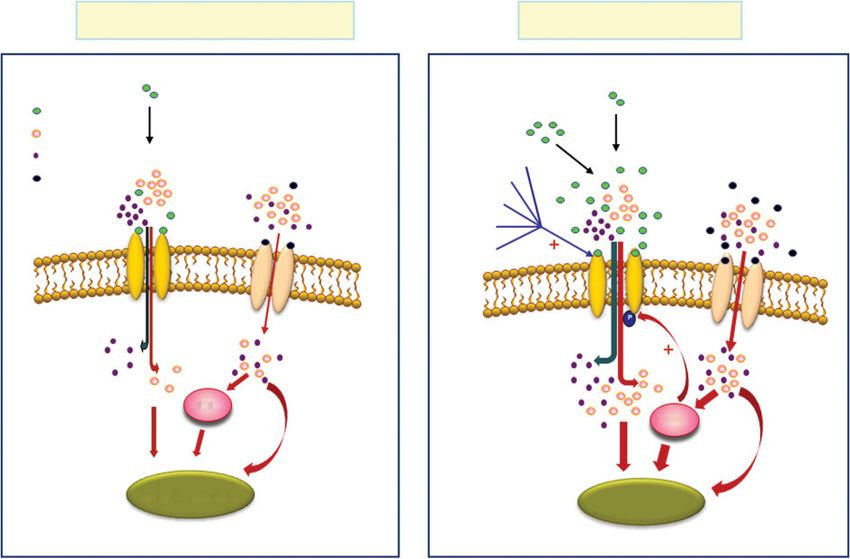
Non-ischemic condition Ischemic condition
Pre synaptic release Pre synaptic release
H+ Lactic acid
Ca2+
Na+ Arachidonic acid
Glu Ca2+ Glu Dynorphines
Glu
Na+ Ca2+ Nitric oxide Ca2+
H+ Na+
Protease
H+ Ca2+
ASIC 1a GluR Spermine ASIC 1a
GluR
CaMKII
CaMKII
Synaptic plasticity Cell death
FIGURE 1 | Activation of ASIC1a channels and outcomes in non-ischemic and ischemic conditions. In non-ischemic condition, activation of
ASIC1a channels and slight increase of cellular Ca2+ is involved in synaptic plasticity. In ischemic condition, ASIC1a activation is dramatically
potentiated by various factors. Increased concentration of H+ per se activates more current. In addition, various ischemia-related signals not only
increased the amplitude but also reduced the desensitization of the ASIC responses. Overload of neurons with Ca2+ leads to cell death.
Volume 1, September/October 2012 © 2012 WILEY-VCH Verlag GmbH & Co. KGaA, Weinheim. 657Focus Article wires.wiley.com/mts
addition to being involved in cellular signaling as a CaMKII phosphorylation of ASIC1a with KN-93, or
lipid second messenger,28 AA plays important roles in mutation of ASIC1a at Ser478 and Ser479, produced
pathological conditions including brain ischemia.28,29 neuroprotection. Thus, phosphorylation of ASIC1a by
Following brain ischemia, the rise of [Ca2+ ]i leads CaMKII deteriorates ASIC-mediated neuronal injury
to activation of phospholipase A2, resulting in in ischemia.
increased production of lipid mediators including
AA. Although the exact mechanisms are unclear, high
concentrations of lipid mediators are known to cause Dynorphins
neurotoxicity.28 Dynorphins are endogenous neuropeptides abun-
On the basis of demonstrated effects of AA dantly expressed in the central nervous system (CNS).
on a variety of voltage-gated and ligand-gated They are involved in a variety of physiologic func-
ion channels, for example, potentiation of NMDA tions. Under pathophysiological conditions where
channel currents,30 Allen and Attwell tested the effect their levels are substantially elevated, these peptides
of AA on ASICs. In rat cerebellar Purkinje cells, bath can be neurotoxic, partially mediated through gluta-
perfusion of 5 or 10 μM AA produced a large increase mate receptors.35 Recently, Sherwood and Askwith
in the amplitude of the ASIC current. In addition reported that, at high concentrations dynorphins such
to potentiating the peak amplitude, AA enhanced as big dynorphin potentiate acid-activated currents in
or induced an additional sustained component of mouse cortical neurons and in CHO cells expressing
the current.31 In heterologous expression systems, homomeric ASIC1a channels.36 The potentiation of
AA potentiates both homomeric ASIC1a and ASIC2a the ASIC1a activity was mediated through a reduction
channels.32 Thus, promoting the activation of ASIC1a of the steady-state desensitization of these channels.
channels could be one of the mechanisms mediating In the absence of big dynorphine, pre-exposing neu-
the neurotoxicity of AA. rons to conditioning pH of 7.0 completely desensitizes
the channels, resulting in no responses to subsequent
larger decrease in pH (e.g., to 5.0). In the presence of
big dynorphine, however, ASIC1a currents were read-
CaMKII
ily activated. As expected, big dynorphine enhanced
Ca2+ /calmodulin (CaM)-dependent protein kinase II
ASIC1a-mediated neuronal injury during prolonged
(CaMKII) is the most abundant kinase isoform in
acidosis.
the brain and a major mediator of the function of
excitatory glutamate receptors. Influx of intracellular
Ca2+ through glutamate receptors triggers an Lactate
autophosphorylation of CaMKII and activation of Back in 2001, Immke and McCleskey demonstrated
the enzyme. Although activation of CaMKII plays that, in sensory neurons that innervate the heart and
a prominent role in synaptic plasticity, increased COS-7 cells transfected with AIC1a channels, addition
CaMKII activity has been implicated in the regulation of lactate, at the level seen in ischemia, dramatically
of neuronal cell death after cerebral ischemia.33 increased the amplitude of the ASIC current activated
Studies by Gao et al. demonstrated a link by a moderate pH drop to ∼7.0.37 The increase of the
between CaMKII activation and acidotoxic neuronal current amplitude was accompanied by a reduced
death.34 They showed that global ischemia in rats current desensitization. Applications of lactate at
results in an increased phosphorylation of ASIC1a pH values that do not activate ASICs caused no
at Ser478 and Ser479 by CaMKII. This phosphory- response. Thus, lactate acted by potentiating but not
lation sensitizes the channel to low pH, exacerbat- activating the ASICs. The effect of lactate persisted
ing cell death by allowing prolonged and increased in excised membrane patches indicating the lack of
Ca2+ entry. second messenger involvement. As lactate has the
Consistent with the report by Xiong and ability to chelate the divalent cations, which have
colleagues,15 Gao and colleagues observed an a modulatory role for various membrane receptors
enhancement of ASIC currents by OGD. Inhibition and ion channels,38–40 it was logical to hypothesize
of CaMKII with KN-93 or CaMKIINtide abolished that potentiation of the ASIC currents could be due
the enhancement of ASIC currents, indicating an to a chelation of Ca2+ and Mg2+ in the solution.
involvement of CaMKII. They further demonstrated Indeed, adjusting the concentrations of Ca2+ and
an increased phosphorylation of ASIC1a protein Mg2+ eliminated the potentiating effect of lactate.37
after transient global ischemia, which was blocked Similar to the cardiac sensory neurons, potentiation
by intracerebroventricular administration of KN-93 of the ASIC current by lactate has been reported in
or CaMKIINtide. Pharmacological inhibition of other neurons.31
658 © 2012 WILEY-VCH Verlag GmbH & Co. KGaA, Weinheim. Volume 1, September/October 2012WIREs Membrane Transport and Signaling Role of ASICs in cerebral ischemia
TABLE 1 Ischemia-related Endogenous Modulators Known to Potentiate ASIC-mediated Responses
Neurons or ASIC subunit
Modulator tested effective Modulation site(s) Functional outcome References
Arachidonic acid Cerebellar Purkinje cells Unknown Increased amplitude 32, 60
Sensory neuron Reduced desensitization
ASIC1a
ASIC3
CaMK II Hippocampal neurons Intracellular Increased amplitude 34
ASIC1a S478 and S479 Increased injury
Dynorphine Cortical neurons Extracellular Reduced steady-state desensitization 36
Hippocampal neurons PcTX1 site Increased acid-injury
ASIC1a
ASIC1b
Lactate Sensory neuron, Extracellular Increased amplitude 37, 60
Cerebellar Purkinje neurons Reduced desensitization
ASIC1a
ASIC3
Nitric oxide DRG neurons Extracellular Increased current amplitude 45, 61
Neuro2A cells Increased acid-injury
ASIC1a
ASIC1b
ASIC2a
ASIC3
Protease Hippocampal neurons Extracellular Enhanced current amplitude activated 48
ASIC1a PcTX1 site from a baseline pH of 7.0
Increased recovery from desensitization
Spermine Cortical neurons Extracellular Increased amplitude 56, 57
Hippocampal neurons PcTX1 site Reduced desensitization
ASIC1a E219 and E242 Reduced steady-state desensitization
Increased recovery from desensitization
Increased acid-injury
Nitric oxide ASICs, probably through oxidization of cysteine
Nitric oxide (NO) is an important reactive residues.45
oxygen/nitrogen species which has a variety of
physiological and pathological functions.41 During
ischemia, intracellular Ca2+ overload leads to Proteases
activation of the Ca2+ -dependent neuronal form of Brain ischemia is accompanied by increased protease
nitric oxide synthase (nNOS), resulting in an increased activity.46 Following ischemia, blood-derived pro-
production of NO.42,43 NO can also be released by teases have access to the interstitial space from a com-
activated microglia.44 Excessive NO production is promised blood–brain barrier.46,47 Studies by Poirot
known to increase neuronal injury.44 Although the and colleagues demonstrated an ASIC1a-specific mod-
formation of a strong oxidant of peroxynitrite is ulation of the ASIC activity by serine proteases.48
likely involved in cell injury, other mechanisms cannot Exposure of cells to trypsin, for example, leads to a
be excluded. Cadiou and colleagues reported that decreased ASIC1a current if the channel is activated
NO donor S-nitroso-N-acetylpenicillamine (SNAP) by a pH drop from pH 7.4. However, if acidification
potentiates ASIC currents in DRG neurons and in occurs from a lower basal pH (e.g., 7.0), a con-
CHO cells expressing ASIC subunits. Modulators dition pertinent to brain ischemia, protease exposure
of the cGMP/PKG pathway had no effect on the increases, rather than decreases, the ASIC1a activity.48
potentiation, but in excised patches from CHO Further studies demonstrate that trypsin modulates
cells expressing ASIC2a, the potentiation could be the ASIC1a function by cleaving this subunit at Arg-
reversed by externally applying reducing agents. 145, which is in the N-terminus of the extracellular
NO, therefore, has a direct external effect on loop overlapping with the PcTX1 binding site.49
Volume 1, September/October 2012 © 2012 WILEY-VCH Verlag GmbH & Co. KGaA, Weinheim. 659Focus Article wires.wiley.com/mts
Spermine CONCLUSION
Spermine is a polyvalent cation involved in var- Stroke or cerebral ischemia is a leading health problem
ious physiological processes. High concentration worldwide. Unfortunately, there is still no effec-
of spermine can induce neuronal depolarization
tive treatment for stroke patients. Searching for
and cytoplasmic Ca2+ overload, which may lead
new brain injury mechanisms and effective thera-
to neuronal damage.50 Following ischemia, the
peutic strategies is a major challenge and priority.
activity of ornithine decarboxylase (ODC), a rate-
Acidosis is a primary feature associated with cere-
limiting enzyme responsible for polyamine synthesis is
bral ischemia and is known to cause brain injury.
enhanced, leading to elevated level of spermine.51
The finding that activation of ASICs contributes
Although the modulation of NMDA receptor
to acidosis- and ischemia-induced intracellular Ca2+
function52,53 might explain its neurotoxicity, several
accumulation and neuronal injury has suggested that
studies have yielded inconsistent results.54,55
these channels may represent potential new thera-
Babini and colleagues demonstrated that sper-
peutic targets for stroke intervention. In additional
mine potentiates the activities of ASICs.56 More
to developing pharmacological agents directly target-
recently, Duan and colleagues showed that extra-
ing ASICs, alternative strategies can be considered
cellular spermine exacerbated ischemic neuronal
by targeting ischemia-related signals known to poten-
injury through sensitization of ASIC1a channels to
tiate the activity of these channels (Table 1). Alter-
acidosis.57 In addition to increasing channel activa-
native neuroprotective strategies may also consider
tion, spermine reduced channel desensitization and
targeting the mechanisms and pathways that control
accelerated recovery from desensitization in response
the expression level of total protein and/or surface
to repeated acid stimulation. Thus, extracellular
ASIC1a.58,59
spermine contributes to ischemic neuronal injury,
at least in part, by enhancing the activity of ASIC1a
channels.57
ACKNOWLEDGMENT
The work in Z.G.X.’s lab is supported in part by NIH R01NS047506, R01NS066027, UL1 RR025008, U54
RR026137, AHA 0840132N, and ALZ IIRG-10-173350.
REFERENCES
1. Weintraub MI. Thrombolysis (tissue plasminogen acti- traumatic brain injury? Lancet Neurol 2002, 1:
vator) in stroke: a medicolegal quagmire. Stroke 2006, 383–386.
37:1917–1922. 8. Siesjo BK, Katsura K, Kristian T. Acidosis-related dam-
2. Choi DW. Calcium-mediated neurotoxicity: relation- age. Adv Neurol 1996, 71:209–233.
ship to specific channel types and role in ischemic 9. Tombaugh GC, Sapolsky RM. Evolving concepts
damage. Trends Neurosci 1988, 11:465–469. about the role of acidosis in ischemic neuropathology.
3. Lee JM, Zipfel GJ, Choi DW. The changing landscape J Neurochem 1993, 61:793–803.
of ischaemic brain injury mechanisms. Nature 1999, 10. Nedergaard M, Kraig RP, Tanabe J, Pulsinelli WA.
399:A7–A14. Dynamics of interstitial and intracellular pH in evolving
4. Simonian NA, Coyle JT. Oxidative stress in neurode- brain infarct. Am J Physiol 1991, 260:R581–R588.
generative diseases. Annu Rev Pharmacol Toxicol 1996, 11. Rehncrona S. Brain acidosis. Ann Emerg Med 1985,
36:83–106. 14:770–776.
5. Coyle JT, Puttfarcken P. Oxidative stress, glutamate, 12. Wemmie JA, Price MP, Welsh MJ. Acid-sensing ion
and neurodegenerative disorders. Science 1993, 262: channels: advances, questions and therapeutic opportu-
689–695. nities. Trends Neurosci 2006, 29:578–586.
6. Polster BM, Fiskum G. Mitochondrial mechanisms 13. Swanson RA, Farrell K, Simon RP. Acidosis causes
of neural cell apoptosis. J Neurochem 2004, 90: failure of astrocyte glutamate uptake during hypoxia.
1281–1289. J Cereb Blood Flow Metab 1995, 15:417–424.
7. Ikonomidou C, Turski L. Why did NMDA recep- 14. McDonald JW, Bhattacharyya T, Sensi SL, Lobner D,
tor antagonists fail clinical trials for stroke and Ying HS, Canzoniero LMT, Choi DW. Extracellular
660 © 2012 WILEY-VCH Verlag GmbH & Co. KGaA, Weinheim. Volume 1, September/October 2012WIREs Membrane Transport and Signaling Role of ASICs in cerebral ischemia
acidity potentiates AMPA receptor-mediated cortical 28. Farooqui AA, Horrocks LA. Phospholipase A2-
neuronal death. J Neurosci 1998, 18:6290–6299. generated lipid mediators in the brain: the good, the
15. Xiong ZG, Zhu XM, Chu XP, Minami M, Hey J, bad, and the ugly. Neuroscientist 2006, 12:245–260.
Wei WL, MacDonald JF, Wemmie JA, Price MP, 29. Siesjo BK, Katsura K. Ischemic brain damage: focus on
Welsh MJ, et al. Neuroprotection in ischemia: block- lipids and lipid mediators. Adv Exp Med Biol 1992,
ing calcium-permeable Acid-sensing ion channels. Cell 318:41–56.
2004, 118:687–698. 30. Miller B, Sarantis M, Traynelis SF, Attwell D. Potenti-
16. Waldmann R, Champigny G, Bassilana F, Heurteaux C, ation of NMDA receptor currents by arachidonic acid.
Lazdunski M. A proton-gated cation channel involved Nature 1992, 355:722–725.
in acid-sensing. Nature 1997, 386:173–177. 31. Allen NJ, Attwell D. Modulation of ASIC channels
17. Mari Y, Katnik C, Cuevas J. ASIC1a channels are in rat cerebellar Purkinje neurons by ischemia-related
activated by endogenous protons during ischemia and signals. J Physiol (Lond) 2002, 543:521–529.
contribute to synergistic potentiation of intracellular 32. Smith ES, Cadiou H, McNaughton PA. Arachidonic
Ca(2+) overload during ischemia and acidosis. Cell acid potentiates acid-sensing ion channels in rat sen-
Calcium 2010, 48:70–82. sory neurons by a direct action. Neuroscience 2007,
18. Yermolaieva O, Leonard AS, Schnizler MK, Abboud 145:686–698.
FM, Welsh MJ. Extracellular acidosis increases neu- 33. Coultrap SJ, Vest RS, Ashpole NM, Hudmon A, Bayer
ronal cell calcium by activating acid-sensing ion channel KU. CaMKII in cerebral ischemia. Acta Pharmacol Sin
1a. Proc Natl Acad Sci U S A 2004, 101:6752–6757. 2011, 32:861–872.
19. Sherwood TW, Lee KG, Gormley MG, Askwith CC. 34. Gao J, Duan B, Wang D-G, Deng X-H, Zhang
Heteromeric acid-sensing ion channels (ASICs) com- G-Y, Xu L, Xu T-L. Coupling between NMDA recep-
posed of ASIC2b and ASIC1a display novel channel tor and acid-sensing ion channel contributes to ischemic
properties and contribute to acidosis-induced neuronal neuronal death. Neuron 2005, 48:635–646.
death. J Neurosci 2011, 31:9723–9734. 35. Hauser KF, Foldes JK, Turbek CS. Dynorphin A (1-13)
20. Pignataro G, Simon RP, Xiong ZG. Prolonged activa- neurotoxicity in vitro: opioid and non-opioid mecha-
tion of ASIC1a and the time window for neuroprotec- nisms in mouse spinal cord neurons. Exp Neurol 1999,
tion in cerebral ischaemia. Brain 2007, 130:151–158. 160:361–375.
21. Biegon A, Fry PA, Paden CM, Alexandrovich A, Tsen- 36. Sherwood TW, Askwith CC. Dynorphin opioid pep-
ter J, Shohami E. Dynamic changes in N-methyl-d- tides enhance acid-sensing ion channel 1a activity
aspartate receptors after closed head injury in mice: and acidosis-induced neuronal death. J Neurosci 2009,
implications for treatment of neurological and cogni- 29:14371–14380.
tive deficits. Proc Natl Acad Sci U S A 2004, 101: 37. Immke DC, McCleskey EW. Lactate enhances the acid-
5117–5122. sensing Na+ channel on ischemia-sensing neurons. Nat
22. Li M, Inoue K, Branigan D, Kratzer E, Hansen JC, Chen Neurosci 2001, 4:869–870.
JW, Simon RP, Xiong Z-G. Acid-sensing ion channels 38. Xiong ZG, MacDonald JF. Sensing of extracellular
in acidosis-induced injury of human brain neurons. calcium by neurones. Can J Physiol Pharmacol 1999,
J Cereb Blood Flow Metab 2010, 30:1247–1260. 77:715–721.
23. Hesselager M, Timmermann DB, Ahring PK. pH depen- 39. Hess P, Lansman JB, Tsien RW. Calcium channel selec-
dency and desensitization kinetics of heterologously tivity for divalent and monovalent cations. Voltage
expressed combinations of acid-sensing ion channel and concentration dependence of single channel cur-
subunits. J Biol Chem 2004, 279:11006–11015. rent in ventricular heart cells. J Gen Physiol 1986, 88:
24. Krishtal O. The ASICs: signaling molecules? Modula- 293–319.
tors? Trends Neurosci 2003, 26:477–483. 40. Zhou W, Jones SW. Surface charge and calcium chan-
25. Grunder S, Chen X. Structure, function, and pharma- nel saturation in bullfrog sympathetic neurons. J Gen
cology of acid-sensing ion channels (ASICs): focus on Physiol 1995, 105:441–462.
ASIC1a. Int J Physiol Pathophysiol Pharmacol 2010, 41. Star RA. Nitric oxide. Am J Med Sci 1993, 306:
2:73–94. 348–358.
26. Bickler PE, Buck LT. Adaptations of vertebrate neu- 42. Schulz JB, Matthews RT, Klockgether T, Dichgans J,
rons to hypoxia and anoxia: maintaining critical Ca2+ Beal MF. The role of mitochondrial dysfunction and
concentrations. J Exp Biol 1998, 201:1141–1152. neuronal nitric oxide in animal models of neurodegen-
27. Chu XP, Papasian CJ, Wang JQ, Xiong ZG. Modulation erative diseases. Mol Cell Biochem 1997, 174:193–197.
of acid-sensing ion channels: molecular mechanisms 43. Bolanos JP, Almeida A. Roles of nitric oxide in brain
and therapeutic potential. Int J Physiol Pathophysiol hypoxia-ischemia. Biochim Biophys Acta 1999, 1411:
Pharmacol 2011, 3:288–309. 415–436.
Volume 1, September/October 2012 © 2012 WILEY-VCH Verlag GmbH & Co. KGaA, Weinheim. 661Focus Article wires.wiley.com/mts
44. Boje KM, Arora PK. Microglial-produced nitric oxide 53. Rock DM, Macdonald RL. Polyamine regulation of
and reactive nitrogen oxides mediate neuronal cell N-methyl-d-aspartate receptor channels. Annu Rev
death. Brain Res 1992, 587:250–256. Pharmacol Toxicol 1995, 35:463–482.
45. Cadiou H, Studer M, Jones NG, Smith E StJ, Ballard A, 54. Johnson TD. Polyamines and cerebral ischemia. Prog
McMahan SB, McNaughton PA. Modulation of acid- Drug Res 1998, 50:193–258.
sensing ion channel activity by nitric oxide. J Neurosci 55. Li J, Doyle KM, Tatlisumak T. Polyamines in the brain:
2007, 27:13251–13260. distribution, biological interactions, and their potential
46. Gingrich MB, Traynelis SF. Serine proteases and brain therapeutic role in brain ischaemia. Curr Med Chem
damage - is there a link? Trends Neurosci 2000, 2007, 14:1807–1813.
23:399–407. 56. Babini E, Paukert M, Geisler HS, Grunder S. Alternative
47. Vivien D, Buisson A. Serine protease inhibitors: novel splicing and interaction with di- and polyvalent cations
therapeutic targets for stroke? J Cereb Blood Flow control the dynamic range of acid-sensing ion channel
Metab 2000, 20:755–764. 1 (ASIC1). J Biol Chem 2002, 277:41597–41603.
57. Duan B, Wang Y-Z, Yang T, Chu X-P, Yu Y, Huang
48. Poirot O, Vukicevic M, Boesch A, Kellenberger S. Selec-
Y, Cao H, Hansen J, Simon RP, et al. Extracellular
tive regulation of acid-sensing ion channel 1 by serine
spermine exacerbates ischemic neuronal injury through
proteases. J Biol Chem 2004, 279:38448–38457.
sensitization of ASIC1a channels to extracellular acido-
49. Vukicevic M, Weder G, Boillat A, Boesch A, Kellen- sis. J Neurosci 2011, 31:2101–2112.
berger S. Trypsin cleaves acid-sensing ion channel 1a in 58. Pignataro G, Cuomo O, Esposito E, Sirabell R, Renzo
a domain that is critical for channel gating. J Biol Chem Di, Annunziato GL. ASIC1a contributes to neuro-
2006, 281:714–722. protection elicited by ischemic preconditioning and
50. Toninello A, Salvi M, Mondovi B. Interaction of bio- postconditioning. Int J Physiol Pathophysiol Pharmacol
logically active amines with mitochondria and their role 2011, 3:1–8.
in the mitochondrial-mediated pathway of apoptosis. 59. Chai S, Li M, Branigan D, Xiong ZG, Simon RP. Activa-
Curr Med Chem 2004, 11:2349–2374. tion of acid-sensing ion channel 1a (ASIC1a) by surface
51. Kindy MS, Hu Y, Dempsey RJ. Blockade of ornithine trafficking. J Biol Chem 2010, 285:13002–13011.
decarboxylase enzyme protects against ischemic brain 60. Allen NJ, Attwell D. Modulation of ASIC channels
damage. J Cereb Blood Flow Metab 1994, 14: in rat cerebellar Purkinje neurons by ischaemia-related
1040–1045. signals. J Physiol 2002, 543:521–529.
52. Benveniste M, Mayer ML. Multiple effects of sper- 61. Jetti SK, Swain SM, Majumder S, Chatterjee S, Poorn-
mine on N-methyl-d-aspartic acid receptor responses ima V , Bera AK. Evaluation of the role of nitric oxide
of rat cultured hippocampal neurones. J Physiol 1993, in acid sensing ion channel mediated cell death. Nitric
464:131–163. Oxide 2010, 22:213–219.
662 © 2012 WILEY-VCH Verlag GmbH & Co. KGaA, Weinheim. Volume 1, September/October 2012You can also read