Chemical Science - RSC Publishing

 
CONTINUE READING
Chemical Science - RSC Publishing
Volume 12
                                                                                  Number 25

Chemical
                                                                                  7 July 2021
                                                                                  Pages 8573–8932

Science
rsc.li/chemical-science

                          ISSN 2041-6539

                          EDGE ARTICLE
                          Do-Hyeon Kim, Sung Ho Ryu et al.
                          Blue-conversion of organic dyes produces artifacts in
                          multicolor fluorescence imaging
Chemical Science - RSC Publishing
Chemical
                                                                                               Science
                                                                                                                                                                                                                               View Article Online
                                                                                                   EDGE ARTICLE                                                                                                                View Journal | View Issue

                                                                                                                                             Blue-conversion of organic dyes produces artifacts
         This article is licensed under a Creative Commons Attribution 3.0 Unported Licence.

                                                                                               Cite this: Chem. Sci., 2021, 12, 8660
                                                                                                                                             in multicolor fluorescence imaging†
                                                                                                  All publication charges for this article   Do-Hyeon Kim, ‡*a Yeonho Chang,‡a Soyeon Park,‡a Min Gyu Jeong,b
                                                                                               have been paid for by the Royal Society       Yonghoon Kwon,a Kai Zhou, a Jungeun Noh,a Yun-Kyu Choi,c Triet Minh Hong,a
Open Access Article. Published on 18 2021. Downloaded on 2021/11/5 11:15:04.

                                                                                               of Chemistry
                                                                                                                                             Young-Tae Chang c and Sung Ho Ryu *a

                                                                                                                                             Multicolor fluorescence imaging is a powerful tool visualizing the spatiotemporal relationship among
                                                                                                                                             biomolecules. Here, we report that commonly employed organic dyes exhibit a blue-conversion
                                                                                                                                             phenomenon, which can produce severe multicolor image artifacts leading to false-positive
                                                                                               Received 1st February 2021
                                                                                               Accepted 16th May 2021
                                                                                                                                             colocalization by invading predefined spectral windows, as demonstrated in the case study using EGFR
                                                                                                                                             and   Tensin2.    These     multicolor   image   artifacts   become   much    critical   in   localization-based
                                                                                               DOI: 10.1039/d1sc00612f
                                                                                                                                             superresolution microscopy as the blue-converted dyes are photoactivatable. We provide a practical
                                                                                               rsc.li/chemical-science                       guideline for the use of organic dyes for multicolor imaging to prevent artifacts derived by blue-conversion.

                                                                                               Biomolecules act in concert within subcellular structures to                           dyes.8–10 The cytosolic proteins in live cells have been labeled
                                                                                               generate various biomolecular processes. Multicolor uores-                            with membrane permeable organic dyes.11
                                                                                               cence imaging by labeling biomolecules with several uo-                                  While investigating cell motility dynamics mediated by the
                                                                                               rophores allows the visualization of the spatiotemporal                                epidermal growth factor receptor (EGFR) using live-cell uo-
                                                                                               relationships among them, providing direct evidence for                                rescence microscopy, we observed the unexpected phenomenon
                                                                                               understanding their concerted behaviors and functions.1                                that organic dyes are converted to another species emitting
                                                                                                  Choosing suitable dyes based on the purpose of the experi-                          shorter wavelengths than their original emission wavelength
                                                                                               ment is critical for uorescence microscopy. Multicolor imaging                        aer the imaging. We labeled SNAP-tagged EGFR expressed on
                                                                                               requires uorophores that uoresce at distinct and well-                               COS7 cells with O6-benzylguanine (BG)-conjugated Alexa Fluor
                                                                                               separated spectra.2 Superresolution localization microscopy                            647 (A647). Aer imaging the A647-EGFR on the plasma
                                                                                               requires photoactivatable/-switchable uorophores to achieve                           membrane using total internal reection uorescence (TIRF)
                                                                                               the blinking necessary to distinguish individual uorophores                           microscopy through the far-red channel (654–870 nm) with
                                                                                               within the diffraction limit.3,4 Long-term imaging requires                             642 nm laser illumination, we spotted unanticipated uores-
                                                                                               highly photostable yet sufficiently bright uorophores.5                                 cence in the red channel (572–624 nm) upon excitation by
                                                                                                  Organic dyes are commonly utilized due to their advanta-                            561 nm laser illumination, which did not exist before the
                                                                                               geous characteristics, including a broad range of excitation and                       imaging of the A647-EGFR. It was formerly reported that far-red
                                                                                               emission spectra, high brightness and photostability, and                              cyanine dyes, including A647 and Cy5, contain near-red uo-
                                                                                               photoactivatable/-switchable properties.4,6,7 The specic                              rescent impurities.12 However, the amount of this newly
                                                                                               labeling of a target protein with an organic dye has been ach-                         appeared uorescence in the red channel was strongly corre-
                                                                                               ieved by using the antibodies crosslinked via N-hydrox-                                lated with the amount of photobleached A647-EGFR by 642 nm
                                                                                               ysuccinimide ester or maleimide modied organic dyes or the                            laser illumination (Fig. 1a). We also observed the same
                                                                                               genetic fusion of SNAP- or Halo-tags covalently binding to                             phenomenon when endogenous EGFR was labeled with the
                                                                                               benzylguanine or chloroalkane moieties attached to organic                             A647-conjugated anti-EGFR antibody (Fig. S1†). To conrm this
                                                                                                                                                                                      phenomenon in vitro, nonconjugated A647 dissolved in DMSO
                                                                                                                                                                                      was directly illuminated with a 642 nm laser to induce photo-
                                                                                               a
                                                                                                                                                                                      bleaching on the bulk scale. Surprisingly, instead of becoming
                                                                                                Department of Life Sciences, Pohang University of Science and Technology, Pohang,
                                                                                               37673, Republic of Korea. E-mail: sungho@postech.ac.kr; genesis@postech.ac.kr          colorless as expected, the blue-colored dye turned pink aer
                                                                                               b
                                                                                                Integrative Biosciences and Biotechnology, Pohang University of Science and           sufficient laser illumination (Fig. 1b). This product contained
                                                                                               Technology, Pohang, 37673, Republic of Korea                                           two major substances detectable at 280 nm, one of which
                                                                                               c
                                                                                                 Department of Chemistry, Pohang University of Science and Technology, Pohang,        absorbed 561 nm light (Fig. 1c). Neither substance absorbed
                                                                                               37673, Republic of Korea                                                               647 nm light, indicating that the photobleaching of A647 was
                                                                                               † Electronic supplementary           information    (ESI)   available.   See   DOI:
                                                                                                                                                                                      complete. When the 561 nm-absorbing substance was illumi-
                                                                                               10.1039/d1sc00612f
                                                                                                                                                                                      nated with a 561 nm laser, red uorescence was emitted
                                                                                               ‡ These authors contributed equally to this work.

                                                                                               8660 | Chem. Sci., 2021, 12, 8660–8667                                                    © 2021 The Author(s). Published by the Royal Society of Chemistry
Chemical Science - RSC Publishing
View Article Online

                                                                                               Edge Article                                                                                                               Chemical Science
         This article is licensed under a Creative Commons Attribution 3.0 Unported Licence.
Open Access Article. Published on 18 2021. Downloaded on 2021/11/5 11:15:04.

                                                                                               Fig. 1 Blue-conversion of far-red organic dyes upon photobleaching. (a) TIRF images of A647-EGFR on COS7 cells in the far-red (upper panels)
                                                                                               channel excited at 642 nm and the red (lower panels) channel excited at 561 nm before (left panels) and after (right panels) photobleaching of
                                                                                               A647-EGFR. (b) A647 dissolved in DMSO before (left) and after (right) photobleaching using direct laser illumination. (c) HPLC analysis of A647
                                                                                               after photobleaching with absorbance at 280 nm, 561 nm, and 647 nm. (d) Emission spectra of A647 before (red) and after (green) photo-
                                                                                               bleaching, excited at 647 nm and 561 nm, respectively. (e) Emission spectra of far-red organic dyes before (red) and after (green) photobleaching.
                                                                                               Scale bars, 5 mm.

                                                                                               (Fig. 1d), which coincided with the de novo uorescence of A647-          sulfonated, whereas those peaks are greatly blue-shied as their
                                                                                               EGFR produced by 642 nm laser illumination in the red channel             rings are PEGylated. The blue-shied emission peaks aer the
                                                                                               (Fig. 1a).                                                                blue-conversion of Cy5 (no sulfonation and PEGylation), A647
                                                                                                   A few uorophores based on coumarin and triarylmethane                (sulfonation), DY649P1 (sulfonation), CF647 (PEGylation) and
                                                                                               were previously reported to exhibit this ‘blue-conversion’                CF660C (PEGylation) are at 59 nm, 54 nm, 49 nm, 86 nm, and
                                                                                               phenomenon derived by laser excitation,13,14 which can display            102 nm, respectively. A rhodamine, CF633, showed the lowest
                                                                                               residual crosstalk between multicolor channels in STED                    blue-shi of the emission peak aer the blue-conversion (35
                                                                                               microscopy.15 However, blue-conversion of cyanine-based uo-              nm), while a benzopyrylium hemicyanine, DY654, showed the
                                                                                               rophores remained controversial: while A647 was photo-                    highest blue-shi (166 nm) among the dyes we examined.
                                                                                               converted to near-red upon intense and prolonged illumination             Compared to other chemical families, cyanines tended to
                                                                                               with green/yellow lasers,16 A647 was one of the far-red organic           display multiple blue-converted species. Blue-conversion of
                                                                                               dyes that were not turned into near-red upon two-photon exci-             these far-red dyes was also conrmed in vivo (Fig. S4†). SNAP-
                                                                                               tation.17 Thus, we extensively explored whether blue-conversion           EGFR was labeled to the dyes conjugated with the BG moiety,
                                                                                               derived by an excitation laser as we observed in A647 also occurs         and the blue-conversion of all the examined dyes was observed,
                                                                                               in commonly used and commercially available organic dyes in               except for Atto 655 (Fig. S4†) although the strong blue-
                                                                                               various chemical families for conventional uorescent micros-             conversion of Atto 655 was displayed in vitro (Fig. 1e and S2†).
                                                                                               copy (Table S1†): cyanine (Alexa Fluor 647, Alexa Fluor 680,              This difference between in vitro and in vivo results might be due
                                                                                               CF647, CF660C, Cy5, Cy5.5, Cy3B, Dylight 650, and Dyomics                 to the cleavage of BG-Atto 655 induced by the blue-conversion,
                                                                                               649p1), benzopyrylium hemicyanine (Dyomics 654), rhodamine                making the fragments leave from the BG moiety conjugated to
                                                                                               (CF633, CF660R, and silicon rhodamine), carborhodamine (Atto              a SNAP-tag, which results in the loss of uorescence for SNAP-
                                                                                               647N), and oxazine (Atto 655). All these far-red dyes remained            EGFR aer the blue-conversion. Furthermore, when Cy3B was
                                                                                               opaque and changed colors aer direct illumination with the               photobleached by 561 nm laser illumination, de novo uores-
                                                                                               642 nm laser on a bulk scale, except for initially colorless SiR          cence appeared in the yellow channel (500–549 nm) upon
                                                                                               (Fig. S2†). The color-changed dyes contained one or more                  488 nm laser excitation (Fig. S4†), implying that the blue-
                                                                                               species exhibiting distinct absorption and emission spectra at            conversion of the red dyes also widely occurs, including Lyso-
                                                                                               shorter wavelengths than those of the original species (Fig. 1e           tracker Red DND-99 (Invitrogen) that has been previously re-
                                                                                               and S3†). Interestingly, we observed that the two cyanine dyes            ported to exhibit red-to-green blue-conversion.18 Among the
                                                                                               close to near-IR, Cy5.5 and A680 exhibited their blue-converted           examined dyes with various chemical families, there is no dye
                                                                                               species with emission peaks at 607 nm and 603 nm, respec-                 that did not exhibit blue-conversion at all.
                                                                                               tively, which are almost the same as the blue-converted species              We quantitatively measured the degree of blue-conversion
                                                                                               of their precursor far-red dyes, Cy5 and A647 with emission               from the far-red to the red channel by estimating the number
                                                                                               peaks at 607 nm and 611 nm, respectively (Fig. 1d, e and Table            of dye molecules in each channel using their single-molecule
                                                                                               S1†). We also found that the emission peaks of blue-converted             intensities, which enables us to directly analyze the ratio of
                                                                                               cyanine dyes are less blue-shied as their aromatic rings are             blue-converted molecules without determining the extinction

                                                                                               © 2021 The Author(s). Published by the Royal Society of Chemistry                                         Chem. Sci., 2021, 12, 8660–8667 | 8661
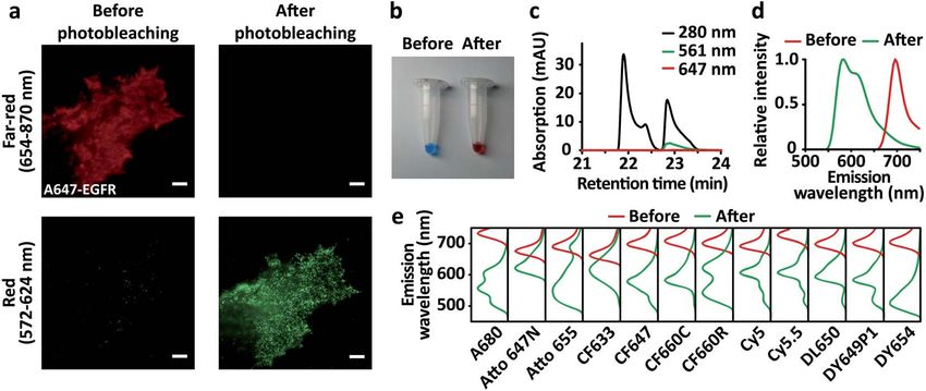
Chemical Science - RSC Publishing
View Article Online

                                                                                               Chemical Science                                                                                                                   Edge Article

                                                                                               coefficients of the unknown blue-converted species (see the                  far-red dyes only, although some uorescent proteins were
                                                                                               details in Methods). The far-red dyes were blue-converted into             previously reported to exhibit the blue-conversion.24–28 Both
                                                                                               the red channel by up to 5.37% (Fig. S5†). We could not observe            A647- and CF660R-Tensin2 displayed patch-like structures
                                                                                               the strong relationship between their chemical structures (or              (Fig. 2a and d) and RFP-EGFR showed a relatively even distri-
                                                                                               families) and the amount of blue-conversion (Table S1 and                  bution over the entire plasma membrane (Fig. 2b and e),
                                                                                               Fig. S5†).                                                                 consistent with the previous reports.29,30 Interestingly, however,
                                                                                                  Multicolor imaging has long been utilized in dichroic                   the distribution of EGFR gradually changed patch-like within
                                                                                               mirrors and emission lters to separate distinctly emitting                1 min of RFP-EGFR imaging in the cells coexpressing A647-
                                                                                               uorophores into desired channels. This technique relies on                Tensin2 (ESI Video S1†), whereas only the uorescence inten-
         This article is licensed under a Creative Commons Attribution 3.0 Unported Licence.

                                                                                               the fact that the excitation and emission spectra of uorophores           sity decreased while the distribution of EGFR did not change as
                                                                                               do not change during the imaging. However, this level of blue-             expected in the cells coexpressing CF660R-Tensin2 (ESI Video
                                                                                               conversion was expected to signicantly invade the predened               S2†). This phenomenon occurred because the 561 nm laser used
                                                                                               spectral channels. To compare the multicolor uorescence
Open Access Article. Published on 18 2021. Downloaded on 2021/11/5 11:15:04.

                                                                                                                                                                          for exciting RFP also excites A647 and CF660R, causing differ-
                                                                                               images produced by blue-conversion sensitive and resistant                 ential levels of blue-conversion depending on their photo-
                                                                                               dyes, we selected A647 and CF660R. Although these uo-                     stability. We conrmed that the signal for RFP-EGFR increased
                                                                                               rophores displayed similar levels of blue-conversion aer                  during the imaging only in the region where A647-Tensin2 was
                                                                                               complete photobleaching, CF660R is substantially more pho-                 located (Fig. 2b and c). Because of its high photostability, the
                                                                                               tostable than A647. Thus, the differential amount of blue-                  amount of blue-conversion of CF660R was not signicant even
                                                                                               conversion of the two dyes can be proportionally induced by                aer RFP was substantially photobleached (Fig. 2e and f). The
                                                                                               the duration of laser illumination.                                        two membrane proteins exhibited apparent colocalization on
                                                                                                  We conducted the colocalization analysis of EGFR and                    the plasma membrane when Tensin2 was labeled with A647
                                                                                               Tensin2 as a case study. ErbB and Tensin families are previously           aer the mild photobleaching that occurred under the
                                                                                               known kinases and phosphatases involved in focal adhesion                  conventional imaging conditions for uorescence microscopy,31
                                                                                               dynamics in cell motility.19–21 The direct association between             but not when Tensin2 was labeled with CF660R (ESI Fig. S6†). It
                                                                                               ErbB and Tensin family members has been reported,22,23 but not             is possible to conclude that EGFR and Tensin2 are colocalized if
                                                                                               between EGFR and Tensin2. BG-A647 or BG-CF660R was labeled                 a popular A647 was simply used, even though in reality these
                                                                                               with SNAP-Tensin2 coexpressed with red uorescent protein                  two proteins have no such relationship, as shown by using
                                                                                               (RFP)-conjugated EGFR in COS7 cells. Because we observed that              CF660R.
                                                                                               red uorescent protein (RFP) exhibited no blue-conversion, RFP                 We further observed that many organic dyes, including A647,
                                                                                               was utilized to examine the effect derived by blue-conversion of            A680, Atto 647N, CF633, CF647, CF660C, CF660R, Cy5, Cy5.5

                                                                                               Fig. 2 Multicolor imaging artifacts produced by blue-conversion. TIRF images of BG-A647- (a) or BG-CF660R- (d) labeled SNAP-Tensin2 in the
                                                                                               far-red channel. TIRF images of RFP-EGFR (b and e) in the red channel with 45 s of continuous imaging. The insets display magnified images of
                                                                                               the selected regions at the same positions with (orange box) or without (blue box) the Tensin2 signal in the far-red channel. (c and f) Time profiles
                                                                                               of the fluorescence intensity in the selected regions. Scale bars, 5 mm (white) and 0.5 mm (orange). The fluorescence intensity in the selected
                                                                                               regions for the red channel kept increasing during the imaging of RFP only in the region where A647-Tensin 2 was located whereas kept
                                                                                               decreasing in the region where A647-Tensin 2 was not located (c). The fluorescence intensity in the selected regions for the red channel kept
                                                                                               decreasing during the imaging of RFP regardless of regions where CF660R-Tensin 2 located or not (f).

                                                                                               8662 | Chem. Sci., 2021, 12, 8660–8667                                         © 2021 The Author(s). Published by the Royal Society of Chemistry
View Article Online

                                                                                               Edge Article                                                                                                                    Chemical Science

                                                                                               DL650, DY649P1, and SiR, blue-converted by laser excitation                  (Fig. 3c and d). Not only the recovered amount of the blue-
                                                                                               initially existed largely in a dark state, which could be recovered          converted species in a bright state by UV photoactivation, but
                                                                                               to a bright state by UV illumination (Fig. S4a–c, e–l and n†).               also the single-molecule brightness of the blue-converted
                                                                                               Because superresolution techniques based on single-molecule                  species contributed to the production of the artifact in the
                                                                                               localization rely on the UV-induced photoactivatability of uo-              reconstructed images. The blue-converted A647 was only
                                                                                               rophores, we explored the effect of blue-conversion on multi-                 slightly brighter than mEos3.2, making it almost impossible to
                                                                                               color superresolution imaging. By replacing the RFP with                     distinguish between the two uorophores. However, the blue-
                                                                                               mEos3.2 for the conjugation of EGFR, we performed colocali-                  converted CF660R was signicantly dimmer than mEos3.2,
                                                                                               zation analysis of EGFR and Tensin2 using single-molecule                    allowing the automatic rejection (SNR < 3) during the image
         This article is licensed under a Creative Commons Attribution 3.0 Unported Licence.

                                                                                               localization microscopy. The reconstructed superresolution                   reconstruction.
                                                                                               image of mEos3.2-EGFR showed apparent patch-like structures                      The demonstrated colocalization artifact derived by blue-
                                                                                               colocalized with A647-Tensin2 at a subdiffraction resolution                  conversion of organic dyes is an exemplary case where the
Open Access Article. Published on 18 2021. Downloaded on 2021/11/5 11:15:04.

                                                                                               (Fig. 3a and b), whereas no signicant colocalization was                    expression level of Tensin2 is an order-of-magnitude higher
                                                                                               observed in the superresolution imaging with CF660R-Tensin2                  than that of EGFR. Even below 2% of the blue-conversion can

                                                                                               Fig. 3 Multicolor superresolution imaging artifacts produced by blue-conversion. TIRF images of BG-A647- or BG-CF660R-labeled Tensin2 in
                                                                                               the far-red channel (left panels in (a) and (c)). Reconstructed superresolution images of mEos3.2-EGFR in the red channel (middle panels in (a)
                                                                                               and (c)). Overlay of the images from the two channels (right panels in (a) and (c)). (b) and (d) Intensity profiles of the indicated lines (white) in the
                                                                                               overlaid images. Scale bars, 5 mm.

                                                                                               © 2021 The Author(s). Published by the Royal Society of Chemistry                                             Chem. Sci., 2021, 12, 8660–8667 | 8663
View Article Online

                                                                                               Chemical Science                                                                                                        Edge Article

                                                                                               produce a detrimental artifact in multi-color biomolecule           (Fig. 1d, e, and Table S1†). The modications of Cy5.5 and A680
                                                                                               imaging in which the asymmetric amount of labeling between          were introduced into their aromatic rings of the precursors
                                                                                               different channels is frequently encountered. Numerous cases         while the length of their polymethine chain was preserved.
                                                                                               with differential expression levels can be found in the human        Furthermore, other cyanine dyes with the same length of poly-
                                                                                               proteome.32 Furthermore, labeling efficiency varies from              methine chains exhibited a similar range of emission peaks
                                                                                               different targets and methods. For instance, the affinity of           (579 to 632 nm) (Table S1†), implying that a primary factor
                                                                                               antibodies used to label biomolecules varies signicantly from      contributing to the blue-conversion of cyanine dyes is the length
                                                                                               target-to-target and clone-to-clone. Besides, blue-conversion is    of their polymethine chains. This interpretation is also sup-
                                                                                               accompanied by photobleaching of the labeled dye with expo-         ported by that Cy3B with the shorter length of the polymethine
         This article is licensed under a Creative Commons Attribution 3.0 Unported Licence.

                                                                                               nential kinetics. Thus, not only the increase of blue-converted     chain exhibited its blue-converted uorescence mainly in the
                                                                                               uorescence but also the exponential decrease of the labeled        yellow channel (500 to 549 nm) (Fig. S4o†).
                                                                                               dye reduces their signal gap. Finally, colocalization analysis          We provide a practical guideline for the use of organic dyes
                                                                                                                                                                   for multicolor uorescence imaging to prevent artifacts derived
Open Access Article. Published on 18 2021. Downloaded on 2021/11/5 11:15:04.

                                                                                               depends on the local spatial correlation rather than the signal
                                                                                               amplitude between the uorescence detected in two channels.         by blue-conversion. First, unnecessary excitation of the dye
                                                                                               Thus, even the small bleed-through caused by blue-conversion        should be minimized because it not only diminishes the orig-
                                                                                               can be critical in colocalization analysis if the targets exhibit   inal uorescence intensity but also produces de novo uores-
                                                                                               spatial patterns.                                                   cence in other channels for shorter emission spectra. Second,
                                                                                                  Previously, it has been reported that triarylmethane dyes and    highly photostable dyes should be adopted. If photosensitive
                                                                                               coumarin dyes were blue-converted by photooxidative N-deal-         dyes must be utilized for specic applications, an oxygen scav-
                                                                                               kylation.13,33–35 Furthermore, N,N0 -di-tert-alkylrhodamine uo-    enging system should be included because oxygen is critical to
                                                                                               rophores have been recently developed to prevent the blue-          the process of blue-conversion (Fig. S7†), although its use may
                                                                                               conversion based on its mechanism.14 However, the chemical          limit live-cell applications. The photostabilizer-conjugated u-
                                                                                               mechanism behind the blue-conversion of cyanine dyes (A647,         orophores or recently developed blue-conversion-resistant tri-
                                                                                               A680, Cy5, Cy5.5, CF647, CF660C, DL650, and DY649P1) is not         arylmethane uorophores could also be utilized.14,37 Third,
                                                                                               fully understood although a photooxidative reaction is essential    when the expression levels of two proteins are substantially
                                                                                               to their blue-conversion (Fig. S7†). When we depleted oxygen        different, the protein with the higher expression should be
                                                                                               using PCA/PCD, the blue-conversion of A647 signicantly             labeled with a dye with a shorter wavelength or partially labeled
                                                                                               reduced (Fig. S7a†). In particular, singlet oxygen is likely        to compensate for the expression level. We also provide a step-
                                                                                               involved in the blue-conversion process because the amount of       by-step procedure for checking and troubleshooting for the
                                                                                               blue-conversion showed a positive correlation when we modu-         artifacts derived by blue-conversion of organic dyes in multi-
                                                                                               lated the level of singlet oxygen (Fig. S7b and c†). Recently,      color uorescence imaging (ESI†).
                                                                                               a study36 published during the revision of our manuscript re-           Although we introduced the immediate negative effect of the
                                                                                               ported that Cy5 becomes dioxetane species (emmax ¼ 434 nm),         blue-conversion of organic dyes, this new photoconversion
                                                                                               photooxidative cleavage products (emmax ¼ 210 nm, 345 nm,           pathway of cyanine dyes can bring the advantages of uores-
                                                                                               370 nm), and Cy3 (emmax ¼ 540 nm) aer the blue-conversion,         cence imaging applications. The super-resolution techniques
                                                                                               although the mechanism behind its blue-conversion has not           require the photoactivation (or photoswitching) of organic dyes
                                                                                               been fully described. However, unlike their study, we observed      where an oxygen scavenging system and a primary thiol are
                                                                                               that Cy5 produced the blue-converted species with their emis-       typically required to induce their photoactivatability, which
                                                                                               sion peaks majorly at 607 nm, 567 nm, and 523 nm (Fig. 1e).         possibly exerts an adverse effect in live cell imaging. However,
                                                                                               Especially, the blue-converted species at 607 nm was respon-        the photoactivation of the blue-converted species occurs
                                                                                               sible for more than 50% of the emission spectrum of the blue-       without requiring any buffer conditions as it occurs in water,
                                                                                               converted Cy5 (Fig. 1e) and this species was primarily detected     PBS, and DMEM, aiding the super-resolution microscopy for
                                                                                               both in vitro and in vivo in the red channel (572 to 624 nm)        live cells in their physiological conditions. Although we
                                                                                               (Fig. S4†), but the recent study entirely lacks this principal      demonstrated the artifact of super-resolution images derived by
                                                                                               species. Although the authors claimed that these species were       the blue-conversion of A647 (Fig. 3a) as a limitation, this
                                                                                               not observed lower than 0.10 kW cm2 where super-resolution         demonstration can be interpreted as an advantage of super-
                                                                                               microscopy works, our blue-conversion data for Cy5 were ob-         resolution microscopy using the blue-conversion of A647
                                                                                               tained aer the bleaching at 0.05 kW cm2 and clearly               working without requiring any buffer conditions.
                                                                                               demonstrated the practical super-resolution imaging that the            Care must be taken in multicolor imaging applications,
                                                                                               blue-converted species was detected in the red channel (Fig. 3).    including colocalization, uorescence resonance energy trans-
                                                                                               Interestingly, we found that Cy5.5 close to near-IR (emmax ¼ 694    fer, uorescence correlation spectroscopy, single-particle
                                                                                               nm) and its precursor, Cy5 (emmax ¼ 666 nm), exhibited their        tracking, or screenings using uorescence-based multi-well
                                                                                               blue-converted species with the same emission peaks at              plate format assays, to prevent false positives produced by
                                                                                               607 nm. Another case was also observed with another cyanine         blue-conversion of organic dyes.38 Controls are essentially
                                                                                               dye close to near-IR, A680 (emmax ¼ 702 nm), and its precursor,     required by performing each single labeling for all the multi-
                                                                                               A647 (emmax ¼ 665 nm), the blue-converted species of which          color channels utilized or using the blue-conversion resistant
                                                                                               showed the emission peaks at 603 and 611 nm, respectively           dyes to repeat the results. In addition to the blue-conversion,

                                                                                               8664 | Chem. Sci., 2021, 12, 8660–8667                                 © 2021 The Author(s). Published by the Royal Society of Chemistry
View Article Online

                                                                                               Edge Article                                                                                                    Chemical Science

                                                                                               the red-conversion of organic dyes such as Hoechst 33258,             Primer   2:    50 -GGACTTCATTGAATTCTGTGAGGGA
                                                                                               DAPI, and Bodipy FL from green to yellow, orange, red or near-      CCCAGCCCAGGCTTGCCCAG.
                                                                                               red has been previously reported,39–41 which also should be
                                                                                               carefully considered to avoid the multicolor imaging artifacts.     Sample preparation
                                                                                                                                                                   COS7 cells (American Type Culture Collection, ATCC) were
                                                                                               Methods                                                             cultured in Dulbecco's modied Eagle's medium (DMEM, 12-
                                                                                                                                                                   604F, Lonza) supplemented with 10% (v/v) FBS (Gibco) at 37  C,
                                                                                               Reagents
                                                                                                                                                                   5% CO2, and 95% humidity in a 6-well plate. The transient
                                                                                               Benzylguanine-conjugated Alexa Fluor 647 (#S9136S) and
         This article is licensed under a Creative Commons Attribution 3.0 Unported Licence.

                                                                                                                                                                   expression of SNAP-EGFR alone or the coexpression of SNAP-
                                                                                               benzylguanine-conjugated Dyomics 649P1 (#S9159S) were               Tensin2 and RFP-EGFR or SNAP-Tensin2 and mEos3.2-EGFR
                                                                                               purchased from New England BioLabs. Alexa Fluor 647                 was achieved by plasmid transfection using Lipofectamine
                                                                                               (#A20006), Alexa Fluor 680 (#A20008) and DyLight 650 (#62265)       LTX (#15338100, Invitrogen) according to the manufacturer's
Open Access Article. Published on 18 2021. Downloaded on 2021/11/5 11:15:04.

                                                                                               conjugated with succinimidyl ester (NHS ester) were purchased       protocol. Aer 36 h, the cells were treated with 1 mM BG-organic
                                                                                               from Thermo Scientic. Atto 647N NHS ester (#18373) and Atto        dyes for 30 min and then washed three times with PBS. The cells
                                                                                               655 NHS ester (#76245) were purchased from ATTO-TEC. Cy3B           were detached with 1 mM EDTA and then seeded onto a 25 mm
                                                                                               NHS ester (#PA63101), Cy5 NHS ester (#PA15101), and Cy5.5           glass coverslip in phenol red-free DMEM with 10% FBS. For cell
                                                                                               NHS ester (#PA15601) were purchased from GE Healthcare.             xation, the phenol red-free DMEM was removed, and the
                                                                                               CF633 (#92133), CF647 (#92135), CF660C (#92137), and CF660R         coverslips were rinsed with PBS. Then, the cells were xed with
                                                                                               (#92134) conjugated with NHS ester were purchased from Bio-         4% paraformaldehyde and 0.2% glutaraldehyde in PBS for
                                                                                               tium. Dyomics 654 conjugated with NHS ester (#654-01) was           15 min at 37  C and washed three times with PBS.
                                                                                               purchased from Dyomics (Jena). Each dye with NHS ester was
                                                                                               reacted with BG-NH2 (#S9148S, New England Biolabs) in               Blue-conversion of organic dyes
                                                                                               anhydrous dimethylformamide (DMF, #227056, Sigma-Aldrich)
                                                                                                                                                                   For blue-conversion of organic dyes in vitro, the nonconjugated
                                                                                               at 30  C overnight according to the manufacturer's instructions
                                                                                                                                                                   far-red organic dyes were dissolved in anhydrous DMSO or
                                                                                               (New England Biolabs).
                                                                                                                                                                   distilled water at 500 mM to 5 mM according to their solubility.
                                                                                                                                                                   Then, 100 mL aliquots of the dyes were transferred to e-tubes
                                                                                               Microscope setup
                                                                                                                                                                   and directly illuminated with a 642 nm laser with a beam size
                                                                                               Multicolor imaging was performed on a homemade objective-           greater than the size of the e-tube and a ux of 10 kW cm2. The
                                                                                               type total internal reection uorescence (TIRF) microscope         completion of blue-conversion required 24 h to 72 h depending
                                                                                               built on an inverted microscope (IX-81, Olympus) equipped with      on the photostability of the dye. Photographs of the original
                                                                                               an XYZ automated stage (MS-2000, Applied Scientic Instru-          dyes were taken at 100 mM to match the color saturation of the
                                                                                               mentation). A 405 nm laser (DL-405-120, Crystal Laser),             blue-converted dyes for comparison under white LED light.
                                                                                               a 488 nm laser (35-LAL-415-220R, Melles Griot), a 561 nm laser         For blue-conversion of far-red organic dyes in vivo, BG-
                                                                                               (YLK 6150T, Lasos) and a 642 nm laser (2RU-VFL-P-1000-642,          conjugated far-red organic dyes were utilized to label SNAP-
                                                                                               MPB Communications) were aligned with an oil-immersion              EGFR expressed on COS7 cells. The labeled dyes on the cells
                                                                                               TIRF objective lens (APON 100XOTIRF/1.49, Olympus). The             were almost completely photobleached by the epi-illumination
                                                                                               uorescence from multiple uorophores was separated using           of the 642 nm laser at 50–500 W cm2 within 10 s to 1 min
                                                                                               a dichroic mirror (ZTUV-405/488/561/647RPC, Chroma) and             depending on the photostability of the dyes until individual
                                                                                               emission lters (ET595/50m and T635lpxr, Chroma) and                uorophores could be resolved. Typically, cyanine dyes such as
                                                                                               collected by two electron multiplying charge-coupled device         A647, A680, Cy5, Cy5.5, and DY654 were photobleached quickly,
                                                                                               (EM-CCD) cameras (iXon Ultra 897, Andor Technology) in an           while rhodamine dyes or non-cyanine dyes such as Atto 647N,
                                                                                               adaptor (TuCam, Andor Technology). A 1.6 amplier and              Atto 655, CF633, CF660C, CF660R, and SiR were photobleached
                                                                                               a 1.43 tube lens were used for higher magnication. All            slowly. For specic cases, DY654 was photobleached at 50 W
                                                                                               instrument operation and data acquisition were controlled by        cm2 approximately for 10 s, whereas Atto 655 was photo-
                                                                                               MetaMorph (Molecular Devices) and custom plug-ins written in        bleached at 500 W cm2 approximately for 1 min. These ranges
                                                                                               MATLAB (MathWorks).                                                 of laser power and exposure duration did not induce any
                                                                                                                                                                   noticeable damage to live cells. For photoactivation of the blue-
                                                                                               Plasmid DNA                                                         converted organic dyes, the blue-converted organic dyes on the
                                                                                               RFP-EGFR was kindly gied by Dr Thorsten Wohland (National          cells were illuminated through TIR using a 405 nm laser with an
                                                                                               University of Singapore). To install Tensin2 at the N-terminus of   intensity of 0.6 W cm2 for 20 s.
                                                                                               SNAP, the SNAP-tagged gene from the pSNAPf vector (N9183S,
                                                                                               New England Biolabs) was subcloned into pEGFP-C1/Tensin2            Multicolor uorescence imaging
                                                                                               with the following primers 1–2. SNAP-EGFR and mEos3.2-              SNAP-EGFR labeled with organic dyes on live (Fig. 1, S1, and
                                                                                               EGFR were prepared as previously described.38                       S4†) or xed (Fig. 2 and 3) COS7 cells was TIR illuminated using
                                                                                                  Primer              1:              50 -CGCTAGCGCTACCGGT         a 642 nm laser with an intensity of 0.2 W cm2 and an exposure
                                                                                               CGCCACCATGGACAAAGACTGCGAA.                                          time of 500 ms in the far-red channel and using a 561 nm laser

                                                                                               © 2021 The Author(s). Published by the Royal Society of Chemistry                               Chem. Sci., 2021, 12, 8660–8667 | 8665
View Article Online

                                                                                               Chemical Science                                                                                                         Edge Article

                                                                                               with an excitation intensity of 10–20 W cm2 with an exposure        molecules before photobleaching in the far-red channel divided
                                                                                               time of 500 ms in the red channel.                                   by the absolute number of blue-converted molecules aer
                                                                                                  For colocalization analysis, multispectral uorescent beads       photobleaching in the red channel.
                                                                                               (TetraSpeck Microspheres, 0.2 mm, Thermo Scientic) were
                                                                                               imaged at the single-molecule level in both channels immedi-
                                                                                                                                                                    Superresolution uorescence imaging
                                                                                               ately aer cell imaging. To register the images from the two EM-
                                                                                               CCDs, an affine transformation matrix was calculated using the         Glass coverslips (25 mm, #0111580, Marienfeld Laboratory
                                                                                               bead images.                                                         Glassware, Lauda-Konigshofen) were cleaned by sonication in
                                                                                                                                                                    a water bath (1510R-DTH, Branson) with deionized water for
         This article is licensed under a Creative Commons Attribution 3.0 Unported Licence.

                                                                                               Fluorescence spectroscopy of the blue-converted organic dyes         5 min, then in acetone (#A0097, Samchun Chemical) for 30 min,
                                                                                                                                                                    and in 1% hydrouoric acid (#695068, Sigma-Aldrich) for
                                                                                               The absorption spectra of the original and blue-converted
                                                                                                                                                                    15 min. Then, the coverslips were rinsed 20 times with deion-
                                                                                               organic dyes were measured using a NanoDrop 2000 (Thermo
                                                                                                                                                                    ized water. Next, the coverslips were sterilized in ethanol
Open Access Article. Published on 18 2021. Downloaded on 2021/11/5 11:15:04.

                                                                                               Scientic) according to the manufacturer's instructions. The
                                                                                                                                                                    (#1.00983.1011, Merck) under UV light for more than 30 min
                                                                                               emission spectra of the dyes were measured using a NanoDrop
                                                                                                                                                                    and washed three times with PBS. The coverslips were coated
                                                                                               3000 (Thermo Scientic). The wavelength of the absorption
                                                                                                                                                                    with bronectin (#F2006, Sigma-Aldrich) (100 mg mL1) dis-
                                                                                               maximum of each dye was used as the excitation wavelength as
                                                                                                                                                                    solved in PBS for 1 h, prior to seeding the COS7 cells coex-
                                                                                               follows: Alexa Fluor 647 at 561 nm; Alexa Fluor 680 at 527 nm;
                                                                                                                                                                    pressing mEos3.2-EGFR and SNAP-Tensin2 labeled with BG-
                                                                                               Atto 647N at 573 nm; Atto 655 at 474 nm; CF633 at 510 nm;
                                                                                                                                                                    Alexa Fluor 647 or BG-CF660R. Aer the seeded cells were
                                                                                               CF647 at 497 nm; CF660C at 525 nm and 555 nm; CF660R at
                                                                                                                                                                    attached and spread on the coverslip for 2 h, single-molecule
                                                                                               594 nm; Cy5 at 564 nm; Cy5.5 at 540 nm; Dyomics 649P1 at
                                                                                                                                                                    localization microscopy was performed as previously
                                                                                               565 nm; Dyomics 654 at 510 nm; and DyLight 650 at 566 nm.
                                                                                                                                                                    described.42
                                                                                               HPLC analysis of the blue-converted organic dye
                                                                                               The liquid chromatographic separation of the blue-converted          Data availability
                                                                                               dyes was conducted using a C18 column (250 mm  21.2
                                                                                               mm, 5 mm, Phenomenex) with an HPLC instrument (Promi-                All relevant data supporting the key ndings of this study are
                                                                                               nence, Shimadzu Scientic Instruments). A gradient solvent           available within the article and its ESI† or from the corre-
                                                                                               system of 10 : 90 to 95 : 5 acetonitrile : water with 0.1% TFA was   sponding author upon reasonable request.
                                                                                               used with a run time of 60 min. The ow rate was 4 mL min1
                                                                                               with a mobile phase consisting of 0.1% triuoroacetic acid
                                                                                               (TFA) in water and 0.1% TFA in acetonitrile under binary             Author contributions
                                                                                               gradient conditions. The absorbance was monitored at 280 nm,         D.-H. K. contributed conceptualization, methodology, investi-
                                                                                               561 nm and 647 nm using a photodiode array system. The result        gation, analysis, manuscript preparation, funding acquisition,
                                                                                               of HPLC analysis is displayed in Fig. 1c.                            and supervision. Y. C. contributed conceptualization, method-
                                                                                                                                                                    ology, investigation, analysis, and manuscript preparation. S. P.
                                                                                               Quantication of the blue-conversion level                           contributed conceptualization, methodology, investigation, and
                                                                                               The measurement of the extinction coefficients of the blue-            analysis. M. J. contributed investigation. Y. K. contributed
                                                                                               converted organic dyes for quantication was challenging             investigation. K. Z. contributed investigation. J. N. contributed
                                                                                               because of issues associated with low yield and difficulty in the      investigation. Y.-K. C. contributed investigation. T. M. H.
                                                                                               purication of the multiple blue-converted species derived from      contributed investigation. Y.-T. C. contributed resources. S. R.
                                                                                               their original form. Instead, we used an alternative quantica-      contributed funding acquisition and supervision.
                                                                                               tion method utilizing single-molecule intensity, which enables
                                                                                               us to estimate the degree of blue-conversion from its original
                                                                                               channel to the other channels without determining the extinc-        Conflicts of interest
                                                                                               tion coefficients of each blue-converted species.
                                                                                                                                                                    The authors declare no competing interest.
                                                                                                  The total intensity of a far-red organic dye labeled on SNAP-
                                                                                               EGFR on COS7 cells was measured in the far-red channel. Then,
                                                                                               the labeled dye was photobleached up to the density at which         Acknowledgements
                                                                                               individual uorophores could be resolved. The average single-
                                                                                               molecule intensity was measured from hundreds of individual          This work was supported by the Global Research Laboratory
                                                                                               uorophores with the same imaging conditions to obtain a total       (GRL) Program through the National Research Foundation of
                                                                                               intensity measurement. The absolute amount of the organic dye        Korea (NRF) funded by the Ministry of Science and ICT (No.
                                                                                               was calculated by dividing the total intensity by the single-        NRF-2016K1A1A2912722) and National Research Foundation of
                                                                                               molecule intensity. Then, this procedure was repeated for the        Korea (NRF) grant funded by the Ministry of Education Science
                                                                                               blue-converted dye in the red channel. The level of blue-            and    Technology     of   Korea     (MEST)     (No.    NRF-
                                                                                               conversion was determined using the absolute number of dye           2019R1A2C2002152).

                                                                                               8666 | Chem. Sci., 2021, 12, 8660–8667                                  © 2021 The Author(s). Published by the Royal Society of Chemistry
View Article Online

                                                                                               Edge Article                                                                                                       Chemical Science

                                                                                                                                                                     21 M. Katz, I. Amit, A. Citri, T. Shay, S. Carvalho, S. Lavi,
                                                                                               References
                                                                                                                                                                        F. Milanezi, L. Lyass, N. Amariglio, J. Jacob-Hirsch, N. Ben-
                                                                                                1 C. P. Toseland, J. Chem. Biol., 2013, 6, 85–95.                       Chetrit, G. Tarcic, M. Lindzen, R. Avraham, Y. C. Liao,
                                                                                                2 M. S. Gunewardene, F. V. Subach, T. J. Gould,                         P. Trusk, A. Lyass, G. Rechavi, N. L. Spector, S. H. Lo,
                                                                                                  G. P. Penoncello, M. V. Gudheti, V. V. Verkhusha and                  F. Schmitt, S. S. Bacus and Y. Yarden, Nat. Cell Biol., 2007,
                                                                                                  S. T. Hess, Biophys. J., 2011, 101, 1522–1528.                        9, 961–969.
                                                                                                3 M. Zhang, H. Chang, Y. Zhang, J. Yu, L. Wu, W. Ji, J. Chen,        22 R. B. Jones, A. Gordus, J. A. Krall and G. MacBeath, Nature,
                                                                                                  B. Liu, J. Lu, Y. Liu, J. Zhang, P. Xu and T. Xu, Nat.                2006, 439, 168–174.
                                                                                                  Methods, 2012, 9, 727–729.                                         23 Y. Cui, Y. C. Liao and S. H. Lo, Mol. Cancer Res., 2004, 2, 225–
         This article is licensed under a Creative Commons Attribution 3.0 Unported Licence.

                                                                                                4 G. T. Dempsey, J. C. Vaughan, K. H. Chen, M. Bates and                232.
                                                                                                  X. Zhuang, Nat. Methods, 2011, 8, 1027–1036.                       24 S. Habuchi, M. Cotlet, T. Gensch, T. Bednarz, S. Haber-
                                                                                                5 T. A. Tsunoyama, Y. Watanabe, J. Goto, K. Naito, R. S. Kasai,         Pohlmeier, J. Rozenski, G. Dirix, J. Michiels,
Open Access Article. Published on 18 2021. Downloaded on 2021/11/5 11:15:04.

                                                                                                  K. G. N. Suzuki, T. K. Fujiwara and A. Kusumi, Nat. Chem.             J. Vanderleyden, J. Heberle, F. C. De Schryver and
                                                                                                  Biol., 2018, 14, 497–506.                                             J. Hoens, J. Am. Chem. Soc., 2005, 127, 8977–8984.
                                                                                                6 L. D. Lavis and R. T. Raines, ACS Chem. Biol., 2014, 9, 855–       25 G. J. Kremers, K. L. Hazelwood, C. S. Murphy,
                                                                                                  866.                                                                  M. W. Davidson and D. W. Piston, Nat. Methods, 2009, 6,
                                                                                                7 D. Maurel, S. Banala, T. Laroche and K. Johnsson, ACS Chem.           355–358.
                                                                                                  Biol., 2010, 5, 507–516.                                           26 G. Valentin, C. Verheggen, T. Piolot, H. Neel, M. Coppey-
                                                                                                8 S. van de Linde, A. Löschberger, T. Klein, M. Heidbreder,            Moisan and E. Bertrand, Nat. Methods, 2005, 2, 801.
                                                                                                  S. Wolter, M. Heilemann and M. Sauer, Nat. Protoc., 2011,          27 S. E. Verrier and H. D. Soling, Nat. Methods, 2006, 3, 491–492,
                                                                                                  6, 991–1009.                                                          author reply 492–493.
                                                                                                9 A. Keppler, S. Gendreizig, T. Gronemeyer, H. Pick, H. Vogel        28 C. Thaler, S. S. Vogel, S. R. Ikeda and H. Chen, Nat. Methods,
                                                                                                  and K. Johnsson, Nat. Biotechnol., 2003, 21, 86–89.                   2006, 3, 491, author reply 492–493.
                                                                                               10 G. V. Los, L. P. Encell, M. G. McDougall, D. D. Hartzell,          29 J. W. Yam, F. C. Ko, C. Y. Chan, D. Y. Jin and I. O. Ng, Cancer
                                                                                                  N. Karassina, C. Zimprich, M. G. Wood, R. Learish,                    Res., 2006, 66, 8367–8372.
                                                                                                  R. F. Ohana, M. Urh, D. Simpson, J. Mendez,                        30 F. S. Wouters and P. I. Bastiaens, Curr. Biol., 1999, 9, 1127–
                                                                                                  K. Zimmerman, P. Otto, G. Vidugiris, J. Zhu, A. Darzins,              1130.
                                                                                                  D. H. Klaubert, R. F. Bulleit and K. V. Wood, ACS Chem.            31 N. C. Shaner, M. Z. Lin, M. R. McKeown, P. A. Steinbach,
                                                                                                  Biol., 2008, 3, 373–382.                                              K. L. Hazelwood, M. W. Davidson and R. Y. Tsien, Nat.
                                                                                               11 J. B. Grimm, B. P. English, J. Chen, J. P. Slaughter, Z. Zhang,       Methods, 2008, 5, 545–551.
                                                                                                  A. Revyakin, R. Patel, J. J. Macklin, D. Normanno,                 32 J. Malmström, M. Beck, A. Schmidt, V. Lange, E. W. Deutsch
                                                                                                  R. H. Singer, T. Lionnet and L. D. Lavis, Nat. Methods,               and R. Aebersold, Nature, 2009, 460, 762–765.
                                                                                                  2015, 12, 244–250.                                                 33 N. Evans, J. Soc. Dyers Colour., 1970, 86, 174–177.
                                                                                               12 M. B. Stone and S. L. Veatch, Chemphyschem, 2014, 15, 2240–        34 N. Evans, Text. Res. J., 1973, 43, 697–700.
                                                                                                  2246.                                                              35 X. Li, G. Liu and J. Zhao, New J. Chem., 1999, 23, 1193–1196.
                                                                                               13 B. Winters, H. Mandelberg and W. Mohr, Appl. Phys. Lett.,          36 D. A. Helmerich, G. Beliu, S. S. Matikonda, M. J. Schnermann
                                                                                                  1974, 25, 723–725.                                                    and M. Sauer, Nat. Methods, 2021, 18, 253–257.
                                                                                               14 A. N. Butkevich, M. L. Bossi, G. Lukinavičius and S. W. Hell,     37 P. Tinnefeld and T. Cordes, Nat. Methods, 2012, 9, 426–427,
                                                                                                  J. Am. Chem. Soc., 2019, 141, 981–989.                                author reply 427–428.
                                                                                               15 F. R. Winter, M. Loidolt, V. Westphal, A. N. Butkevich,            38 D. H. Kim, S. Park, D. K. Kim, M. G. Jeong, J. Noh, Y. Kwon,
                                                                                                  C. Gregor, S. J. Sahl and S. W. Hell, Sci. Rep., 2017, 7, 46492.      K. Zhou, N. K. Lee and S. H. Ryu, PLoS Biol., 2018, 16,
                                                                                               16 L. Dirix, K. Kennes, E. Fron, Z. Debyser, M. Van der                  e2006660.
                                                                                                  Auweraer, J. Hoens and S. Rocha, ChemPhotoChem, 2018,             39 D. Zurek-Biesiada, S. Kedracka-Krok and J. W. Dobrucki,
                                                                                                  2, 433–441.                                                           Cytometry, Part A, 2013, 83, 441–451.
                                                                                               17 S. J. Kwok, M. Choi, B. Bhayana, X. Zhang, C. Ran and              40 T. J. Karg and K. G. Golic, Chromosoma, 2018, 127, 235–245.
                                                                                                  S. H. Yun, Sci. Rep., 2016, 6, 23866.                              41 E. Sezgin, G. Chwastek, G. Aydogan, I. Levental, K. Simons
                                                                                               18 E. C. Freundt, M. Czapiga and M. J. Lenardo, Cell Res., 2007,         and P. Schwille, Chembiochem, 2013, 14, 695–698.
                                                                                                  17, 956–958.                                                       42 E. Betzig, G. H. Patterson, R. Sougrat, O. W. Lindwasser,
                                                                                               19 M. E. Feigin and S. K. Muthuswamy, Exp. Cell Res., 2009, 315,         S. Olenych, J. S. Bonifacino, M. W. Davidson, J. Lippincott-
                                                                                                  707–716.                                                              Schwartz and H. F. Hess, Science, 2006, 313, 1642–1645.
                                                                                               20 Y. Pylayeva and F. G. Giancotti, Nat. Cell Biol., 2007, 9, 877–
                                                                                                  879.

                                                                                               © 2021 The Author(s). Published by the Royal Society of Chemistry                                  Chem. Sci., 2021, 12, 8660–8667 | 8667
You can also read