Adenomyosis as a Potential Risk Factor for Adverse Pregnancy Outcomes: A Multicenter Case-Control Study - J-Stage
←
→
Page content transcription
If your browser does not render page correctly, please read the page content below
Tohoku J. Exp. Med., 2020, 251, 231-239 Adenomyosis and Pregnancy Outcomes                                                              231

        Adenomyosis as a Potential Risk Factor for Adverse Pregnancy
        Outcomes: A Multicenter Case-Control Study
             Satoshi Shinohara,1 Yasuhiko Okuda,1 Shuji Hirata1 and Kohta Suzuki2
               1
                 Department of Obstetrics and Gynecology, Faculty of Medicine, University of Yamanashi, Chuo, Yamanashi,
                 Japan
               2
                 Department of Health and Psychosocial Medicine, Aichi Medical University School of Medicine, Nagakute,
                 Aichi, Japan

   As the number of women who postpone their first pregnancy until their late 30s or early 40s is increasing,
   adenomyosis is more frequently encountered by obstetricians. Some studies have reported on the
   relationship between adenomyosis and pregnancy complications. We aimed to investigate the effect of
   adenomyosis on pregnancy complications and outcomes and associations between adenomyosis type and
   pregnancy outcomes. This multicenter retrospective 1:4 case-control study included 61 women with
   singleton pregnancies diagnosed with adenomyosis. The control group included women with singleton
   pregnancies without adenomyosis; these women were matched to those with adenomyosis using
   propensity scores. The incidence of obstetric complications, delivery, and neonatal outcomes were
   compared. The adenomyosis group (n = 61) had significantly higher incidence of preterm delivery (21.3%
   vs. 9.4%), hypertensive disorders of pregnancy (13.1% vs. 5.3%), cesarean delivery (46.0% vs. 20.9%),
   and postpartum hemorrhage (57.3% vs. 36.8%) than the control group (n = 244). Subgroup analysis by the
   adenomyosis type revealed that the diffuse adenomyosis group (n = 41) was significantly more likely to
   experience preterm labor (29.3% vs. 7.3%), hypertensive disorders of pregnancy (17.0% vs. 5.5%), severe
   hypertensive disorders of pregnancy (12.2% vs. 1.8%), preterm premature rupture of membranes (12.2%
   vs. 2.4%), cesarean delivery (61.3% vs. 18.9%), and postpartum hemorrhage (70.7% vs. 44.5%) than the
   control group (n = 164). The focal adenomyosis (n = 20) group was not statistically different from the
   control group (n = 80) with respect to obstetric complications. Women with diffuse adenomyosis require
   more careful perinatal management than previously thought.

   Keywords: adenomyosis; hypertensive disorders of pregnancy; postpartum hemorrhage; pregnancy outcome; preterm
   labor
   Tohoku J. Exp. Med., 2020 July, 251 (3), 231-239.

                                                                          term delivery (Juang et al. 2007; Mochimaru et al. 2015;
                         Introduction                                     Tamura et al. 2017; Hashimoto et al. 2018), preterm prema-
      Adenomyosis is defined as the presence of endometrial               ture rupture of membranes (pPROM), fetal growth restric-
glands and stroma deep within the myometrium. Most                        tion (Mochimaru et al. 2015; Hashimoto et al. 2018), hyper-
patients with adenomyosis are diagnosed in the fourth and                 tensive disorders of pregnancy (HDP) (Mochimaru et al.
fifth decades of life (Kunz et al. 2007). However, as the                 2015; Tamura et al 2017; Hashimoto et al. 2018), placental
number of women who are postponing their first pregnancy                  malposition (Hashimoto et al. 2018), and fetal malpresenta-
until their late 30s or early 40s is increasing, adenomyosis              tion (Mochimaru et al. 2015). However, these studies had
is being more frequently encountered by obstetricians.                    the following limitations: (1) small sample size; (2) single-
Moreover, the number of pregnancies complicated by ade-                   center setting or tertiary care setting; (3) inconsistent popu-
nomyosis has increased because of advanced infertility                    lation between the case and control groups; and (4) inability
treatments (Kunz et al. 2007; Harada et al. 2016). A limited              to prove causality because of the use of descriptive
number of studies have reported an increased risk of pre-                 research.

        Received May 26, 2020; revised and accepted June 24, 2020. Published online July 17, 2020; doi: 10.1620/tjem.251.231.
        Correspondence: Satoshi Shinohara, Department of Obstetrics and Gynecology, Faculty of Medicine, University of Yamanashi, 1110
  Shimokato, Chuo, Yamanashi 409-3898, Japan.
        e-mail: shinohara617@gmail.com
  ©2020 Tohoku University Medical Press. This is an open-access article distributed under the terms of the Creative Commons
  Attribution-NonCommercial-NoDerivatives 4.0 International License (CC-BY-NC-ND 4.0). Anyone may download, reuse, copy, reprint, or
  distribute the article without modifications or adaptations for non-profit purposes if they cite the original authors and source properly.
  https://creativecommons.org/licenses/by-nc-nd/4.0/

                                                                    231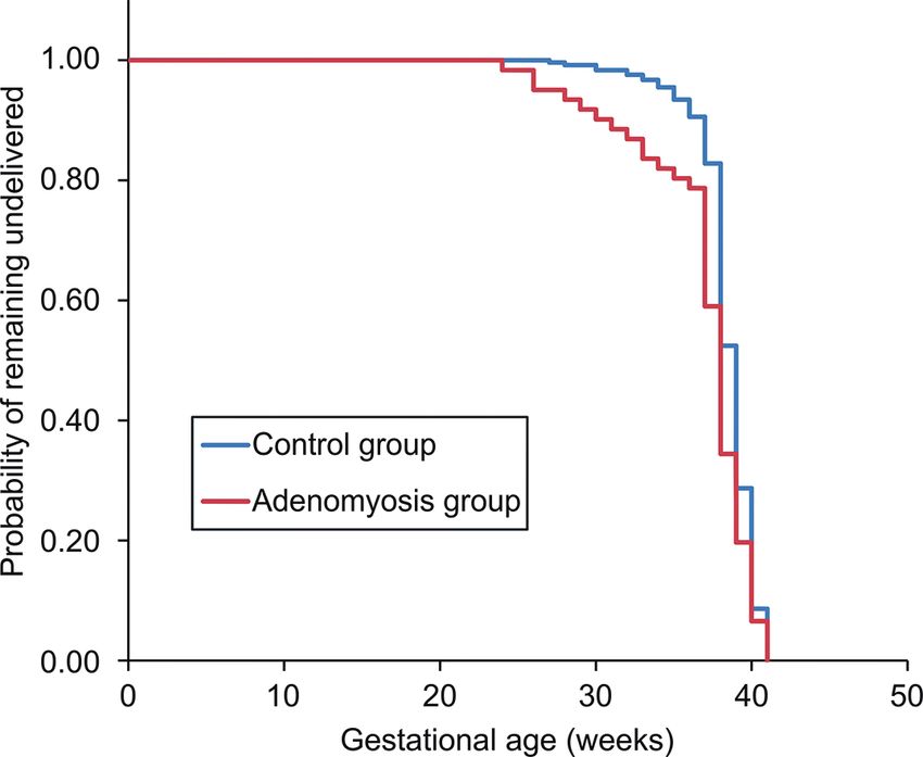
232                                                     S. Shinohara et al.

     Adenomyosis can be classified into the following two           (Sakhel and Abuhamad 2012).
categories: (1) diffuse adenomyosis, the extensive form of                Patients with a coexisting uterine myoma were
the disease characterized by foci of endometrial mucosa             included if they satisfied the aforementioned diagnostic cri-
scattered throughout the uterine musculature; and (2) focal         teria for adenomyosis. Transvaginal USG findings were
adenomyosis, in which the area of the hypertrophic and dis-         used to identify the type (focal or diffuse) of uterine adeno-
torted endometrium and myometrium is restricted (Byun et            myosis based on a previous study that reported that non-
al. 1999; Bergeron et al. 2006; Tamura et al. 2017).                invasive diagnosis of adenomyosis was possible with
However, little attention has been paid to the association          sufficiently high accuracy using both transvaginal USG and
between the specific type of adenomyosis and pregnancy              MRI and that the difference in accuracy between the two
outcomes.                                                           methods was not significant (Champaneria et al. 2010).
     Therefore, we performed a multicenter case–control             Additionally, MRI is less widely available, more expensive,
study to clarify the potential adverse effects of adenomyosis       and not well-tolerated by all patients compared to USG. In
on pregnancy and to improve perinatal prognosis. The pri-           this study, MRI was performed in only six patients.
mary objective of this multicenter case-control study was to              The control group was selected from among 2,331
investigate the effect of adenomyosis on pregnancy compli-          women who did not meet the exclusion criteria (a history of
cations and outcomes. Our secondary objective was to ana-           surgery for uterine myoma or adenomyosis, uterine malfor-
lyze the association between the type of adenomyosis and            mation, and multiple gestations), underwent transvaginal
pregnancy outcomes.                                                 USG in the first trimester, and were confirmed to be free of
                                                                    adenomyosis with uterine enlargement. These women were
                 Materials and Methods                              randomly selected from those with singleton pregnancies
Study design and population                                         who delivered after 22 weeks’ gestation at the University of
     In this case–control study, data on women with single-         Yamanashi Hospital, Yamanashi Prefectural Hospital, Kofu
ton pregnancies who delivered after 22 weeks’ gestation             Municipal Hospital, Kofu-Kyoritsu Hospital, National
and had adenomyosis were obtained from the records at the           Hospital Organization Kofu National Hospital, and
University of Yamanashi Hospital, Yamanashi Prefectural             Fujiyoshida Municipal Medical Center between January
Hospital, Kofu Municipal Hospital, Kofu-Kyoritsu                    2008 and February 2019. In particular, when selecting each
Hospital, National Hospital Organization Kofu National              participant for the control group from among the 2,331
Hospital, and Fujiyoshida Municipal Medical Center                  women, we used propensity score (PS) matching to adjust
between January 2008 and February 2019. Magnetic reso-              for potential confounders, including maternal age at deliv-
nance imaging (MRI) and transvaginal ultrasonography                ery, parity, and use of assisted reproductive technology
(USG) are highly accurate diagnostic tools for detecting            (ART), because these factors have been shown to be con-
adenomyosis (MRI: sensitivity, 70.0-89.0%; specificity,             founding factors for several obstetric complications
86.0-92.5%, transvaginal USG: sensitivity, 72.0-86.0%;              (Shevell et al. 2005; Takemura et al. 2013; Toshimitsu et al.
specificity, 81.0-92.8%) (Reinhold et al. 1996; Bazot et al.        2014). For this study, the following three control groups
2001; Champaneria et al. 2010). The inclusion criteria for          were set up: control A, for which 61 pregnant women com-
adenomyosis consisted of obvious uterine enlargement and            plicated with adenomyosis; control B, for which 41 preg-
the presence of specific features on MRI or transvaginal            nant women complicated with diffuse adenomyosis; and
USG before or early in pregnancy.                                   control C, for which 20 pregnant women complicated with
     The MRI criteria for diagnosing adenomyosis included           focal adenomyosis.
(1) a myometrial mass with indistinct margins of primarily                All procedures in this study were conducted in accor-
low signal intensity or (2) diffuse or focal thickening of the      dance with the ethical standards of the Human Subjects
junctional zone with formation of an ill-defined area of low        Review Committees of the University of Yamanashi
signal intensity on T2-weighted images (Dueholm and                 Hospital (reference number: 1881) and in conformance with
Lundorf 2007; Hashimoto et al. 2018). The transvaginal              the guidelines of the Declaration of Helsinki, as revised in
USG criteria for diagnosing adenomyosis included (1) myo-           Tokyo 2004. The study protocol was reviewed and
metrial anterior-posterior asymmetry and/or (2) thickening          approved by the Human Subjects Review Committees of
of the anterior and posterior myometrial walls, with either         the University of Yamanashi Hospital, Yamanashi
increased or decreased echogenicity (Bazot et al. 2001;             Prefectural Hospital, Kofu Municipal Hospital, Kofu-
Hashimoto et al. 2018). In addition to these criteria, the          Kyoritsu Hospital, National Hospital Organization Kofu
transvaginal USG criteria for diagnosing diffuse adenomyo-          National Hospital, and Fujiyoshida Municipal Medical
sis included no distinction of the endometrial-myometrial           Center. Informed consent was not obtained from patients
junction and subendometrial myometrial striations (Hanafi           owing to the retrospective study design, and patient ano-
2013). The transvaginal USG criteria for diagnosing focal           nymity was preserved. However, patients were provided
adenomyosis included focal area of myometrical thicken-             with the opportunity to refuse the use of their data through
ing, which presented as a heterogeneous mildly echogenic            the university’s website.
focal nodule with indistinct margins and cystic spaces
Adenomyosis and Pregnancy Outcomes 233
Data collection and definitions of variables to test for statistical differences in the gestational age at
The baseline demographic data were collected from delivery between the diffuse and focal adenomyosis groups.
the medical records of the aforementioned six hospitals. Finally, the Mann-Whitney U test was used to analyze con-
The demographic and medical data included maternal age tinuous variables, and the chi-square test (or Fisher’s exact
at delivery, pregestational weight, gestational age, parity, test when the expected frequency was < 5) was used to ana-
delivery method (vaginal or cesarean delivery), and use of lyze categorical variables, such as maternal age and the
ART (in vitro fertilization or intracytoplasmic sperm injec- incidence of obstetrical complications, between the diffuse
tion). In addition, HDP, preterm delivery, cesarean deliv- adenomyosis group and control group B and between the
ery, gestational diabetes mellitus (GDM), pPROM, placen- focal adenomyosis group and control group C. The signifi-
tal malposition, fetal malpresentation, postpartum cance level was set at P < 0.05. All analyses were per-
hemorrhage (PPH), and small for gestational age (SGA) formed using Bell Curve for Excel (Social Survey Research
infants were considered obstetric complications. Information Co., Ltd, Tokyo) and IBM SPSS Statistics 25
Gestational age was determined based on the mother’s last (IBM Corp., Armonk, NY).
menstrual period. If gestational age according to the moth-
er’s last menstrual period differed from that determined Results
based on USG at < 11 weeks by more than 7 days, the latter Effects of adenomyosis on pregnancy outcomes
was used to calculate gestational age. In the first case-control study, the data for 61 women
When we compared the frequency of cesarean deliv- with adenomyosis and 244 women without adenomyosis
ery, we excluded cesarean deliveries performed owing to a (control group A) were extracted after matching for age,
previous cesarean delivery or previous uterine surgery in parity, and ART. Table 1 provides an overview of the base-
the case and control groups because in such cases, cesarean line characteristics of the adenomyosis and control groups.
deliveries were performed with or without perinatal compli- Maternal age (35.2 ± 4.5 years vs. 35.2 ± 4.6 years, P =
cations. HDP was defined as blood pressure ≥ 140/90 0.91) and the rates of nulliparity (34.4% vs. 40.2%, P =
mmHg on at least two occasions during pregnancy 0.42) and ART (32.8% vs. 33.2%, P = 0.95) were similar in
(Ohkuchi et al. 2017). Moreover, severe HDP was defined both groups. Other characteristics between the adenomyo-
as blood pressure ≥ 160/110 mmHg (Ohkuchi et al. 2017). sis and control groups were similar, except for birth weight;
GDM was diagnosed if at least one abnormal plasma glu- the median gestational age at delivery was lower in the ade-
cose value (≥ 92, 180, and 153 mg/dl for fasting, one-hour, nomyosis group (36.8 ± 3.9 weeks vs. 38.4 ± 2.0 weeks, P
and two-hour plasma glucose concentrations, respectively) = 0.001; Table 2).
was noted after a 75-g oral glucose tolerance test (Wendland Kaplan-Meier analysis (Fig. 1) showed a significant
et al. 2012). Placental malposition was defined as placenta difference in the gestational duration between the adenomy-
previa or a low-lying placenta. Fetal malpresentation was osis and control groups (log-rank test, P = 0.03). The inci-
defined as any fetal presentation other than cephalic presen- dence of obstetrical complications and perinatal outcomes
tation. PPH was defined as active bleeding, including are presented in Table 2. Preterm labor was noted for 13 of
amniotic fluid exceeding 500 mL in a vaginal delivery or 61 (21.3%) women in the adenomyosis group and 23 of 244
1000 mL in a cesarean delivery within 24 hours of delivery (9.4%) women in the control group; thus, preterm labor was
(Minakami et al. 2014). SGA was defined as infants with a significantly more frequently noted in the adenomyosis
weight below the 10th percentile in each gestational week group than in the control group (odds ratio [OR], 2.60; 95%
(Itabashi et al. 2010). Pregestational body mass index was confidence interval [CI], 1.23-5.50).
calculated according to the World Health Organization stan- HDP developed in 8 of 61 (13.1%) women in the ade-
dard (bodyweight [kg]/height [m2]). nomyosis group and 13 of 244 (5.3%) women in the control
group; thus, HDP was noted significantly more frequently
Statistical analysis in the adenomyosis group (OR, 2.68; 95% CI, 1.06-6.80).
The Mann-Whitney U test was used to analyze contin- Moreover, the incidence of severe HDP was higher in the
uous variables, such as maternal age, and the chi-square test adenomyosis group (9.8%) than in the control group
(or Fisher’s exact test when the expected frequency was < (3.6%); however, the difference was not statistically signifi-
5) was used to analyze categorical variables, such as the cant (OR, 2.84; 95% CI, 0.97-8.34). In the adenomyosis
incidence of obstetric complications. We used Kaplan- group, 23 of 50 (46.0%) women underwent cesarean deliv-
Meier analysis and the log-rank test for examining the sta- ery; this rate was significantly higher than that in the con-
tistical differences in the gestational age at delivery between trol group (42/201 women [20.9%]; OR, 3.22; 95% CI,
the case group (patients with adenomyosis) and control 1.68-6.19).
group A. Next, to analyze whether different types of adeno- PPH developed in 35 of 61 (57.3%) women in the ade-
myosis affect the pregnancy complications, patients with nomyosis group and 90 of 244 (36.8%) women in the con-
adenomyosis were divided into two groups (focal adeno- trol group; thus, PPH was significantly more frequently
myosis group or diffuse adenomyosis group). noted in the adenomyosis group (OR, 2.30; 95% CI, 1.30-
We used Kaplan-Meier analysis and the log-rank test 4.07). Furthermore, the incidence of PPH was examined234 S. Shinohara et al.
Table 1. Baseline characteristics of the study population.
Adenomyosis group Control group
Variable P value
n = 61 n = 244
Maternal age, years 35.2 ± 4.5 35.2 ± 4.6 0.88
Nulliparity 21 (34.4) 98 (40.2) 0.42
IVF 20 (32.8) 81 (33.2) 0.95
Pre-pregnancy BMI, kg/m2 21.3 ± 2.6 21.5 ± 3.3 0.82
Male sex 31 (50.8) 119 (48.8) 0.86
Birth weight, g 2,639 ± 706.5 2,947 ± 520.0 0.003
Values are presented as mean ± standard deviation or as number (%).
BMI, body mass index; IVF, in vitro fertilization.
Table 2. Comparison of the pregnancy outcomes between the adenomyosis group and control group A.
Adenomyosis group Control group A Odds ratio
P value
n = 61 n = 244 (95% CI)
Preterm labor 13 (21.3) 23 (9.4) 0.01 2.60 (1.23-5.50)
Gestational age at delivery 36.8 ± 3.9 38.4 ± 2.0 0.001 –
HDP 8 (13.1) 13 (5.3) 0.045 2.68 (1.06-6.80)
Severe HDP 6 (9.8) 9 (3.6) 0.09 2.84 (0.97-8.34)
GDM 9 (14.8) 21 (8.6) 0.15 1.83 (0.80-4.25)
pPROM 5 (8.2) 11 (4.5) 0.33 1.89 (0.63-5.66)
Placental malposition 5 (8.2) 8 (3.3) 0.14 2.63 (0.83-8.36)
Fetal malpresentation 5 (8.2) 8 (3.3) 0.14 2.63 (0.83-8.36)
Cesarean delivery† 23/50 (46.0) 42/201 (20.9) < 0.001 3.22 (1.68-6.19)
PPH 35 (57.3) 90 (36.8) 0.004 2.30 (1.30-4.07)
PPH in cesarean delivery† 19/23 (82.6) 15/42 (35.7) < 0.001 8.55 (2.45-29.8)
PPH in vaginal delivery 11/27 (40.7) 60/159 (37.7) 0.76 1.13 (0.49-2.61)
SGA infant 8 (13.1) 24 (9.8) 0.45 1.38 (0.59-3.25)
UA pH 7.31 ± 0.07 7.30 ± 0.06 0.50 –
Values are presented as number (%) or as mean ± standard deviation.
CI, confidence interval; HDP, hypertensive disorders of pregnancy; GDM, gestational diabetes mellitus; pPROM, preterm
premature rupture of membranes; PPH, postpartum hemorrhage; SGA, small for gestational age; UA, umbilical artery.
†Cesarean delivery due to a previous cesarean delivery or previous uterine surgery were excluded.
separately for cesarean delivery and vaginal delivery. In groups were similar, except for gestational age at delivery
cesarean delivery cases, PPH developed in 19 of 23 (82.6%) in the diffuse adenomyosis group. The median gestational
women in the adenomyosis group and 15 of 42 (35.7%) age at delivery was smaller for women in the diffuse adeno-
women in the control group; hence, PPH was significantly myosis group than for those in the focal adenomyosis group
more frequently reported in the adenomyosis group (OR, (35.8 ± 4.3 vs. 38.7 ± 1.4 weeks, P = 0.004).
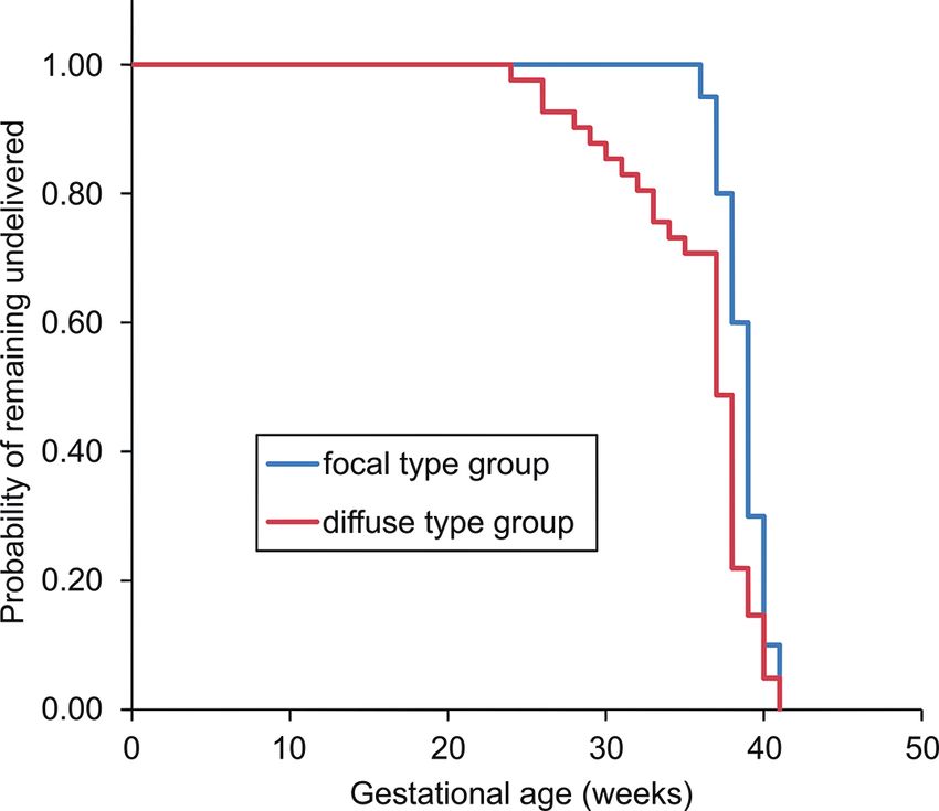
8.55; 95% CI, 2.45-29.8). Conversely, in cases of vaginal Kaplan-Meier analysis (Fig. 2) indicated a significant
delivery, PPH developed in 11 of 27 (40.7%) women in the difference in the gestational duration between the diffuse
adenomyosis group and 60 of 159 (37.7%) women in the and focal adenomyosis groups (log-rank test, P = 0.01). A
control group; there was no difference in the frequency in second case–control analysis (diffuse vs. control B or focal
the adenomyosis group (OR, 1.13; 95% CI, 0.49-2.61). vs. control C) was performed to determine whether differ-
ent types of adenomyosis affect the pregnancy-related com-
Effects of diffuse or focal adenomyosis on pregnancy plications.
outcomes To examine the effects of diffuse adenomyosis on the
Subsequently, the patients were categorized into the pregnancy complications, the data for 41 women with dif-
following two groups according to the type of adenomyo- fuse adenomyosis and 164 controls (group B) were
sis: 41 women with diffuse adenomyosis and 20 women extracted after matching for age, parity, and ART. The
with focal adenomyosis. Table 3 presents the clinical char- maternal age (36.1 ± 3.8 vs. 35.9 ± 3.8, P = 0.76) and rates
acteristics of women with diffuse and focal adenomyosis. of nulliparity (31.7% vs. 32.9%, P = 0.88) and ART (36.6%
The characteristics of the diffuse and focal adenomyosis vs. 35.3%, P = 0.88) were similar in both groups (data notAdenomyosis and Pregnancy Outcomes 235
Fig. 1. Kaplan-Meier analysis of the gestational duration
between the adenomyosis and control groups. Fig. 2. Kaplan-Meier curve for the gestational age between
A significant difference in the gestational duration is not- the focal and diffuse adenomyosis groups.
ed between the adenomyosis (n = 61) and control A (n = A significant difference in the gestational duration is not-
244) groups. ed between the focal (n = 20) and diffuse adenomyosis (n
= 41) groups.
shown). The other characteristics, except for birth weight trol group; thus PPH was significantly more frequently
(2499 ± 779.5 g vs. 2997 ± 487.3 g, P < 0.001), were simi- noted in the adenomyosis group (OR, 16.1; 95% CI, 3.01-
lar between the diffuse adenomyosis group and control 85.6). In the vaginal delivery cases, PPH developed in 7 of
group B. 12 (58.3%) women in the diffuse adenomyosis group and
The diffuse adenomyosis group was significantly more 51 of 111 (45.9%) women in the control group; however,
likely to experience preterm labor (OR, 5.24; 95% CI, 2.15- the differences in frequency were not statistically significant
12.8), HDP (OR, 3.54; 95% CI, 1.23-10.2), severe HDP in the adenomyosis group (OR, 1.64; 95% CI, 0.49-5.50;
(OR, 7.45; 95% CI, 1.70-32.6), pPROM (OR, 5.56; 95% Table 4). Although the difference in the incidence of pla-
CI, 1.42-21.7), cesarean delivery (non-reassuring fetal sta- cental malposition was not statistically significant (OR,
tus, n = 5; placental malposition, n = 4; fetal malpresenta- 3.43; 95% CI, 0.88-13.4), the incidence of placental malpo-
tion, n = 3; intrauterine infection, n = 1; maternal disorders, sition was also greater in the diffuse adenomyosis group
n = 1, labor arrest, n = 1, other causes, n = 4; OR, 6.76; than in the control group (Table 4).
95% CI, 2.92-15.6), and PPH (OR, 3.01; 95% CI, 1.44- Finally, to examine the effects of focal adenomyosis
6.31) than the control group. on the pregnancy complications, the data of 20 women with
PPH development was examined for cesarean delivery focal adenomyosis and 80 controls (group C) were extracted
and vaginal delivery separately. In cesarean delivery cases, after matching for age, parity, and ART. The maternal age
PPH developed in 17 of 19 (89.5%) women in the diffuse (33.6 ± 5.5 vs. 33.6 ± 5.6, P = 1.00) and rates of nulliparity
adenomyosis group and 9 of 26 (34.6%) women in the con- (40.0% vs. 40.0%, P = 1.00) and ART (25.0% vs. 25.0%, P
Table 3. Baseline characteristics of the patients with diffuse and focal adenomyosis.
Patients with diffuse adenomyosis Patients with focal adenomyosis
Variable P value
n = 41 n = 20
Maternal age, years 36.1 ± 3.8 33.6 ± 5.3 0.10
Gestational age at delivery 35.8 ± 4.3 38.7 ± 1.4 0.004
Nulliparity 13 (31.7) 32 (40.0) 0.52
IVF 15 (36.6) 20 (25.0) 0.40
Pre-pregnancy BMI, kg/m2 21.0 ± 2.0 24.3 ± 2.9 0.70
Male sex 24 (58.5) 38 (47.5) 0.06
Birth weight, g 2,499 ± 779.5 2,976 ± 507.8 0.06
Values are presented as mean ± standard deviation or as number (%).
BMI, body mass index; IVF, in vitro fertilization.236 S. Shinohara et al.
Table 4. Comparison of the pregnancy outcomes between the diffuse adenomyosis group and control group B.
Diffuse adenomyosis group Control group B Odds ratio
P value
n = 41 n = 164 (95% CI)
Preterm labor 12 (29.3) 12 (7.3) < 0.001 5.24 (2.15-12.8)
HDP 7 (17.0) 9 (5.5) 0.02 3.54 (1.23-10.2)
Severe HDP 5 (12.2) 3 (1.8) 0.009 7.45 (1.70-32.6)
GDM 7 (17.0) 25 (15.2) 0.78 1.14 (0.46-2.87)
pPROM 5 (12.2) 4 (2.4) 0.02 5.56 (1.42-21.7)
Placental malposition 4 (9.8) 5 (3.0) 0.08 3.43 (0.88-13.4)
Fetal malpresentation 3 (7.3) 9 (5.5) 0.71 1.36 (0.35-5.27)
Cesarean delivery† 19/31 (61.3) 26/137 (18.9) < 0.001 6.76 (2.92-15.6)
PPH 29 (70.7) 73 (44.5) 0.003 3.01 (1.44-6.31)
SGA infant 6 (14.6) 14 (8.5) 0.25 1.84 (0.66-5.12)
PPH in cesarean† delivery 17/19 (89.5) 9/26 (34.6) < 0.001 16.1 (3.01-85.6)
PPH in vaginal delivery 7/12 (58.3) 51/111 (45.9) 0.54 1.64 (0.49-5.50)
UA pH 7.31 ± 0.07 7.30 ± 0.06 0.60 –
Values are presented as mean ± standard deviation or as number (%).
CI, confidence interval; HDP, hypertensive disorders of pregnancy; GDM, gestational diabetes mellitus; pPROM, preterm
premature rupture of membranes; PPH, postpartum hemorrhage; SGA, small for gestational age; UA, umbilical artery.
†Cesarean deliveries due to a previous cesarean delivery or previous uterine surgery were excluded.
Table 5. Comparison of the pregnancy outcomes between the focal adenomyosis group and control group C.
Focal adenomyosis group Control group C
P value Odds ratio (95% CI)
n = 20 n = 80
Preterm labor 1 (5.0) 7 (8.8) 1.00 0.55 (0.06-4.74)
HDP 1 (5.0) 2 (2.5) 0.49 2.05 (0.18-23.8)
Severe HDP 1 (5.0) 0 (0.0) 0.20 –
GDM 2 (10.0) 8 (10.0) 1.00 1.00 (0.20-5.12)
pPROM 0 (0.0) 1 (1.3) 1.00 –
Placental malposition 1 (5.0) 3 (3.8) 1.00 1.35 (0.13-13.7)
Fetal malpresentation 2 (10.0) 3 (3.8) 0.27 2.85 (0.44-18.3)
Cesarean delivery† 4/17 (23.5) 9/69 (13.0) 0.27 2.05 (0.55-7.69)
PPH 6 (30.0) 30 (37.5) 0.61 0.71 (0.25-2.06)
PPH in cesarean† delivery 2/4 (50.0) 4/9 (44.4) 1.00 1.25 (0.12-13.2)
PPH in vaginal delivery 4/13 (30.8) 23/60 (38.3) 0.76 0.71(0.20-2.59)
SGA infant 2 (10.0) 6 (7.5) 0.66 1.37 (0.26-7.36)
UA pH 7.31 ± 0.07 7.31 ± 0.06 0.65 –
Values are presented as mean ± standard deviation or as number (%).
CI, confidence interval; HDP, hypertensive disorders of pregnancy; GDM, gestational diabetes mellitus; pPROM, preterm
premature rupture of membranes; PPH, postpartum hemorrhage; SGA, small for gestational age; UA, umbilical artery.
†Cesarean deliveries due to a previous cesarean delivery or previous uterine surgery were excluded.
= 1.00) were similar in both groups (data not shown). The adenomyosis was associated preterm labor, HDP, severe
incidence of obstetric complications and perinatal outcomes HDP, pPROM, cesarean delivery, and PPH. In contrast,
were not significantly different between the focal adenomy- focal adenomyosis may not be associated with poor preg-
osis group and the control group (Table 5). nancy outcomes. To the best of our knowledge, this is the
first study to clarify the association between adenomyosis,
Discussion especially the diffuse type, and poor pregnancy outcomes in
The main results of this study are, as followed: first, women in a multicenter setting.
women with adenomyosis were more likely to be associated An increase in the incidence of HDP in the adenomyo-
with preterm labor, HDP, cesarean delivery, and PPH. sis group was an important finding. During pregnancy,
Second, among women with uterine adenomyosis, diffuse invasion of the trophoblasts in the endometrium and myo-Adenomyosis and Pregnancy Outcomes 237
metrial junctional zone induces decidualization and unique more common in the adenomyosis group than in the control
vascular changes. Impaired decidualization of the myome- group.
trial spiral arteries is a predisposing factor for failed intra- Another important finding was the increased incidence
vascular trophoblast invasion (Brosens et al. 2010, 2013; of PPH in the adenomyosis group. The presence of adeno-
Tamura et al. 2017). Defective deep placentation has been myosis could have impaired the functionality of the gravid
associated with HDP (Brosens et al. 2010, 2013; Tamura et uterus, thereby, increasing uterine atony and leading to PPH
al. 2017). Our result showing an increased risk of HDP in development (Vlahos et al. 2017). In addition, the high fre-
pregnant women with adenomyosis may be due to the detri- quency of HDP, which is a risk factor for PPH (von Schmidt
mental effects of adenomyosis on this process. Although auf Altenstadt et al. 2013; Minakami et al. 2014), in the
previous studies have clarified the association between HDP adenomyosis group may be responsible for this result. In
and SGA infants (Verlohren et al. 2012; Figueras and particular, the amount of blood loss during cesarean deliv-
Gratacós 2014), our study showed no significant difference ery was significantly higher in the adenomyosis group than
in the SGA infant rates between the adenomyosis and con- in the control group (1203 ± 493 mL vs. 936 ± 509 mL, P =
trol groups. However, a type II error may have occurred 0.008; data not shown). Therefore, it may be important to
because of the sample size. prepare for autologous blood transfusion in the late third
Another important finding of our study was that cesar- trimester for women with adenomyosis, and at the time of
ean delivery was significantly more common in the adeno- cesarean section, it may be effective to consider using Bakri
myosis group than in the control group. Several studies balloon tamponade since the Bakri device is more effective
have reported that the possibility of cesarean delivery in managing postpartum hemorrhage, if inserted early after
increases in women with adenomyosis (Mochimaru et al. delivery (Vintejoux et al. 2015).
2015; Hashimoto et al. 2018). The following reasons may To date, previous studies have not evaluated whether
be considered. First, placental malposition was more fre- the type of adenomyosis affects the pregnancy complica-
quently observed in women with adenomyosis than in those tions. According to previous reports, diffuse adenomyosis
without. A plausible mechanism is that the adenomyosis is more common than the focal type. In a study, the preva-
lesion in the uterine body has a detrimental effect on the lence of diffuse and focal types of adenomyosis was 81.7%
normal implantation process and disturbs the implantation and 18.3%, respectively (Sofic et al. 2016). In another
site, resulting in the development of placental malposition study, the prevalence was 66.7% and 33.3%, respectively
(Hashimoto et al. 2018). (Byun et al. 1999). In the present study, 65.6% (40/61) of
Second, several studies have reported that pregnant pregnancies involved diffuse adenomyosis. Previous stud-
women with adenomyosis had higher rates of pPROM than ies claimed that adenomyosis is associated with poor preg-
those without (Juang et al. 2007; Mochimaru et al. 2015). nancy outcomes (Juang et al. 2007; Mochimaru et al. 2015;
Prostaglandin has been implicated as a risk factor for Tamura et al. 2017; Hashimoto et al. 2018). Here, we dem-
pPROM as it causes uterine irritability and collagen degra- onstrated that uterine adenomyosis, especially the diffuse
dation within the fetal membranes (Tjugum and Norström type, was associated with high-risk perinatal cases.
1985; Juang et al. 2007). Previous studies have reported an Considering the increased incidence of HDP, pPROM,
increased level of prostaglandin in women with adenomyo- placental malposition, fetal malpresentation, and PPH,
sis (Koike et al. 1994; Juang et al. 2007). Although the women with diffuse adenomyosis, which is marked by
underlying pathophysiological pathways should be investi- widespread lesions compared to the focal type, may be
gated, prostaglandin may play a role in the association prone to developing pregnancy complications. We believe
between adenomyosis and pPROM. As the number of cases that this study provides obstetricians with useful informa-
of intrauterine infection and fetal distress owing to pPROM tion for improving prenatal management and counseling for
have increased, the frequency of cesarean deliveries in the patients with adenomyosis in the clinical setting.
preterm period increased. There are certain limitations of this study. First, it may
Third, cesarean delivery as the indication of fetal mal- be difficult to extrapolate our results to the general popula-
presentation was more common in women with adenomyo- tion because of the relatively small sample size. Therefore,
sis than in those without. This is because adenomyosis nar- a large-scale multicenter prospective cohort study is
rows the intrauterine cavity and decreases the uterine required to confirm these results in the general population.
extensibility, like a uterine myoma (Mochimaru et al. 2015). Second, the data on gestational weight gain, intake of alco-
We found no significant differences with respect to the pla- hol and caffeine, antiphospholipid, thyroid disease, family
cental malposition, pPROM, and fetal malpresentation history, and socioeconomic status, which may affect the
between the adenomyosis and control groups. However, as pregnancy complications, were not considered in this study
with HDP, a type II error may have occurred because of the (Harita et al. 2012; Räisänen et al. 2013; Minakami et al.
sample size. Women in the adenomyosis group tended to 2014). The women in the present study may have been
have higher complication rates than those in the control affected by the aforementioned risk factors.
groups; however, the difference was not significant. Owing Although some limitations exist, the strength of this
to this tendency, we concluded that cesarean delivery was study is the selection of case and control groups from mul-238 S. Shinohara et al.
tiple institutions to reduce the effect of selection bias. This Hashimoto, A., Iriyama, T., Sayama, S., Nakayama, T., Komatsu,
is the first study to examine the causality between adeno- A., Miyauchi, A., Nishii, O., Nagamatsu, T., Osuga, Y. & Fujii,
T. (2018) Adenomyosis and adverse perinatal outcomes:
myosis and pregnancy in a multicenter case-control study. increased risk of second trimester miscarriage, preeclampsia,
In conclusion, although studies with a larger sample and placental malposition. J. Matern. Fetal Neonatal Med.,
size are required, it appears that adenomyosis, especially 31, 364-369.
the diffuse type, is significantly associated with perinatal Itabashi, K., Fujimura, M. & Kusuda, S. (2010) Introduction of
new neonatal standard anthropometric measurements. Nihon
adverse outcomes. Our findings provide valuable evidence Shonika Gakkai Zasshi, 114, 1271-1293.
that women with diffuse adenomyosis require more careful Juang, C.M., Chou, P., Yen, M.S., Twu, N.F., Horng, H.C. & Hsu,
perinatal management than previously thought. W.L. (2007) Adenomyosis and risk of preterm delivery.
BJOG, 114, 165-169.
Acknowledgments Koike, H., Ikenoue, T. & Mori, N. (1994) Studies on prostaglandin
production relating to the mechanism of dysmenorrhea in
We thank the study subjects for allowing the use of endometriosis. Nihon Naibunpi Gakkai Zasshi, 70, 43-56.
their personal data. We would also like to thank Editage Kunz, G., Herbertz, M., Beil, D., Huppert, P. & Leyendecker, G.
(www.editage.com) for English language editing. The (2007) Adenomyosis as a disorder of the early and late human
authors did not receive a specific grant for this research reproductive period. Reprod. Biomed. Online, 15, 681-685.
Minakami, H., Maeda, T., Fujii, T., Hamada, H., Iitsuka, Y.,
from any funding agency in the public, commercial, or not- Itakura, A., Itoh, H., Iwashita, M., Kanagawa, T., Kanai, M.,
for-profit sectors. Kasuga, Y., Kawabata, M., Kobayashi, K., Kotani, T., Kudo,
Y., et al. (2014) Guidelines for obstetrical practice in Japan:
Conflict of Interest Japan Society of Obstetrics and Gynecology (JSOG) and
Japan Association of Obstetricians and Gynecologists (JAOG)
The authors declare no conflict of interest. 2014 edition. J. Obstet. Gynaecol. Res., 40, 1469-1499.
Mochimaru, A., Aoki, S., Oba, M.S., Kurasawa, K., Takahashi, T.
References & Hirahara, F. (2015) Adverse pregnancy outcomes associ-
Bazot, M., Cortez, A., Darai, E., Rouger, J., Chopier, J., Antoine, ated with adenomyosis with uterine enlargement. J. Obstet.
J.M. & Uzan, S. (2001) Ultrasonography compared with Gynaecol. Res., 41, 529-533.
magnetic resonance imaging for the diagnosis of adenomyosis: Ohkuchi, A., Hirashima, C., Takahashi, K., Suzuki, H. &
correlation with histopathology. Hum. Reprod., 16, 2427- Matsubara, S. (2017) Prediction and prevention of hyperten-
2433. sive disorders of pregnancy. Hypertens. Res., 40, 5-14.
Bergeron, C., Amant, F. & Ferenczy, A. (2006) Pathology and Räisänen, S., Gissler, M., Sankilampi, U., Saari, J., Kramer, M.R.
physiopathology of adenomyosis. Best Pract. Res. Clin. & Heinonen, S. (2013) Contribution of socioeconomic status
Obstet. Gynaecol., 20, 511-521. to the risk of small for gestational age infants: a population-
Brosens, I., Derwig, I., Brosens, J., Fusi, L., Benagiano, G. & based study of 1,390,165 singleton live births in Finland. Int.
Pijnenborg, R. (2010) The enigmatic uterine junctional zone: J. Equity Health, 12, 28.
the missing link between reproductive disorders and major Reinhold, C., McCarthy, S., Bret, P.M., Mehio, A., Atri, M.,
obstetrical disorders? Hum. Reprod., 25, 569-574. Zakarian, R., Glaude, Y., Liang, L. & Seymour, R.J. (1996)
Brosens, I., Pijnenborg, R. & Benagiano, G. (2013) Defective Diffuse adenomyosis: comparison of endovaginal US and MR
myometrial spiral artery remodelling as a cause of major imaging with histopathologic correlation. Radiology, 199,
obstetrical syndromes in endometriosis and adenomyosis. 151-158.
Placenta, 34, 100-105. Sakhel, K. & Abuhamad, A. (2012) Sonography of adenomyosis.
Byun, J.Y., Kim, S.E., Choi, B.G., Ko, G.Y., Jung, S.E. & Choi, J. Ultrasound Med., 31, 805-808.
K.H. (1999) Diffuse and focal adenomyosis: MR imaging Shevell, T., Malone, F.D., Vidaver, J., Porter, T.F., Luthy, D.A.,
findings. Radiographics, 19, S161-170. Comstock, C.H., Hankins, G.D., Eddleman, K., Dolan, S.,
Champaneria, R., Abedin, P., Daniels, J., Balogun, M. & Khan, Dugoff, L., Craigo, S., Timor, I.E., Carr, S.R., Wolfe, H.M.,
K.S. (2010) Ultrasound scan and magnetic resonance imaging Bianchi, D.W., et al. (2005) Assisted reproductive technology
for the diagnosis of adenomyosis: systematic review and pregnancy outcome. Obstet. Gynecol., 106, 1039-1045.
comparing test accuracy. Acta Obstet. Gynecol. Scand., 89, Sofic, A., Husic-Selimovic, A., Carovac, A., Jahic, E., Smailbe-
1374-1384. govic, V. & Kupusovic, J. (2016) The significance of MRI
Dueholm, M. & Lundorf, E. (2007) Transvaginal ultrasound or evaluation of the uterine junctional zone in the early diagnosis
MRI for diagnosis of adenomyosis. Curr. Opin. Obstet. of adenomyosis. Acta Inform. Med., 24, 103-106.
Gynecol., 19, 505-512. Takemura, Y., Osuga, Y., Fujimoto, A., Oi, N., Tsutsumi, R.,
Figueras, F. & Gratacós, E. (2014) Update on the diagnosis and Koizumi, M., Yano, T. & Taketani, Y. (2013) Increased risk of
classification of fetal growth restriction and proposal of a placenta previa is associated with endometriosis and tubal
stage-based management protocol. Fetal Diagn. Ther., 36, factor infertility in assisted reproductive technology preg-
86-98. nancy. Gynecol. Endocrinol., 29, 113-115.
Hanafi, M. (2013) Ultrasound diagnosis of adenomyosis, leio- Tamura, H., Kishi, H., Kitade, M., Asai-Sato, M., Tanaka, A.,
myoma, or combined with histopathological correlation. J. Murakami, T., Minegishi, T. & Sugino, N. (2017) Complica-
Hum. Reprod. Sci., 6, 189-193. tions and outcomes of pregnant women with adenomyosis in
Harada, T., Khine, Y.M., Kaponis, A., Nikellis, T., Decavalas, G. & Japan. Reprod. Med. Biol., 16, 330-336.
Taniguchi, F. (2016) The impact of adenomyosis on women’s Tjugum, J. & Norström, A. (1985) The influence of prostaglandin
fertility. Obstet. Gynecol. Surv., 71, 557-568. E2 and oxytocin on the incorporation of [3H]proline and [3H]
Harita, N., Kariya, M., Hayashi, T., Sato, K.K., Aoki, T., Naka- glucosamine in the human amnion. Eur. J. Obstet. Gynecol.
mura, K., Endo, G. & Narimoto, K. (2012) Gestational body- Reprod. Biol., 19, 137-143.
weight gain among underweight Japanese women related to Toshimitsu, M., Nagamatsu, T., Nagasaka, T., Iwasawa-Kawai, Y.,
small-for-gestational-age birth. J. Obstet. Gynaecol. Res., 38, Komatsu, A., Yamashita, T., Osuga, Y. & Fujii, T. (2014)
1137-1144. Increased risk of pregnancy-induced hypertension and opera-Adenomyosis and Pregnancy Outcomes 239
tive delivery after conception induced by in vitro fertilization/ Biomed Res. Int., 2017, 5926470.
intracytoplasmic sperm injection in women aged 40 years and von Schmidt auf Altenstadt, J.F., Hukkelhoven, C.W., van Roos-
older. Fertil. Steril., 102, 1065-1070. e1. malen, J. & Bloemenkamp, K.W. (2013) Pre-eclampsia
Verlohren, S., Stepan, H. & Dechend, R. (2012) Angiogenic increases the risk of postpartum haemorrhage: a nationwide
growth factors in the diagnosis and prediction of pre- cohort study in the Netherlands. PLoS One, 8, e81959.
eclampsia. Clin. Sci. (Lond.), 122, 43-52. Wendland, E.M., Torloni, M.R., Falavigna, M., Trujillo, J., Dode,
Vintejoux, E., Ulrich, D., Mousty, E., Masia, F., Marès, P., de M.A., Campos, M.A., Duncan, B.B. & Schmidt, M.I. (2012)
Tayrac, R. & Letouzey, V. (2015) Success factors for Bakri Gestational diabetes and pregnancy outcomes: a systematic
balloon usage secondary to uterine atony: a retrospective, review of the World Health Organization (WHO) and the
multicentre study. Aust. NZ J. Obstet. Gynaecol., 55, 572-577. International Association of Diabetes in Pregnancy Study
Vlahos, N.F., Theodoridis, T.D. & Partsinevelos, G.A. (2017) Groups (IADPSG) diagnostic criteria. BMC Pregnancy Child-
Myomas and adenomyosis: impact on reproductive outcome. birth, 12, 23.You can also read